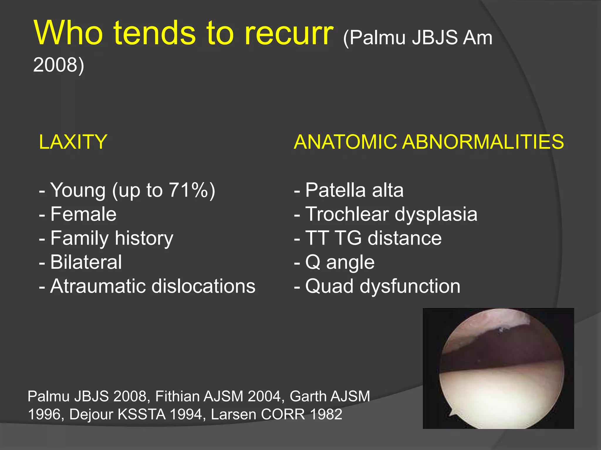
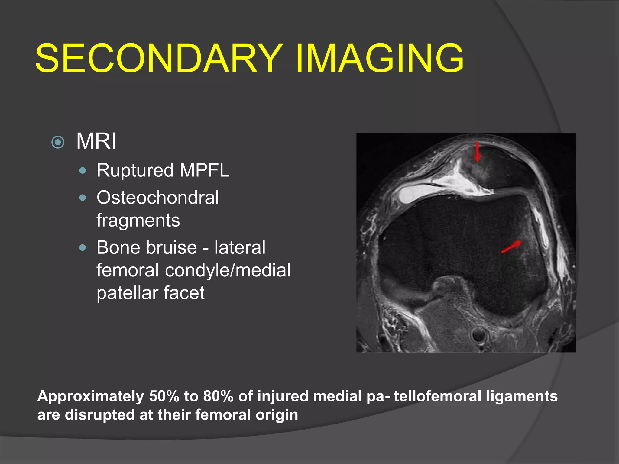
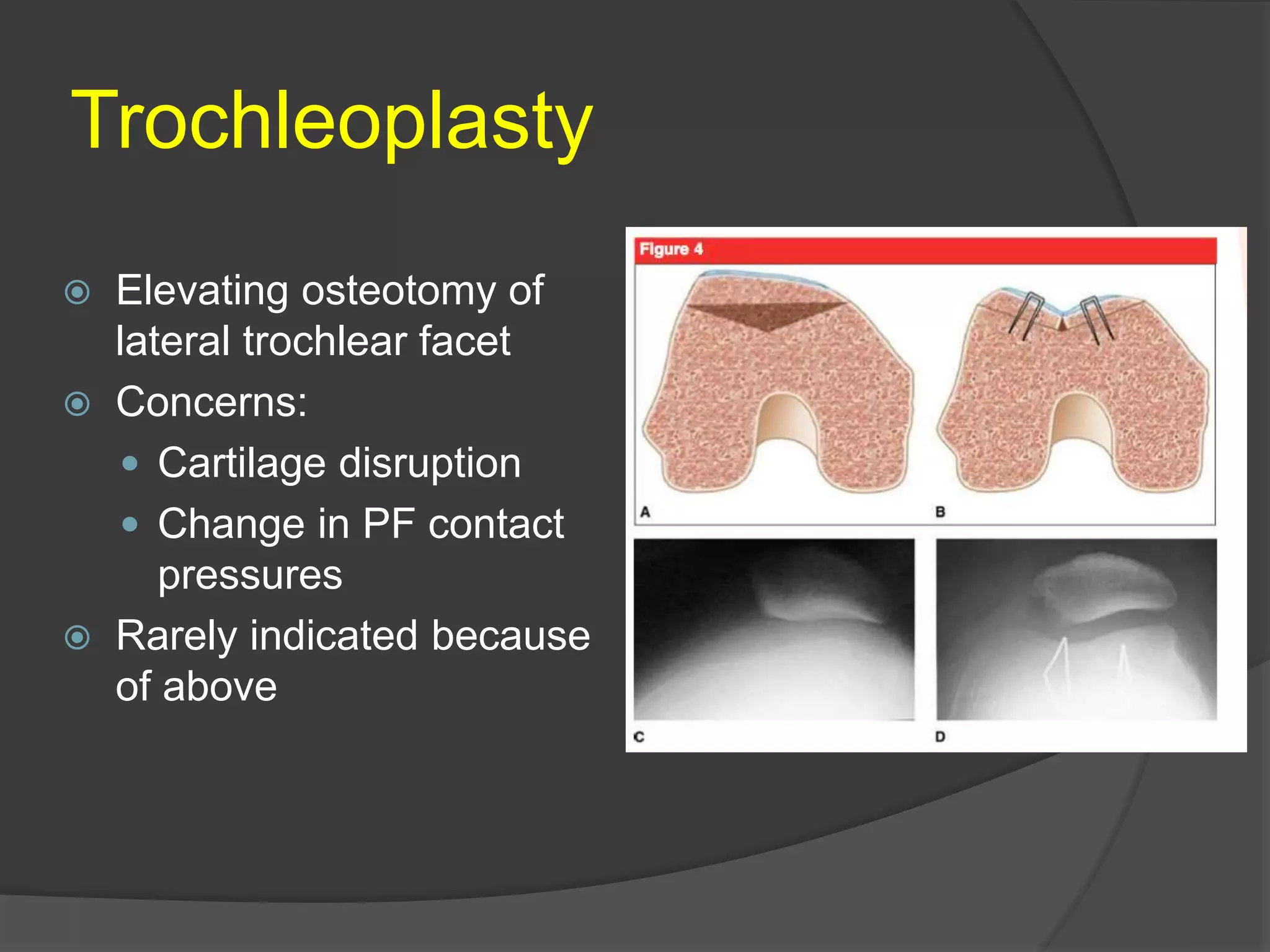
Gonzalo Samitier MD
DIAGNOSIS
 LUX or SUBLUX PATELLOFEMORAL
 PAIN wo INSTABILITY
DIAGNOSIS
1st EPISODE (15-44% recurrence Hawkins RJ
AJSM 1986)
 TRAUMATIC (20% recurrence Cash and
Hughston AJSM 1988 )
 ATRAUMATIC (43% recurrence Cash and
Hughston AJSM 1988)
 COMBINATION
RECURRENT (50% recurrence chance if
second episode Fithian AJSM 2004)
Surgical treatment??
Surgical treatment
ETIOLOGY
- Patella alta
- Throclear dysplasia
- Alignment
- Rotational (Torsion). Femoral and tibial
- Genu Valgum
- Q angle
- Hyperlaxity
- Muscular weakness (VMO)
- Tight lateral structures
- Traumatic MPFL insufficiency
- Iatrogenic
Who tends to recurr (Palmu JBJS Am
2008)
LAXITY
- Young (up to 71%)
- Female
- Family history
- Bilateral
- Atraumatic dislocations
ANATOMIC ABNORMALITIES
- Patella alta
- Trochlear dysplasia
- TT TG distance
- Q angle
- Quad dysfunction
Palmu JBJS 2008, Fithian AJSM 2004, Garth AJSM
1996, Dejour KSSTA 1994, Larsen CORR 1982
IMPORTANT ANTOMY
 TROCHLEAR GROOVE (primary bony stabilizer)
 MEDIAL PATELLOFEMORAL
LIGAMENT (primary soft tissue stabilizer) 60% lateral
restraint (Desio AJSM 1998)
 QUADRICEPS (VMO) (Dynamic stabilizer)
PHYSICAL EXAM
- Gait
- Standing alignment
- Q angle
- J sign (indicates patela alta)
- Laxity (N less than 50% or 2 Q Carson et al Clin Orthop
1984)
- Prone rotational alignment (femoral anteversion
and tibial torsion N<20º)
Others: patelar tilt, aprehension test, grinding,
ROM and tracking
IMAGING
- Patella alta
- Patellar tilt
- Patelar morphology
- Trochlear dysplasia
IMAGING
- Patella alta
- Patellar tilt
- Patelar morphology
- Trochlear dysplasia
(Dejour et al. Sport Med Arthroscopy
2007)
SECONDARY IMAGING
CT Scan
TT-TG distance
(Tibial tubercle -
trochlear groove >20mm
abnormal
Hip to ankle long
standing film
CT Torsional profile
 MRI
 Ruptured MPFL
 Osteochondral
fragments
 Bone bruise - lateral
femoral condyle/medial
patellar facet
SECONDARY IMAGING
Approximately 50% to 80% of injured medial pa- tellofemoral ligaments
are disrupted at their femoral origin
TREATMENT
 CONS
 SURGICAL
Non-operative Treatment
 PT
 Stretching
 Strengthening (esp VMO and Gluteal)
 Closed chain/WB (Stensdotter et al. Escamilla et al.)
 Weight loss (reduce PF loads)
 Inmobilization vs Bracing (Palumbo)
Maenpaa and Lehto (AJSM 1997),
Operative Treatment
- Soft tissue procedures:
- Lateral release
- Medial repair
- Proximal realignment
- MPFL reconstruction
Bone Procedures
- Trochleoplasty
- Distal Femoral Osteotomies
- Distal realignment
- Rotational Osteotomies
Operative Treatment
- Soft tissue procedures:
- Lateral release
- Medial repair
- Proximal realignment
- MPFL reconstruction
Bone Procedures
- Trochleoplasty
- Distal Femoral Osteotomies
- Distal realignment
- Rotational Osteotomies
Lateral release
Not recommended to be done in isolation for instability
- Contribute to 10% of the restraining force to lateral translation
(Desio et al AJSM 1998)
- Results decline from initial 80% to 29%-74% satisfactory rating
at 4 years (LattermannC et al. Med Arthroscopy 2007)
Medial repair
- Used often in the acute setting. Open or
arthroscopic
- Doesn’t address tears of the femoral side
- Limitations: can cause over medialization and tilt
- Prospective Studies: No difference medial vs non
op treatment (Nikku Acta Orthop 2005, 1997, Palmu JBJS 2008,
Sillanpaa PJ AJSM 2008)
- Cases series showing acceptable results
(Ali S Arthroscopy 2007, SchottlePG Arthroscopy 2006, HallbruchtJL
Arthroscopy 2001, Ahmad CS AJSM 2000, Boring TH CORR 1978
MPFL Reconstruction
- Used for incopetent medial structures
- Goals: re-create MPFL anatomy
- Re-establish a stable checkrein 0-30º
- Concerns: overtightening, patella fracture
- Controversial (graft choice, graft fixation, graft positioning,
Graft tension, Dynamic reconstruction)
Systematic review Bucket C et al. AJSM
2009
14 studies including Level III/IV
Stability and Clinical Outcomes encouraging
but current studies are small
Donald C Fithian Dr., MD
Kaiser Permanente Med Ctr Ortho
Operative Treatment
- Soft tissue procedures:
- Lateral release
- Medial repair
- Proximal realignment
- MPFL reconstruction
Bone Procedures
- Trochleoplasty
- Distal Femoral Osteotomies
- Distal realignment
- Rotational Osteotomies
Trochleoplasty
 Elevating osteotomy of
lateral trochlear facet
 Concerns:
 Cartilage disruption
 Change in PF contact
pressures
 Rarely indicated because
of above
Fulkerson Procedure
(Anteromedial Tibial Tubercle AMZ
Transfer)
 Hallmark indication: Increased TT-TG distance
 Corrects Q angle by medializing tubercle
(~0.5-1cm)
 Used only when patella not tracking in center
of trochlea
 Tubercle move anterior (~1cm) as well to
unload PF joint and move point of contact
proximal throughout flexion (distal pole of
patella common source of pain)
 Re-attached with multiple screw fixation
 NWB 6-8 weeks
PATELLAR INSTABILITY
ALGORITHM
Severe Alignment Abnormalities
Genu Valgum
- Distal Femoral OT
- Guided Growth
Torsion >20º above normal
- Femoral rotational OT >35º
- Tibial rotational OT 40º
Increased TT-TG >20mm
Q Angle > 15-20º
Distal Re-Alignment
- If Alta then distalize
Incompetent Medial
restraint
MPFL Reconstruction
IF UNSUCCESSFUL, LOOK FOR ABNORMALITIES NOT INITIALLY APPRECIATED
BOTH?
NOYES
CONCLUSIONS
 There are multiple causes for Patellofemoral
instability
 Good evidence for the non-operative treatment of
an acute patellar dislocation, most of the current
surgical treatments for chronic patellar instability
are based on Level-IV evidence
 Customize your treatment based in the problem
 Be familiar with MPFL reconstruction technique
 Tubercle osteotomy should not be performed if
there is associated medial or proximal patellar
chondrosis
 Watch the alignment
Gracias …

Patellofemoral instability

  • 1.
  • 2.
    DIAGNOSIS  LUX orSUBLUX PATELLOFEMORAL  PAIN wo INSTABILITY
  • 3.
    DIAGNOSIS 1st EPISODE (15-44%recurrence Hawkins RJ AJSM 1986)  TRAUMATIC (20% recurrence Cash and Hughston AJSM 1988 )  ATRAUMATIC (43% recurrence Cash and Hughston AJSM 1988)  COMBINATION RECURRENT (50% recurrence chance if second episode Fithian AJSM 2004) Surgical treatment?? Surgical treatment
  • 4.
    ETIOLOGY - Patella alta -Throclear dysplasia - Alignment - Rotational (Torsion). Femoral and tibial - Genu Valgum - Q angle - Hyperlaxity - Muscular weakness (VMO) - Tight lateral structures - Traumatic MPFL insufficiency - Iatrogenic
  • 5.
    Who tends torecurr (Palmu JBJS Am 2008) LAXITY - Young (up to 71%) - Female - Family history - Bilateral - Atraumatic dislocations ANATOMIC ABNORMALITIES - Patella alta - Trochlear dysplasia - TT TG distance - Q angle - Quad dysfunction Palmu JBJS 2008, Fithian AJSM 2004, Garth AJSM 1996, Dejour KSSTA 1994, Larsen CORR 1982
  • 6.
    IMPORTANT ANTOMY  TROCHLEARGROOVE (primary bony stabilizer)  MEDIAL PATELLOFEMORAL LIGAMENT (primary soft tissue stabilizer) 60% lateral restraint (Desio AJSM 1998)  QUADRICEPS (VMO) (Dynamic stabilizer)
  • 7.
    PHYSICAL EXAM - Gait -Standing alignment - Q angle - J sign (indicates patela alta) - Laxity (N less than 50% or 2 Q Carson et al Clin Orthop 1984) - Prone rotational alignment (femoral anteversion and tibial torsion N<20º) Others: patelar tilt, aprehension test, grinding, ROM and tracking
  • 8.
    IMAGING - Patella alta -Patellar tilt - Patelar morphology - Trochlear dysplasia
  • 9.
    IMAGING - Patella alta -Patellar tilt - Patelar morphology - Trochlear dysplasia (Dejour et al. Sport Med Arthroscopy 2007)
  • 10.
    SECONDARY IMAGING CT Scan TT-TGdistance (Tibial tubercle - trochlear groove >20mm abnormal Hip to ankle long standing film CT Torsional profile
  • 11.
     MRI  RupturedMPFL  Osteochondral fragments  Bone bruise - lateral femoral condyle/medial patellar facet SECONDARY IMAGING Approximately 50% to 80% of injured medial pa- tellofemoral ligaments are disrupted at their femoral origin
  • 12.
  • 13.
    Non-operative Treatment  PT Stretching  Strengthening (esp VMO and Gluteal)  Closed chain/WB (Stensdotter et al. Escamilla et al.)  Weight loss (reduce PF loads)  Inmobilization vs Bracing (Palumbo) Maenpaa and Lehto (AJSM 1997),
  • 14.
    Operative Treatment - Softtissue procedures: - Lateral release - Medial repair - Proximal realignment - MPFL reconstruction Bone Procedures - Trochleoplasty - Distal Femoral Osteotomies - Distal realignment - Rotational Osteotomies
  • 15.
    Operative Treatment - Softtissue procedures: - Lateral release - Medial repair - Proximal realignment - MPFL reconstruction Bone Procedures - Trochleoplasty - Distal Femoral Osteotomies - Distal realignment - Rotational Osteotomies
  • 16.
    Lateral release Not recommendedto be done in isolation for instability - Contribute to 10% of the restraining force to lateral translation (Desio et al AJSM 1998) - Results decline from initial 80% to 29%-74% satisfactory rating at 4 years (LattermannC et al. Med Arthroscopy 2007)
  • 17.
    Medial repair - Usedoften in the acute setting. Open or arthroscopic - Doesn’t address tears of the femoral side - Limitations: can cause over medialization and tilt - Prospective Studies: No difference medial vs non op treatment (Nikku Acta Orthop 2005, 1997, Palmu JBJS 2008, Sillanpaa PJ AJSM 2008) - Cases series showing acceptable results (Ali S Arthroscopy 2007, SchottlePG Arthroscopy 2006, HallbruchtJL Arthroscopy 2001, Ahmad CS AJSM 2000, Boring TH CORR 1978
  • 19.
    MPFL Reconstruction - Usedfor incopetent medial structures - Goals: re-create MPFL anatomy - Re-establish a stable checkrein 0-30º - Concerns: overtightening, patella fracture - Controversial (graft choice, graft fixation, graft positioning, Graft tension, Dynamic reconstruction) Systematic review Bucket C et al. AJSM 2009 14 studies including Level III/IV Stability and Clinical Outcomes encouraging but current studies are small
  • 20.
    Donald C FithianDr., MD Kaiser Permanente Med Ctr Ortho
  • 21.
    Operative Treatment - Softtissue procedures: - Lateral release - Medial repair - Proximal realignment - MPFL reconstruction Bone Procedures - Trochleoplasty - Distal Femoral Osteotomies - Distal realignment - Rotational Osteotomies
  • 22.
    Trochleoplasty  Elevating osteotomyof lateral trochlear facet  Concerns:  Cartilage disruption  Change in PF contact pressures  Rarely indicated because of above
  • 24.
    Fulkerson Procedure (Anteromedial TibialTubercle AMZ Transfer)  Hallmark indication: Increased TT-TG distance  Corrects Q angle by medializing tubercle (~0.5-1cm)  Used only when patella not tracking in center of trochlea  Tubercle move anterior (~1cm) as well to unload PF joint and move point of contact proximal throughout flexion (distal pole of patella common source of pain)  Re-attached with multiple screw fixation  NWB 6-8 weeks
  • 27.
    PATELLAR INSTABILITY ALGORITHM Severe AlignmentAbnormalities Genu Valgum - Distal Femoral OT - Guided Growth Torsion >20º above normal - Femoral rotational OT >35º - Tibial rotational OT 40º Increased TT-TG >20mm Q Angle > 15-20º Distal Re-Alignment - If Alta then distalize Incompetent Medial restraint MPFL Reconstruction IF UNSUCCESSFUL, LOOK FOR ABNORMALITIES NOT INITIALLY APPRECIATED BOTH? NOYES
  • 28.
    CONCLUSIONS  There aremultiple causes for Patellofemoral instability  Good evidence for the non-operative treatment of an acute patellar dislocation, most of the current surgical treatments for chronic patellar instability are based on Level-IV evidence  Customize your treatment based in the problem  Be familiar with MPFL reconstruction technique  Tubercle osteotomy should not be performed if there is associated medial or proximal patellar chondrosis  Watch the alignment
  • 29.