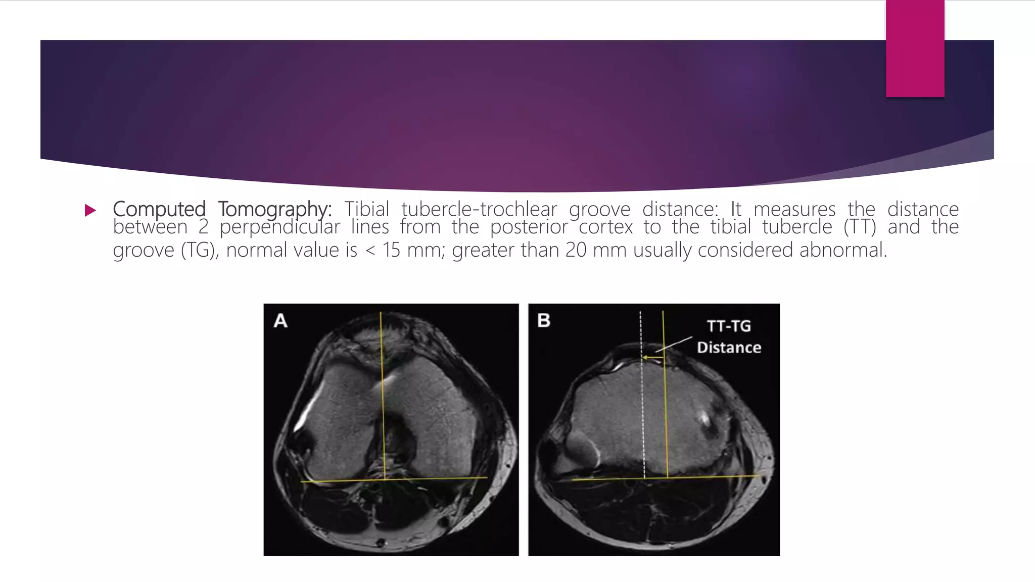
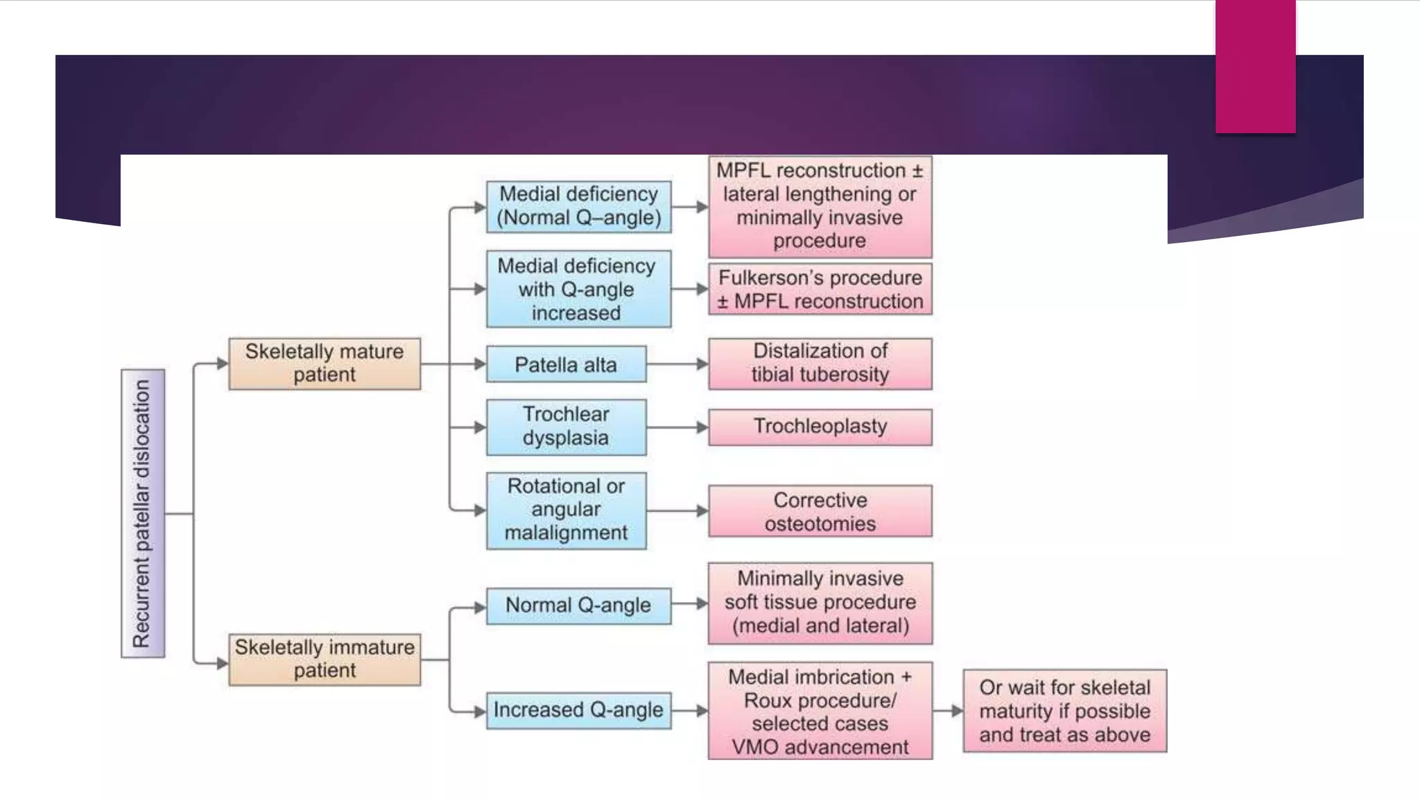
Patellar Instability
DR DITESH JAIN
DEPT OF ORTHOPAEDIC SURGERY
LILAVATI HOSPITAL AND RESEARCH INSTITUTE
 When patella shift or slips out from trochlear groove.
 It can be of two types:
 Subluxation: when patella slips out but immediately goes back in trochlear
groove; there is never an instance of complete separation of articular surface.
 Dislocation: When patella shift completely out of trochlear groove towards lateral
lateral side following complete separation of articular surface.
 Acute Dislocation: Traumatic following mostly sports injuries. 2nd m/c cause of
knee haemarthrosis.
 Chronic Dislocation: may follows primary traumatic event or any associated
misalignment.
 Recurrent Dislocation: Patella dislocate with minimal stress during flexion; may
relocate spontaneously. Usually not associated with haemarthrosis.
 Habitual Dislocate: Patella dislocate with almost every movement of flexion and
reduce spontaneously; Often congenital form of patellar instability.
Recurrent Patella Dislocate
 When patella dislocate more than once usually follows traumatic event.
 After one episode chance of having another episode is 45%.
 While after two episode the chances of further episode rises to 70 – 80%.
Biomechanics of Patella Stabliztion:
 Static Stabilizers: Maintain patella in position (in trochlea)
A. Medial Patellofemoral ligament:
• Primary restraint to lateral translation in 20 – 30 deg flexion.
• Its fibres blend with VMO giving it dynamic component also.
• M/C site of avulsion is femoral attachment.
B. Medial Patellotibial and Medial Patellomeniscal Ligaments: Confers medial
inferior stability.
C. Lateral Retinaculum and ITB.
D. Bony Conformity: Trochlear groove morphology.
 Dynamic Stabilisers: Stabilise the patella during tracking
1. Vastus Medialis obliquus: Pull the patella in 55 deg medially, maintain medial tug
during tacking. It counter Q – angle lateral force vector on patella.
2. Compressive Dynamic Force: Once engaged in trochlea; after medial and lateral
tug neutralise each other, the main patellofemoral force is of compression
provided by increasing force of quadriceps and patellar tendon.
Predisposing Factors:
 Previous Dislocation
 Soft Tissue Factor:
1) Ligament Laxity (EDS)
2) Dysplastic VMO
3) Patellofemoral ligament Insufficiency
4) ITB or Lateral retinacular contracture
 Bony Factors:
1. Small Patella
2. Lateral Femoral Condyle hypoplasia
2. Trochlear Dysplasia: Dejour classification (MRI)
 Alignment of Limb:
1. Familial lateral disposition of patella
2. Increased TT:TG distance (Normal = <15mmmm, >19mm pathological )
3. Excessive lateral patellar tilt
4. Increased femoral anteversion
5. Genu valgum
6. Miserable Malalignment Syndrome: Femoral anteversion, genu valgum, External
Tibial Torsion (All increases Q Angle)
7. Increased Q Angle:
 Male: 10 – 12 deg
 Female : 15 – 18 deg
8. Patella Alta: when patella is located higher than
normal position;
• Insall Salavati = PT/LP (0.8 – 1.2)
• Modified IS = PT/LaP (N = <2)
• Caton Deschamps = distance b/w upper tibia and
lower most articular point/LaP (06 – 1.3)
• Blackburne peel = distance b/w tangent to upper
tibia surface and lower most articular point/LaP (0.5 -
1)
• Morphology Index = PL/LaP (N < 1.5)
• Blumensaat`s Line: Extend to inferior pole of patella
in 30 deg flexion.
Clinical Features:
 Symptoms:
 Recurrent instability at the knee
 C/o knee giving away or slipping out
 May c/o of knee pain
 Clinical Signs:
 Gait Change : Valgus thrust due to MPFL Weakness
 Increased Q angle
 Patella Alta
 Extensor Lag
 Patellofemoral Crepitus
 Apprehension test [Fulkerson relocation Test]:
 Increased Passive patellar lateral translation [Patella Glide test]:
 J sign: Indicate poor patella tracking.
 Ely`s Test: For Rectus Tightness  Ober`s Test: For ITB Tightness
Radiological Features
 AP view: To evaluate Limb Alignment,
version
 Lateral view:
• Articular fracture
• Patella Subluxation
• Patellofemoral arthrosis
• Trochlear Dysplasis: Crossing Sign,
Double contour sign
 Tangential view of patella:
 Sunrise View:
• Lateral patellar tilt: Patellar tilt was supposed to be due to quadriceps weakness
(VMO) or dysplasia (Dejour) but others have related this to MPFL insufficiency
(Beasley and Vidal).
• Depth of femoral intercondylar sulcus greater than 5 mm.
• Patellar tilt: Angle between axis of patella and bicondylar femoral line. Normal
is less than 20°.
• Femoropatellar joint space should be less than 5 mm. Distance more than this
indicates effusion.
• Congruence angle (normal is -6°)
• Patellofemoral index is the ratio of medial patellofemoral joint space to lateral
patellofemoral joint space; normal is less than 1.6.
• Lateral trochlear inclination.
• Bisect offset
• Sulcus Angle: normal 126 – 150; >150 deg indicate trochlear dysplasia
• The sulcus angle is a measure of
trochlear depth (angle AOB here).
• OC is the bisector of this angle. OD
is a line that passes through the
lowest point on the median ridge of
patella forming angle COD which is
the congruence angle. Toward the
medial femoral condyle, the
congruence angle is denoted
negative (–) while if it crosses
toward lateral femoral condyle, the
angle will gain a positive (+) sign.
 Computed Tomography: Tibial tubercle-trochlear groove distance: It measures the distance
between 2 perpendicular lines from the posterior cortex to the tibial tubercle (TT) and the
groove (TG), normal value is < 15 mm; greater than 20 mm usually considered abnormal.
 Magnetic Resonance Imaging:
 To evaluate trochlear dysplasias.
 Osteochondral Lesions
 MPFL tear etc.
Management:
 Non Surgical Approach: This is to be done in only
• Infrequent dislocators.
• If the surgeon feels that the patellar mechanics are able to accommodate the
rehabilitation process.
• VMO training, pelvifemoral rehabilitation, orthotic support can be tried with
experience.
• These measures often serve only as an interim measure till some surgical
intervention is undertaken.
 Surgical Management: Following are the essential assessment before deciding
surgical intervention:-
 Skeletal Maturity
 Q Angle
 Reticular tightness
 Patellar tilt and height
 TT : TG distance
 Trochlear Dysplasia
 Proximal Realignment procedures classically done in skeletal immature patient with normal
TT : TG, Q angle, medial facet and patellar height with or without trochlear dysplasis.
 Distal realignment procedures performed in patient with TT :TG > 20mm, Insall Salavati
index > 1.2.
 Arthroscopic procedures only for removal of loose body or in some case lateral
patellofemoral release.
 Fulkerson Procedure:
 Indication TT : TG > 20mm
 Combination of Elmslie – trillatt (Direct
(Direct medicalization transverse
osteotomy of TT) and Maquett
Procedure.
 Involves both anteriorization and
medicalization of TT correcting both Q
angle and unloading patellofemoral
joint.
 TT – TG corrected upto 10 – 15mm.
 Trocheoplasty: for deeping anterior patellofemoral sulcus and congruous
articulation.
Albee osteotomy Dejour Osteotomy
Recurrent Patellar instability

Recurrent Patellar instability

  • 1.
    Patellar Instability DR DITESHJAIN DEPT OF ORTHOPAEDIC SURGERY LILAVATI HOSPITAL AND RESEARCH INSTITUTE
  • 2.
     When patellashift or slips out from trochlear groove.  It can be of two types:  Subluxation: when patella slips out but immediately goes back in trochlear groove; there is never an instance of complete separation of articular surface.  Dislocation: When patella shift completely out of trochlear groove towards lateral lateral side following complete separation of articular surface.
  • 3.
     Acute Dislocation:Traumatic following mostly sports injuries. 2nd m/c cause of knee haemarthrosis.  Chronic Dislocation: may follows primary traumatic event or any associated misalignment.  Recurrent Dislocation: Patella dislocate with minimal stress during flexion; may relocate spontaneously. Usually not associated with haemarthrosis.  Habitual Dislocate: Patella dislocate with almost every movement of flexion and reduce spontaneously; Often congenital form of patellar instability.
  • 4.
    Recurrent Patella Dislocate When patella dislocate more than once usually follows traumatic event.  After one episode chance of having another episode is 45%.  While after two episode the chances of further episode rises to 70 – 80%.
  • 5.
    Biomechanics of PatellaStabliztion:  Static Stabilizers: Maintain patella in position (in trochlea) A. Medial Patellofemoral ligament: • Primary restraint to lateral translation in 20 – 30 deg flexion. • Its fibres blend with VMO giving it dynamic component also. • M/C site of avulsion is femoral attachment. B. Medial Patellotibial and Medial Patellomeniscal Ligaments: Confers medial inferior stability.
  • 6.
    C. Lateral Retinaculumand ITB. D. Bony Conformity: Trochlear groove morphology.  Dynamic Stabilisers: Stabilise the patella during tracking 1. Vastus Medialis obliquus: Pull the patella in 55 deg medially, maintain medial tug during tacking. It counter Q – angle lateral force vector on patella. 2. Compressive Dynamic Force: Once engaged in trochlea; after medial and lateral tug neutralise each other, the main patellofemoral force is of compression provided by increasing force of quadriceps and patellar tendon.
  • 8.
    Predisposing Factors:  PreviousDislocation  Soft Tissue Factor: 1) Ligament Laxity (EDS) 2) Dysplastic VMO 3) Patellofemoral ligament Insufficiency 4) ITB or Lateral retinacular contracture  Bony Factors: 1. Small Patella 2. Lateral Femoral Condyle hypoplasia
  • 9.
    2. Trochlear Dysplasia:Dejour classification (MRI)
  • 10.
     Alignment ofLimb: 1. Familial lateral disposition of patella 2. Increased TT:TG distance (Normal = <15mmmm, >19mm pathological ) 3. Excessive lateral patellar tilt 4. Increased femoral anteversion 5. Genu valgum 6. Miserable Malalignment Syndrome: Femoral anteversion, genu valgum, External Tibial Torsion (All increases Q Angle)
  • 11.
    7. Increased QAngle:  Male: 10 – 12 deg  Female : 15 – 18 deg
  • 12.
    8. Patella Alta:when patella is located higher than normal position; • Insall Salavati = PT/LP (0.8 – 1.2) • Modified IS = PT/LaP (N = <2) • Caton Deschamps = distance b/w upper tibia and lower most articular point/LaP (06 – 1.3) • Blackburne peel = distance b/w tangent to upper tibia surface and lower most articular point/LaP (0.5 - 1) • Morphology Index = PL/LaP (N < 1.5) • Blumensaat`s Line: Extend to inferior pole of patella in 30 deg flexion.
  • 13.
    Clinical Features:  Symptoms: Recurrent instability at the knee  C/o knee giving away or slipping out  May c/o of knee pain  Clinical Signs:  Gait Change : Valgus thrust due to MPFL Weakness  Increased Q angle  Patella Alta  Extensor Lag  Patellofemoral Crepitus
  • 14.
     Apprehension test[Fulkerson relocation Test]:
  • 15.
     Increased Passivepatellar lateral translation [Patella Glide test]:
  • 16.
     J sign:Indicate poor patella tracking.
  • 17.
     Ely`s Test:For Rectus Tightness  Ober`s Test: For ITB Tightness
  • 18.
    Radiological Features  APview: To evaluate Limb Alignment, version  Lateral view: • Articular fracture • Patella Subluxation • Patellofemoral arthrosis • Trochlear Dysplasis: Crossing Sign, Double contour sign
  • 19.
  • 20.
     Sunrise View: •Lateral patellar tilt: Patellar tilt was supposed to be due to quadriceps weakness (VMO) or dysplasia (Dejour) but others have related this to MPFL insufficiency (Beasley and Vidal). • Depth of femoral intercondylar sulcus greater than 5 mm. • Patellar tilt: Angle between axis of patella and bicondylar femoral line. Normal is less than 20°. • Femoropatellar joint space should be less than 5 mm. Distance more than this indicates effusion. • Congruence angle (normal is -6°) • Patellofemoral index is the ratio of medial patellofemoral joint space to lateral patellofemoral joint space; normal is less than 1.6. • Lateral trochlear inclination. • Bisect offset • Sulcus Angle: normal 126 – 150; >150 deg indicate trochlear dysplasia
  • 21.
    • The sulcusangle is a measure of trochlear depth (angle AOB here). • OC is the bisector of this angle. OD is a line that passes through the lowest point on the median ridge of patella forming angle COD which is the congruence angle. Toward the medial femoral condyle, the congruence angle is denoted negative (–) while if it crosses toward lateral femoral condyle, the angle will gain a positive (+) sign.
  • 22.
     Computed Tomography:Tibial tubercle-trochlear groove distance: It measures the distance between 2 perpendicular lines from the posterior cortex to the tibial tubercle (TT) and the groove (TG), normal value is < 15 mm; greater than 20 mm usually considered abnormal.
  • 23.
     Magnetic ResonanceImaging:  To evaluate trochlear dysplasias.  Osteochondral Lesions  MPFL tear etc.
  • 24.
    Management:  Non SurgicalApproach: This is to be done in only • Infrequent dislocators. • If the surgeon feels that the patellar mechanics are able to accommodate the rehabilitation process. • VMO training, pelvifemoral rehabilitation, orthotic support can be tried with experience. • These measures often serve only as an interim measure till some surgical intervention is undertaken.
  • 25.
     Surgical Management:Following are the essential assessment before deciding surgical intervention:-  Skeletal Maturity  Q Angle  Reticular tightness  Patellar tilt and height  TT : TG distance  Trochlear Dysplasia
  • 27.
     Proximal Realignmentprocedures classically done in skeletal immature patient with normal TT : TG, Q angle, medial facet and patellar height with or without trochlear dysplasis.  Distal realignment procedures performed in patient with TT :TG > 20mm, Insall Salavati index > 1.2.  Arthroscopic procedures only for removal of loose body or in some case lateral patellofemoral release.
  • 28.
     Fulkerson Procedure: Indication TT : TG > 20mm  Combination of Elmslie – trillatt (Direct (Direct medicalization transverse osteotomy of TT) and Maquett Procedure.  Involves both anteriorization and medicalization of TT correcting both Q angle and unloading patellofemoral joint.  TT – TG corrected upto 10 – 15mm.
  • 29.
     Trocheoplasty: fordeeping anterior patellofemoral sulcus and congruous articulation. Albee osteotomy Dejour Osteotomy