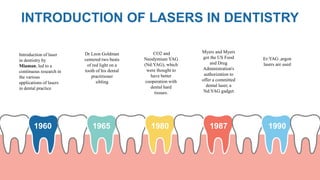
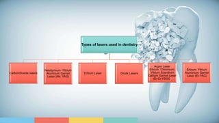
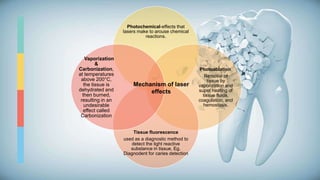
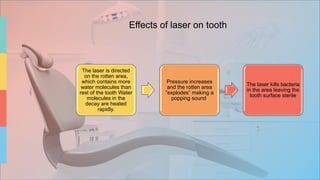
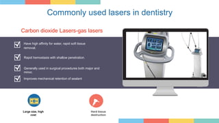
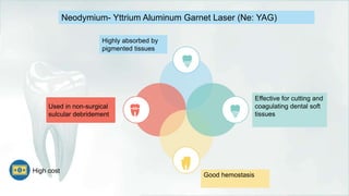
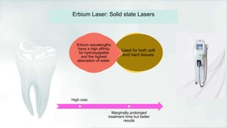
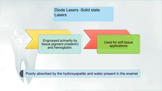
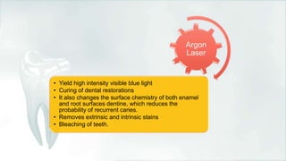
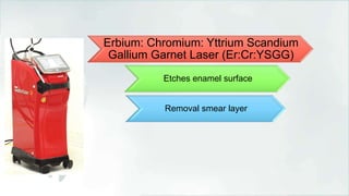
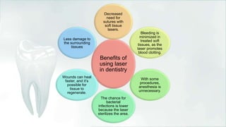
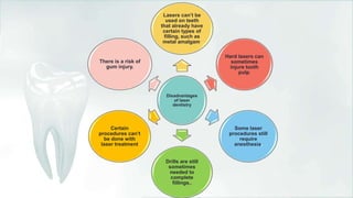
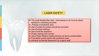
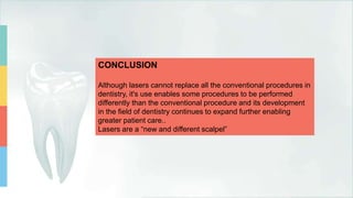
LASERS
IN
DENTISTRY
E.KAROLINEKERSIN
ASSISTANT PROFESSOR
CONTENT
INTRODUCTION
MECHANISM OF LASER ACTION
TYPES OF LASERS
SOFT TISSUE APPLICATIONS
HARD TISSUE APPLICATIONS
DIAGNOSTIC APPLICATIONS
PRONS AND CONS
LASER SAFETY
CONCLUSION
INTRODUCTION OF LASERS IN DENTISTRY
1960 1965 1980 1987 1990
Dr Leon Goldman
centered two beats
of red light on a
tooth of his dental
practitioner
sibling
CO2 and
Neodymium YAG
(Nd:YAG), which
were thought to
have better
cooperation with
dental hard
tissues.
Myers and Myers
got the US Food
and Drug
Administration's
authorization to
offer a committed
dental laser, a
Nd:YAG gadget
Er:YAG ,argon
lasers are used
Introduction of laser
in dentistry by
Miaman, led to a
continuous research in
the various
applications of lasers
in dental practice
Types of lasers used in dentistry
Carbondioxide lasers
Neodymium- Yttrium
Aluminum Garnet
Laser (Ne: YAG)
Erbium Laser Diode Lasers
Argon Laser
Erbium: Chromium:
Yttrium Scandium
Gallium Garnet Laser
(Er:Cr:YSGG)
Erbium: Yttrium
Aluminium Garnet
Laser (Er:YAG)
Mechanism of laser action
• Laser light is a monochromatic light and consists
of a single wavelength of light.
• It consists of three principal parts: An energy
source, an active lasing medium, and two or
more mirrors that form an optical cavity or
resonator.
• For amplification to occur, energy is supplied to
the laser system by a pumping mechanism, such
as, a flash-lamp strobe device, an electrical
current, or an electrical coil.
• This energy is pumped into an active medium
contained within an optical resonator, producing
a spontaneous emission of photons
• Subsequently, amplification by stimulated emission takes place as the photons are reflected
back and forth through the medium by the highly reflective surfaces of the optical resonator,
prior to their exit from the cavity via the output coupler .
• In dental lasers, the laser light is delivered from the laser to the target tissue via a fiberoptic
cable, hollow waveguide, or articulated arm Focusing lenses, a cooling system, and other
controls complete the system.
• The wavelength and other properties of the laser are determined primarily by the composition
of an active medium, which can be a gas, a crystal, or a solid-state semiconductor.
• The light energy produced by a laser can have four different interactions with a target tissue:
Reflection, Transmission, Scattering, and Absorption.
• When a laser is absorbed, it elevates the temperature and produces photochemical effects
depending on the water content of the tissues.
• When a temperature of 100°C is reached, vaporization of the water within the tissue occurs,
a process called ablation.
• At temperatures below 100°C, but above approximately 60°C, proteins begin to denature,
without vaporization of the underlying tissue.
• Conversely, at temperatures above 200°C, the tissue is dehydrated and then burned,
resulting in an undesirable effect called carbonization.
• Absorption requires an absorber of light, termed chromophores, which have a certain affinity
for specific wavelengths of light.
• The primary chromophores in the intraoral soft tissue are Melanin, Hemoglobin, and Water,
and in dental hard tissues, Water and Hydroxyapatite.
• Different laser wavelengths have different absorption coefficients with respect to these
primary tissue components, making the laser selection procedure-dependent
Photochemical-effects that
lasers make to arouse chemical
reactions.
Photoablation.
Removal of
tissue by
vaporization and
super heating of
tissue fluids,
coagulation, and
hemostasis.
Tissue fluorescence
used as a diagnostic method to
detect the light reactive
substance in tissue. Eg.
Diagnodent for caries detection
Vaporization
&
Carbonization,
at temperatures
above 200°C,
the tissue is
dehydrated and
then burned,
resulting in an
undesirable
effect called
Carbonization
Mechanism of laser
effects
The laser is directed
on the rotten area,
which contains more
water molecules than
rest of the tooth Water
molecules in the
decay are heated
rapidly.
Pressure increases
and the rotten area
“explodes” making a
popping sound.
The laser kills bacteria
in the area leaving the
tooth surface sterile
Effects of laser on tooth
Commonly used lasers in dentistry
Have high affinity for water, rapid soft tissue
removal.
Rapid hemostasis with shallow penetration.
Generally used in surgical procedures both major and
minor.
Large size, high
cost
Hard tissue
destruction
Carbon dioxide Lasers-gas lasers
Improves mechanical retention of sealant
Neodymium- Yttrium Aluminum Garnet Laser (Ne: YAG)
Highly absorbed by
pigmented tissues
Effective for cutting and
coagulating dental soft
tissues
Good hemostasis
Used in non-surgical
sulcular debridement
High cost
Used for both soft
and hard tissues
Erbium wavelengths
have a high affinity
for hydroxyapatite
and the highest
absorption of water.
Erbium Laser: Solid state Lasers
High cost.
Marginally prolonged
treatment time but better
results
Diode Lasers -Solid state
Lasers
Engrossed primarily by
tissue pigment (melanin)
and hemoglobin.
Used for soft tissue
applications
Poorly absorbed by the hydroxyapatite and water present in the enamel
Argon
Laser
• Yield high intensity visible blue light
• Curing of dental restorations
• It also changes the surface chemistry of both enamel
and root surfaces dentine, which reduces the
probability of recurrent caries.
• Removes extrinsic and intrinsic stains
• Bleaching of teeth.
Erbium: Chromium: Yttrium Scandium
Gallium Garnet Laser (Er:Cr:YSGG)
Etches enamel surface
Removal smear layer
Removal of inflamed, hypertrophic tissue, and
miscellaneous tissue removal
 Isolated areas of transient tissue hypertrophy can easily be
excised with the diode laser without specialist referral.
 The diode laser is also very useful for a number of isolated
applications, such as, removing tissue that has overgrown mini-
screws, springs and appliances as well as for replacing a tissue
punch if needed ,when placing mini-screws in the unattached
gingiva.
Frenectomies
 A high or prominent labial frenum when indicated, laser assisted
frenectomy is a simple procedure that is best performed after the
diastema is closed as much as possible.
 Ankylosglossia can lead to problems with deglutition, speech,
malocclusion, and potential periodontal problems.
 Frenectomies performed with a laser permit excision of the frena
painlessly, without bleeding, sutures, or surgical packing, and with no
need for special postoperative care.
HARD TISSUE APPLICATIONS
Photochemical effects
 The argon laser produces high intensity visible blue light (488 nm), which is
able to initiate photopolymerization of light-cured dental restorative materials,
which use camphoroquinone as the photoinitiator.
 Argon laser radiation is also able to alter the surface chemistry of both
enamel and root surface dentine,which reduces the probability of recurrent
caries.
 The bleaching effect relies on the specific absorption of a narrow spectral
range of green light (510-540 nm) into the chelate compounds formed
between the apatites, porphyrins, and tetracycline compounds.
 Argon and Potassium Titanyl Phosphate (KTiOPO4, KTP) lasers can achieve
a positive result in cases that are completely unresponsive to conventional
photothermal ‘power’ bleaching.
Cavity preparation, caries, and restorative removal
 Various studies depict the use of Er: YAG, since 1988, for removing caries in the
enamel and dentine by ablation, without the detrimental effect of rise in
temperature on the pulp,even without water-cooling, with low ‘fluences’
 laser (LLLT), similar to air-rotor devices, except that the floor of the cavity is
not as smooth.
 The Er: YAG laser is capable of removing cement, composite resin, and glass
ionomer
Etching
 Laser etching has been evaluated as an alternative to
acid etching of enamel and dentine. Enamel and
dentine surfaces etched with (Er, Cr: YSGG) lasers
show micro-irregularities and no smear layer.
 Adhesion to dental hard tissues after Er: YAG laser
etching is inferior to that obtained after conventional
acid etching.
Treatment of dentinal hypersensitivity
 Dentinal hypersensitivity is one of the most common complaints in
clinical dental practice.
 Comparison of the desensitizing effects of an Er: YAG laser with
those of a conventional desensitizing system on cervically exposed
hypersensitive dentine showed that desensitizing of hypersensitive
dentine with an Er: YAG laser is effective, and maintenance of a
positive result is more prolonged than with other agents
Benefits of
using laser
in dentistry
Decreased
need for
sutures with
soft tissue
lasers.
Bleeding is
minimized in
treated soft
tissues, as the
laser promotes
blood clotting.
With some
procedures,
anesthesia is
unnecessary.
The chance for
bacterial
infections is lower
because the laser
sterilizes the area.
Wounds can heal
faster, and it’s
possible for
tissue to
regenerate.
Less damage to
the surrounding
tissues
Disadvantages
of laser
dentistry
Lasers can’t be
used on teeth
that already have
certain types of
filling, such as
metal amalgam.
Hard lasers can
sometimes
injure tooth
pulp.
Some laser
procedures still
require
anesthesia.
Drills are still
sometimes
needed to
complete
fillings,.
Certain
procedures can’t
be done with
laser treatment
There is a risk of
gum injury.
LASER SAFETY
 The small flexible fiber optic , hand pieces or tip must be steam
sterilized in sterilizing pouches
 Practice of protective wear
 Use of screen & curtains should be promoted
 Use of proper clothing
 Use of anti-fire explosive
 Proper training and courses
 Use of laser filtration masks prevents air borne contamination
 Foot pedal control switch with protective hood
 prevents accidental depression by surgical staff.
CONCLUSION
Although lasers cannot replace all the conventional procedures in
dentistry, it's use enables some procedures to be performed
differently than the conventional procedure and its development
in the field of dentistry continues to expand further enabling
greater patient care..
Lasers are a “new and different scalpel”
REFERENCE
1.A Contemporary Apprise on LASERS and its Applications in
Dentistry Chaitanya Pendyala , Rahul VC Tiwari , Heena Dixit ,
Vaishak Augustine , Queentaj Baruah , Kaveri Baruah
2. Laser in dentistry: An innovative tool in modern dental
practice Sanjeev Kumar Verma, Sandhya Maheshwari, Raj
Kumar Singh, and Prabhat Kumar Chaudhari
THANK YOU

Lasers in dentistry

  • 1.
  • 2.
    CONTENT INTRODUCTION MECHANISM OF LASERACTION TYPES OF LASERS SOFT TISSUE APPLICATIONS HARD TISSUE APPLICATIONS DIAGNOSTIC APPLICATIONS PRONS AND CONS LASER SAFETY CONCLUSION
  • 3.
    INTRODUCTION OF LASERSIN DENTISTRY 1960 1965 1980 1987 1990 Dr Leon Goldman centered two beats of red light on a tooth of his dental practitioner sibling CO2 and Neodymium YAG (Nd:YAG), which were thought to have better cooperation with dental hard tissues. Myers and Myers got the US Food and Drug Administration's authorization to offer a committed dental laser, a Nd:YAG gadget Er:YAG ,argon lasers are used Introduction of laser in dentistry by Miaman, led to a continuous research in the various applications of lasers in dental practice
  • 4.
    Types of lasersused in dentistry Carbondioxide lasers Neodymium- Yttrium Aluminum Garnet Laser (Ne: YAG) Erbium Laser Diode Lasers Argon Laser Erbium: Chromium: Yttrium Scandium Gallium Garnet Laser (Er:Cr:YSGG) Erbium: Yttrium Aluminium Garnet Laser (Er:YAG)
  • 5.
    Mechanism of laseraction • Laser light is a monochromatic light and consists of a single wavelength of light. • It consists of three principal parts: An energy source, an active lasing medium, and two or more mirrors that form an optical cavity or resonator. • For amplification to occur, energy is supplied to the laser system by a pumping mechanism, such as, a flash-lamp strobe device, an electrical current, or an electrical coil. • This energy is pumped into an active medium contained within an optical resonator, producing a spontaneous emission of photons
  • 6.
    • Subsequently, amplificationby stimulated emission takes place as the photons are reflected back and forth through the medium by the highly reflective surfaces of the optical resonator, prior to their exit from the cavity via the output coupler . • In dental lasers, the laser light is delivered from the laser to the target tissue via a fiberoptic cable, hollow waveguide, or articulated arm Focusing lenses, a cooling system, and other controls complete the system. • The wavelength and other properties of the laser are determined primarily by the composition of an active medium, which can be a gas, a crystal, or a solid-state semiconductor.
  • 7.
    • The lightenergy produced by a laser can have four different interactions with a target tissue: Reflection, Transmission, Scattering, and Absorption. • When a laser is absorbed, it elevates the temperature and produces photochemical effects depending on the water content of the tissues. • When a temperature of 100°C is reached, vaporization of the water within the tissue occurs, a process called ablation. • At temperatures below 100°C, but above approximately 60°C, proteins begin to denature, without vaporization of the underlying tissue. • Conversely, at temperatures above 200°C, the tissue is dehydrated and then burned, resulting in an undesirable effect called carbonization.
  • 8.
    • Absorption requiresan absorber of light, termed chromophores, which have a certain affinity for specific wavelengths of light. • The primary chromophores in the intraoral soft tissue are Melanin, Hemoglobin, and Water, and in dental hard tissues, Water and Hydroxyapatite. • Different laser wavelengths have different absorption coefficients with respect to these primary tissue components, making the laser selection procedure-dependent
  • 9.
    Photochemical-effects that lasers maketo arouse chemical reactions. Photoablation. Removal of tissue by vaporization and super heating of tissue fluids, coagulation, and hemostasis. Tissue fluorescence used as a diagnostic method to detect the light reactive substance in tissue. Eg. Diagnodent for caries detection Vaporization & Carbonization, at temperatures above 200°C, the tissue is dehydrated and then burned, resulting in an undesirable effect called Carbonization Mechanism of laser effects
  • 10.
    The laser isdirected on the rotten area, which contains more water molecules than rest of the tooth Water molecules in the decay are heated rapidly. Pressure increases and the rotten area “explodes” making a popping sound. The laser kills bacteria in the area leaving the tooth surface sterile Effects of laser on tooth
  • 11.
    Commonly used lasersin dentistry Have high affinity for water, rapid soft tissue removal. Rapid hemostasis with shallow penetration. Generally used in surgical procedures both major and minor. Large size, high cost Hard tissue destruction Carbon dioxide Lasers-gas lasers Improves mechanical retention of sealant
  • 12.
    Neodymium- Yttrium AluminumGarnet Laser (Ne: YAG) Highly absorbed by pigmented tissues Effective for cutting and coagulating dental soft tissues Good hemostasis Used in non-surgical sulcular debridement High cost
  • 13.
    Used for bothsoft and hard tissues Erbium wavelengths have a high affinity for hydroxyapatite and the highest absorption of water. Erbium Laser: Solid state Lasers High cost. Marginally prolonged treatment time but better results
  • 14.
    Diode Lasers -Solidstate Lasers Engrossed primarily by tissue pigment (melanin) and hemoglobin. Used for soft tissue applications Poorly absorbed by the hydroxyapatite and water present in the enamel
  • 15.
    Argon Laser • Yield highintensity visible blue light • Curing of dental restorations • It also changes the surface chemistry of both enamel and root surfaces dentine, which reduces the probability of recurrent caries. • Removes extrinsic and intrinsic stains • Bleaching of teeth.
  • 16.
    Erbium: Chromium: YttriumScandium Gallium Garnet Laser (Er:Cr:YSGG) Etches enamel surface Removal smear layer
  • 22.
    Removal of inflamed,hypertrophic tissue, and miscellaneous tissue removal  Isolated areas of transient tissue hypertrophy can easily be excised with the diode laser without specialist referral.  The diode laser is also very useful for a number of isolated applications, such as, removing tissue that has overgrown mini- screws, springs and appliances as well as for replacing a tissue punch if needed ,when placing mini-screws in the unattached gingiva.
  • 23.
    Frenectomies  A highor prominent labial frenum when indicated, laser assisted frenectomy is a simple procedure that is best performed after the diastema is closed as much as possible.  Ankylosglossia can lead to problems with deglutition, speech, malocclusion, and potential periodontal problems.  Frenectomies performed with a laser permit excision of the frena painlessly, without bleeding, sutures, or surgical packing, and with no need for special postoperative care.
  • 24.
    HARD TISSUE APPLICATIONS Photochemicaleffects  The argon laser produces high intensity visible blue light (488 nm), which is able to initiate photopolymerization of light-cured dental restorative materials, which use camphoroquinone as the photoinitiator.  Argon laser radiation is also able to alter the surface chemistry of both enamel and root surface dentine,which reduces the probability of recurrent caries.  The bleaching effect relies on the specific absorption of a narrow spectral range of green light (510-540 nm) into the chelate compounds formed between the apatites, porphyrins, and tetracycline compounds.  Argon and Potassium Titanyl Phosphate (KTiOPO4, KTP) lasers can achieve a positive result in cases that are completely unresponsive to conventional photothermal ‘power’ bleaching.
  • 25.
    Cavity preparation, caries,and restorative removal  Various studies depict the use of Er: YAG, since 1988, for removing caries in the enamel and dentine by ablation, without the detrimental effect of rise in temperature on the pulp,even without water-cooling, with low ‘fluences’  laser (LLLT), similar to air-rotor devices, except that the floor of the cavity is not as smooth.  The Er: YAG laser is capable of removing cement, composite resin, and glass ionomer
  • 26.
    Etching  Laser etchinghas been evaluated as an alternative to acid etching of enamel and dentine. Enamel and dentine surfaces etched with (Er, Cr: YSGG) lasers show micro-irregularities and no smear layer.  Adhesion to dental hard tissues after Er: YAG laser etching is inferior to that obtained after conventional acid etching.
  • 27.
    Treatment of dentinalhypersensitivity  Dentinal hypersensitivity is one of the most common complaints in clinical dental practice.  Comparison of the desensitizing effects of an Er: YAG laser with those of a conventional desensitizing system on cervically exposed hypersensitive dentine showed that desensitizing of hypersensitive dentine with an Er: YAG laser is effective, and maintenance of a positive result is more prolonged than with other agents
  • 28.
    Benefits of using laser indentistry Decreased need for sutures with soft tissue lasers. Bleeding is minimized in treated soft tissues, as the laser promotes blood clotting. With some procedures, anesthesia is unnecessary. The chance for bacterial infections is lower because the laser sterilizes the area. Wounds can heal faster, and it’s possible for tissue to regenerate. Less damage to the surrounding tissues
  • 29.
    Disadvantages of laser dentistry Lasers can’tbe used on teeth that already have certain types of filling, such as metal amalgam. Hard lasers can sometimes injure tooth pulp. Some laser procedures still require anesthesia. Drills are still sometimes needed to complete fillings,. Certain procedures can’t be done with laser treatment There is a risk of gum injury.
  • 30.
    LASER SAFETY  Thesmall flexible fiber optic , hand pieces or tip must be steam sterilized in sterilizing pouches  Practice of protective wear  Use of screen & curtains should be promoted  Use of proper clothing  Use of anti-fire explosive  Proper training and courses  Use of laser filtration masks prevents air borne contamination  Foot pedal control switch with protective hood  prevents accidental depression by surgical staff.
  • 31.
    CONCLUSION Although lasers cannotreplace all the conventional procedures in dentistry, it's use enables some procedures to be performed differently than the conventional procedure and its development in the field of dentistry continues to expand further enabling greater patient care.. Lasers are a “new and different scalpel”
  • 32.
    REFERENCE 1.A Contemporary Appriseon LASERS and its Applications in Dentistry Chaitanya Pendyala , Rahul VC Tiwari , Heena Dixit , Vaishak Augustine , Queentaj Baruah , Kaveri Baruah 2. Laser in dentistry: An innovative tool in modern dental practice Sanjeev Kumar Verma, Sandhya Maheshwari, Raj Kumar Singh, and Prabhat Kumar Chaudhari
  • 33.