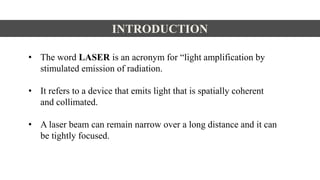
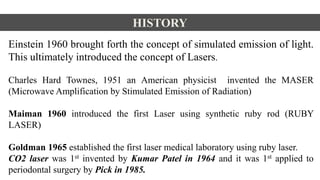
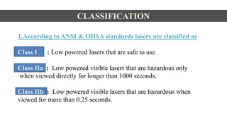
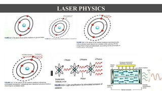
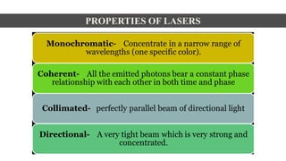
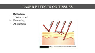
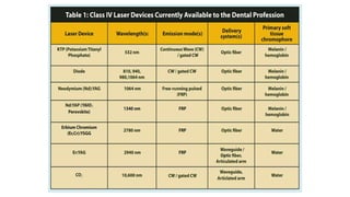
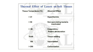
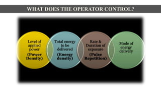
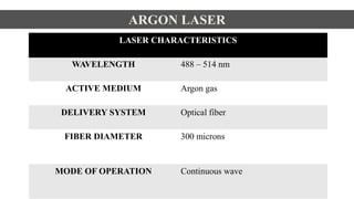
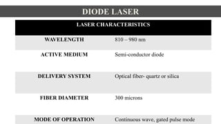
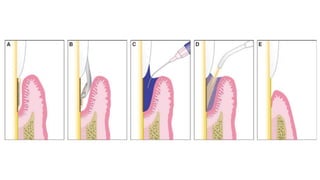
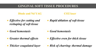
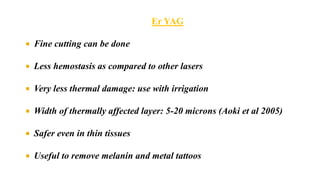
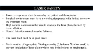
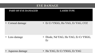
This document discusses lasers used in periodontics. It provides an overview of laser physics, types of lasers including diode, CO2, Nd:YAG and erbium lasers, and their applications in soft tissue procedures and osseous surgery. The benefits of lasers include less pain, better hemostasis and wound healing compared to conventional methods. Safety protocols must be followed when using lasers to prevent eye and tissue damage. Lasers are becoming more widely used in dentistry due to their advantages over traditional techniques.
Periodontal wound healing involves four overlapping phases - exudative, resorptive, proliferative, and regenerative. The proliferative phase includes re-epithelialization, fibroplasia, granulation tissue formation, collagen deposition, angiogenesis, and wound contraction. Growth factors play an important role in regulating periodontal wound healing. Healing after periodontal procedures like scaling and root planing, curettage, ultrasonic curettage, and gingivectomy depends on the extent of tissue disruption and follows a timeline of inflammatory response, epithelial migration, granulation tissue formation, collagen deposition and remodeling.
This document discusses the potential applications of nanotechnology in periodontics. It begins with background on nanotechnology and describes various nanoparticles and how nanoproducts are made. It then discusses the properties of nanomaterials and how they are used for drug delivery, tissue engineering, biofilm studies, tooth repair, dental implants, and bone replacement. The document concludes by describing hypothetical nanorobots that could one day be used to treat periodontal disease at the molecular level through precise, targeted actions guided by external monitoring.
Lasers and its application in periodonticsShilpa Shiv
The document discusses different types of lasers used in periodontology, including their properties, mechanisms of interaction with tissue, safety classifications, and clinical applications. It provides details on lasers such as the argon, diode, Nd:YAG, Er:YAG, and CO2 lasers, covering their wavelengths, active mediums, delivery systems, absorption characteristics, and periodontal uses. The document also examines laser tissue interactions, safety considerations, and the theoretical zones of tissue change caused by laser exposure.
INTRODUCTION
HISTORY
PRINCIPLES OF WORKING OF A LASER
FUNDAMENTALS OF LASER
CHARACTERISTICS OF LASER
CLASSIFICATION OF LASER
EFFECTS OF LASER ON SOFT AND HARD TISSUES
VARIOUS LASERS AVAILABLE FOR PERIDONTAL USE
APPLICATION OF LASER TREATMENT IN PERIODONTAL THERAPY
ADVANTAGES & DISADVANTAGES OF LASER IN PERIODONTAL THERAPY
LASER PRECAUTIONS
LASER HAZARDS
RECENT ADVANCES
CONCLUSION
Surgical gingivectomy involves removing the pocket wall and smoothing root surfaces to eliminate suprabony pockets and provide access for calculus removal. This creates a favorable environment for gingival healing and restoration of a healthy contour. Indications include deep pockets where bone defects cannot be corrected or aesthetic concerns. Contraindications include pockets below the mucogingival junction. The procedure involves exploring each pocket with a probe, removing the pocket wall and granulation tissue, then covering with a surgical pack. Healing occurs over 1-2 months as granulation tissue forms and epithelium migrates over it.
Gingivectomy and gingivoplasty are procedures to remove gum tissue and reshape the gum line. Gingivectomy removes gum tissue from deep pockets, while gingivoplasty contours gum tissue without eliminating pockets. The document outlines the indications, contraindications, techniques, and healing process for gingivectomy. Surgical gingivectomy uses knives and curettes to remove gum tissue in pockets and reshape the gum line. Healing occurs through granulation tissue formation and re-epithelialization over 1-2 weeks. Other techniques include electrosurgery, laser gingivectomy, and chemo surgery.
Resective osseous surgery involves removing tooth-supporting alveolar bone to reshape it and eliminate periodontal pockets. It is indicated for inconsistent bone margins, reverse bone architecture, one-wall defects, and other bone irregularities. The surgery involves using burs and chisels to perform vertical grooving of thick bone, blending any bone ledges on tooth roots, flattening interproximal bone in narrow areas, and gradualizing marginal bone. Flaps are closed to cover the new bony margins. Post-operative maintenance like cleaning and antibiotics is needed to allow healing over 2-3 weeks.
This document provides an overview of resective osseous surgery techniques. It discusses the anatomical forms of bone, osteoplasty and ostectomy procedures, surgical approaches, and techniques. Osteoplasty involves reshaping bone without removing tooth-supporting bone through techniques like grooving and blending. Ostectomy involves removing tooth-supporting bone to eliminate osseous deformities. Specific techniques like horizontal grooving, scribing, and hand instrumentation are described. Post-operative maintenance and expected osseous changes are also summarized.
The document discusses gingival curettage, which involves scraping diseased soft tissue from periodontal pockets. It describes different types of curettage including surgical, chemical, ultrasonic, and laser. Indications for curettage include shallow pockets and as maintenance treatment for recurrent inflammation. Contraindications include acute infections and pockets extending beyond the mucogingival junction. The procedure involves scraping the pocket wall with a curette. Excisional new attachment procedure is also discussed, which uses gingival incision followed by root planing. Healing after curettage involves blood clot formation, leukocyte proliferation, and re-epithelialization within 7 days.
This document summarizes various gingival surgical techniques including gingival curettage. It discusses the history and development of gingival curettage, defines the technique, and outlines the rationale, procedure, indications, contraindications and healing process. Key pioneers in developing gingival curettage techniques are mentioned. Variations of gingival curettage including the excisional new attachment procedure and use of ultrasonic instruments are also summarized.
The gingiva provides three lines of defense against pathogens:
1. The epithelial surface acts as a mechanical and chemical barrier. Tight junctions between keratinocytes and antimicrobial peptides in epithelial layers prevent pathogen entry.
2. Components in saliva and gingival crevicular fluid (GCF) maintain tissue health and help remove debris from the sulcus. GCF is an inflammatory exudate containing enzymes, electrolytes, and host/bacterial compounds.
3. The gingival tissue mounts an innate and adaptive immune response. Langerhans cells and neutrophils present in the junctional epithelium phagocytose pathogens, while T cells and antibodies provide acquired immunity. Together,
This document provides an overview of guided tissue regeneration (GTR). It begins with definitions of periodontal regeneration and GTR. It then discusses the history and development of GTR from the 1970s onwards. The core concept of GTR is explained, which is based on Melcher's hypothesis that only periodontal ligament cells can regenerate the periodontal attachment apparatus. Indications, contraindications, design criteria and objectives of GTR barriers are covered. The document classifies and compares advantages and disadvantages of absorbable versus non-absorbable membranes. Key factors affecting GTR outcomes are discussed. Surgical techniques and the healing of GTR-treated defects are described. The document concludes with additional considerations like complications and the
Scaling and root planing (SRP) is a non-surgical treatment for periodontitis that aims to remove dental plaque and calculus from tooth surfaces. It involves scaling to remove deposits and root planing to smooth root surfaces. The goals are to eliminate periodontitis by removing irritants and restoring a healthy environment for tissue healing. The long-term effectiveness depends on factors like patient compliance, disease severity, and anatomical challenges. Overhanging restorations can interfere with cleaning and disturb the ecological balance, allowing disease-causing bacteria to proliferate.
This document summarizes the process of using free gingival grafts for root coverage. Free gingival grafts are soft tissue grafts that are disconnected from their blood supply when harvested. For survival, they rely on nutrients from the graft bed. To promote survival over avascular root surfaces, the graft bed is extended in size and the graft is made thick to provide capillary channels to transport nutrients to the center. Case examples show grafts harvested from the palate and sutured over denuded root surfaces, with subsequent healing resulting in root coverage and attachment.
This document provides an overview of periodontal flap surgery. It discusses the definitions, historical background, objectives, indications and contraindications of flap surgery. It also covers the advantages and disadvantages, principles of flap design, classification of flaps, properties of an ideal flap, and the main types of incisions used. The document is intended to educate about periodontal flap surgery techniques and factors that influence surgical outcomes.
This document discusses the process and techniques for coronoplasty, which is a selective reduction of occlusal areas to influence mechanical contact situations and neural sensory input patterns. It describes the 10 basic steps for performing coronoplasty, which include adjusting the intercuspal position and retruded contact position, removing prematurities, establishing stable simultaneous contacts, testing and adjusting incisal contacts and excursions, and finishing with polishing rough surfaces. The goal is to establish an optimal occlusion with stable contacts, smooth excursions, and decreased tooth mobility and stress on the teeth and temporomandibular joints.
Gingival Crevicular Fluid (GCF/ Sulcular Fluid)Sk Aziz Ikbal
This document provides an overview of gingival crevicular fluid (GCF). It discusses the history and pioneering researchers in the field. J. Waerhaug in 1950 focused on the anatomy of the gingival sulcus and its transformation during periodontitis. GCF forms via two mechanisms - increased permeability of blood vessels or as an inflammatory exudate. The document outlines the composition of GCF, including cells, electrolytes, proteins, and metabolic products. Finally, it discusses the clinical significance of GCF and how certain components can serve as diagnostic markers for periodontal disease.
A brief description of all topics to recent advances,SDD, host modulation and diabetes, host modulation in smokers, chemically modified tetracyclines, bisphosphonates
This document discusses the historical background and various methods of root biomodification, which involves chemically or mechanically modifying the root surface to promote periodontal regeneration. It describes how citric acid, tetracycline, fibronectin, and EDTA work to demineralize and detoxify the root surface in order to remove the smear layer and expose collagen fibers, making the surface more biocompatible and conducive to new attachment of periodontal tissues. Register and Burdick's 1975 technique using citric acid application for 2-3 minutes is outlined, along with modifications by Miller. The mechanisms and benefits of different agents are explained.
Microscopic features of Gingiva by DR SUHANI GOELdr suhani goel
The document summarizes the microscopic features of gingiva. It describes the different layers of the oral epithelium including the stratum basale, stratum spinosum, stratum granulosum, and stratum corneum. It also discusses the sulcular epithelium and junctional epithelium. The connective tissue of the gingiva contains collagen, reticulin and elastic fibers as well as fibroblasts, macrophages and mast cells. Blood supply to the gingiva is provided by suprapapillary arterioles and arteries from the crest of the interdental septa. Lymphatic drainage occurs through vessels that accompany the blood vessels.
Periodontitis is a chronic inflammatory disease of the tooth-supporting structures. The treatment of this condition is based on the removal of local factors and restoration of the bony architecture. Traditionally osseous surgery has been performed by either manual or motor-driven instruments. However, both these methods have their own advantages and disadvantages. Recently, a novel surgical approach using piezoelectric device has been introduced. It is a promising, meticulous and soft tissue sparing system based on low frequency ultrasonic microvibrations. The absence of macrovibration makes the instrument more manageable and allows greater intraoperative control with an increase in the cutting safety in the more difficult anatomical cutting zone. This presentation emphasizes the mechanism of action, instrumentation, advantages and limitations as well as its applications in periodontology and implantology.
This document discusses the use of lasers in periodontology. It begins by covering laser tissue interaction and the types of lasers available for periodontal applications. The document then summarizes several clinical applications of lasers in periodontology including frenectomies, crown lengthening, biopsies, and treatments for lesions, ulcers, and bleeding disorders. It also discusses uses for guided tissue regeneration, scaling and root planing, and preprosthetic surgery. In general, lasers provide benefits like less bleeding, sterilization of surgical sites, reduced post-op pain and swelling, and faster procedures. The laser-assisted new attachment procedure is described as a method for treating moderate to advanced periodontitis. Both advantages
Vestibuloplasty is a surgical procedure to deepen the oral vestibule by changing the attachments of the soft tissue. There are several types of vestibuloplasty procedures, including mucosal advancement, secondary epithelization, and grafting. Mucosal advancement involves undermining and advancing the oral mucosa, while secondary epithelization uses the oral mucosa to line one side and allows the other side to heal through epithelization. Grafting can use skin, mucosa, or dermis grafts to line the extended vestibule. The document discusses techniques for each type of vestibuloplasty procedure.
The document discusses the junctional epithelium, which forms a collar-like attachment of stratified squamous epithelium connecting the tooth and gingiva. It describes the junctional epithelium's structure, development, functions in protecting the periodontium, and role in gingivitis and pocket formation. The document also reviews regeneration of the junctional epithelium, effects of trauma and implants, and syndromes that can affect it.
INTRODUCTION OF LASERS IN ORAL AND MAXILLOFACIAL SURGERY
INITIAL USE IN ORAL AND MAXILLOFACIAL SURGERY
CHARACTERISTICS OF LASER LIGHT
CLASSIFICATION OF LASERS
TISSUE INTERACTION
TYPES OF LASERS
ADVANTAGES AND DISADVANTAGES
LASER SAFETY IN SURGERY AND ANESTHESIA
PATIENT SELECTION
APPLICATIONS
SURGICAL LASER TECHNIQUE
APPLIED ASPECTS
RECENT ADVANCES
COMPLICATIONS
explained with Limited matter moreover I have included all the images. if you go through a standard textbook and referred to this PPT it will help you so much I hope It helps you
Light is an integral part of our life. Advances in technology are increasing and changing the ways that the patient experience dental treatment. One of the milestones in technological advancements in dentistry is the use of lasers The early 20th century saw one of the greatest inventions in science & technology, in that LASERS which later went on to became a gift to health sciences. Albert Einstein is usually credited for the development of the laser theory. He was the first one to coin the term “Stimulated Emission” in his publication “Zur Quantentheorie der Strahlung”, published in 1917 in the “Physikalische Zeitschrift”
Lasers are devices that produce beams of coherent and very high intensity light. The word LASER is an acronym for “Light Amplification by Stimulated\Emission of Radiation”. A crystal or gas is excited to emit light photons of a characteristic wavelength that are amplified and filtered to make a coherent light beam. The effect of the laser depends upon the power of the beam and the extent to which the beam absorbed. Several types of lasers are available based on the wavelengths. These range from long wavelengths (infrared), to visible wavelengths, to short wavelengths (ultraviolet), to special ultraviolet lasers called excimers. Lasers are used nowadays in many areas in the field of dentistry It is of the most captivating technologies in dental practice. Even though, introduced as an alternative to the traditional halogen curing light, laser now has become the instrument of choice, in many dental applications. Its advancements in the field of dentistry are playing a major role in patient care and well being.
This document discusses lasers used in endodontics. It begins with the history and development of lasers, then classifies dental lasers based on their wavelength and penetrating power. Common lasers used in endodontics are described, including argon, KTP, diode, erbium YAG, erbium chromium, and CO2 lasers. The interaction of laser light with tissue is explained in terms of reflection, absorption, diffusion, and transmission. Laser effects in endodontics include photothermal, photochemical, and photomechanical-acoustic effects. Parameters that influence laser endodontics are discussed, such as continuous wave mode and the importance of chopping or gating emission.
The document discusses lasers used in dentistry. It begins with an introduction to lasers and their history in dentistry. Key topics covered include the mechanism of action of lasers, common dental laser therapies, and safety measures when using lasers. Examples are provided of how different types of lasers like CO2, Nd:YAG, and diode lasers are used for both soft tissue and hard tissue procedures in dentistry.
This document discusses the use of lasers in dentistry. It begins with an introduction and history of lasers, then covers the fundamentals of laser operation and classification of lasers. The main uses of lasers in dentistry include soft tissue procedures like biopsy and surgery. Techniques for ablation, vaporization, and low level laser therapy are described. Benefits are reduced pain and bleeding, while risks include hazards to patients and staff if not used properly. Proper safety protocols and sterilization of laser equipment are emphasized.
The document provides an overview of lasers, including:
1. It defines what a laser is, describing the acronym LASER and how lasers emit a useful form of light energy.
2. It discusses the history and development of lasers, including milestones such as the first laser built in 1960 and early medical uses starting in 1963.
3. It describes the key principles and components of how lasers work, including stimulated emission, the pumping system, and optical cavity that contains the lasing medium.
This document provides an overview of dental calculus and lasers. It discusses the history and development of lasers from Einstein's work in 1917 to current diode lasers. It describes laser physics including stimulated emission and classifications based on gain medium, tissue application, and mode of action. Safety hazards of lasers like ocular injury, tissue damage, fires, and respiratory issues are covered. In conclusion, lasers may become preferred for non-surgical and surgical periodontal therapy in the future.
This document provides an overview of lasers in dentistry. It discusses the history and development of lasers, how lasers are designed and how laser light interacts with tissues. It describes common dental lasers like CO2 and argon lasers, and their applications. CO2 lasers are well absorbed in oral tissues and useful for soft tissue procedures. Argon lasers are absorbed by hemoglobin and melanin, making them good for coagulation. The document outlines the benefits of lasers for various dental procedures.
Lasers have many uses in ophthalmology, both therapeutic and diagnostic. Therapeutically, lasers are used to treat retinal disorders like diabetic retinopathy, macular edema and retinal detachments. They are also used in procedures like laser iridotomy and trabeculoplasty to treat glaucoma. Diagnostically, lasers are used in optical coherence tomography and scanning laser ophthalmoscopy to image the retina. Different types of lasers like argon, Nd:YAG and excimer interact with tissue in various ways such as coagulation, vaporization or ablation, depending on the wavelength and power. While lasers are generally safe, potential complications include pain, elevated pressure, retinal damage and
This document provides an overview of lasers used in dentistry, including their history, mechanisms of action, applications, and safety measures. It discusses how lasers were first developed in the 1960s and introduced to dentistry in the 1990s. The main types of lasers used include CO2, Nd:YAG, Er:YAG, and KTP lasers. Lasers can be used for both hard and soft tissue procedures, such as caries removal, gingivectomies, and lesion removal, with advantages like reduced pain, bleeding, and recovery time compared to traditional techniques. Safety precautions must be followed when using lasers to protect patients and operators.
Different types of lasers and laser delivery systemKrati Gupta
This document discusses different types of lasers and their delivery systems used in ophthalmology. It begins by defining what a laser is and providing a brief history of their development. It then describes the key properties of lasers and the physics behind how they are produced. The document outlines different types of solid state, gas, metal vapor, and other lasers. It discusses the interactions between light and tissue, including photocoagulation, photoablation, photodisruption, and photovaporization. The closing paragraphs cover laser parameters and modes of operation such as continuous wave, pulsed, and Q-switched lasers.
uses of lasers in conservative dentistry and endodonticssucheekiju1
This document discusses the use of lasers in conservative dentistry and endodontics. It provides an introduction to lasers and their classification based on wavelength. The key wavelengths used in dentistry include Nd:YAG, diode, CO2, Er:YAG, and argon lasers. Applications discussed include caries detection and removal, restoration removal, etching, photopolymerization, bleaching, and endodontic uses such as pulp capping, disinfection, and obturation. Lasers provide precision and bloodless operating fields and can improve outcomes for many dental procedures.
1) Lasers have various applications in periodontal and implant dentistry including calculus removal, soft tissue excision and ablation, root decontamination, biostimulation, and bacteria reduction.
2) Studies show lasers may provide benefits like less swelling and pain compared to conventional methods.
3) Different laser wavelengths penetrate tissues to varying depths depending on characteristics, and care must be taken to avoid overheating implants which could damage surfaces.
This document provides an overview of lasers used in ophthalmology. It discusses the basic properties and types of lasers and their applications in treating different parts of the eye, including the skin, anterior chamber, lens, vitreous, and retina. Specific lasers are used to perform procedures like photocoagulation, capsulotomy, trabeculoplasty, and cyclophotocoagulation. The document also covers laser-tissue interactions and lenses used to deliver laser treatment to different ocular structures.
This document discusses the use of lasers in dentistry. It provides a brief history of lasers, describing their development from theoretical concepts in the early 20th century to practical applications starting in the 1960s. It then covers various types of lasers used in dentistry and their wavelengths and interactions with tissue. The main applications of lasers described include uses in diagnostics, endodontics, periodontics, orthodontics, oral surgery, and treatment of conditions like cancers and snoring. Safety measures for laser use are also mentioned.
1. Lasers were first theorized in 1917 and the first laser was demonstrated in 1960. Lasers work by stimulating emission of photons through excited atoms or molecules and optical amplification.
2. Lasers are used commonly in ENT for procedures like stapedotomy, myringotomy, tumor removal, nasal polyp reduction, and turbinate reduction. The most commonly used lasers are CO2, KTP, Nd:YAG, and argon lasers.
3. Laser light can be delivered via articulated arms, fiber optics, and other methods. Tissue interactions include vaporization, coagulation, and incision depending on the laser's wavelength and power parameters. Precise delivery allows for minim
GENETIC MUTATIONS IN PERIODONTAL DISEASE.pptxViola Esther
The document discusses several key concepts in genetics:
1. Genetics is the science of heredity and variation in living organisms, with genes being the basic units of inheritance composed of DNA segments that encode functions.
2. Modern genetics has expanded to study gene functions and behaviors beyond just inheritance.
3. Genetic epidemiology studies the role of genes and their interaction with environmental factors in disease occurrence in human populations.
This document provides an overview of several skin and oral conditions. It discusses the integument and appendages of skin, including hair, hair follicles, glands and nails. It then covers several common skin pathologies like seborrheic keratosis, warts, actinic keratosis and moles. The document also discusses several skin diseases that can present in the oral cavity, such as pemphigus, pemphigoid and lichen planus. Finally, it briefly covers several genetic syndromes and diseases, including Down syndrome, Edwards syndrome, Klinefelter's syndrome and Treacher Collins syndrome.
risk, diagnosis, prognosis and treatment plan.pptxViola Esther
Risk is the probability of developing a disease over a given period of time and can vary between individuals. The document discusses risk factors for periodontal disease including smoking status (current smoker, former smoker, nonsmoker), puberty, pregnancy, and menopause. It also covers clinical parameters used to assess periodontal disease like probing depth, clinical attachment level, mobility, and recession. The document summarizes treatments for periodontal defects such as gingival grafts, guided tissue regeneration, subepithelial connective tissue grafts, and osseous surgery techniques including osteoplasty and ostectomy.
advanced diagnostic aids in periodontics.pptxViola Esther
Advanced periodontal diagnostic techniques provide more information than conventional methods. New clinical tools more accurately measure inflammation, while radiographic advances like digital radiography and cone-beam CT provide 3D imaging. Microbiological tests identify specific pathogens through methods like polymerase chain reaction. Assessing inflammatory mediators and enzymes in gingival crevicular fluid can also characterize the host response beyond traditional measures of attachment loss and bone destruction. However, no single diagnostic has proven able to predict disease progression, so conventional evaluation remains the standard for assessing periodontal status.
This document provides an overview of collagen, including its structure, synthesis, types found in the body, roles in different tissues like the periodontium and tooth structures, degradation and remodeling, and how it is altered in conditions like inflammation, aging and diseases. It discusses 21 types of collagen and their functions, as well as enzymes involved in collagen degradation. Diseases associated with inherited and acquired collagen issues are also mentioned.
The lamina propria of the gingiva contains fibroblasts, mast cells, macrophages, and other cellular elements embedded in an extracellular matrix. The matrix contains collagen, elastin, proteoglycans and glycoproteins that provide structure and regulate cellular functions like adhesion, migration and organization. Blood vessels and nerves innervate the lamina propria. Collagen fibers like dentogingival, alveologingival and circular fibers provide strength and stability to the gingiva. The lamina propria has roles in protection, support and repair of the gingiva.
This document discusses the traditional system of classifying plants, mentioning Eichler and Schimper's contributions. It notes classes of pteridophytes and mentions the dorsal surface of riccia, marchantia, pellia and porella as well as the class of angiosperms.
This document discusses the development of occlusion from the neonatal period through adulthood. It covers the following key points:
1. Occlusion develops through four periods: neonatal, primary dentition, mixed dentition, and permanent dentition. Each period has characteristic features and relationships between the teeth.
2. During the neonatal period, the gum pads have an anterior open bite relationship. As the primary teeth erupt, different molar relationships can form that influence the permanent dentition.
3. In the mixed dentition period, the first permanent molars erupt and can cause early or late shifts to a class I molar relationship through mechanisms like leeway space. Anterior teeth are also exchanged.
This document summarizes key aspects of B cell development and function. It describes how B cells mature through early, pre-B cell and immature B cell stages in the bone marrow. Mature B cells then migrate to secondary lymphoid organs and differentiate into follicular or marginal zone B cells. Upon antigen activation, B cells can differentiate into plasma cells, memory B cells, or plasmablasts. The document also discusses B cell-mediated humoral immunity and antibody production through T cell-dependent and -independent responses.
This document discusses dental amalgam and composites for dental restorations. It indicates that amalgam is used for class I, II lesions and non-aesthetic areas, while composites can be used for all classes of restorations and for aesthetic purposes. Principles of tooth preparation include preserving tooth structure and having walls perpendicular to forces. Different pulp protection materials like liners, sealers and bases are required depending on the depth of the cavity preparation. Glass ionomer cement is introduced which is used for various applications like restorations, liners and fissure sealants. Amalgam carving instruments include discoid, hollenbeck and amalgam knives.
This document outlines endodontic treatment procedures and discusses both non-surgical and surgical endodontic techniques. It lists potential procedural complications for non-surgical endodontics like ledge formation, instrument separation, perforation, and missed canals. The document also mentions surgical endodontics, endodontic microsurgery, and includes references to videos for demonstrations of techniques.
ABDOMINAL TRAUMA in pediatrics part one.drhasanrajab
Abdominal trauma in pediatrics refers to injuries or damage to the abdominal organs in children. It can occur due to various causes such as falls, motor vehicle accidents, sports-related injuries, and physical abuse. Children are more vulnerable to abdominal trauma due to their unique anatomical and physiological characteristics. Signs and symptoms include abdominal pain, tenderness, distension, vomiting, and signs of shock. Diagnosis involves physical examination, imaging studies, and laboratory tests. Management depends on the severity and may involve conservative treatment or surgical intervention. Prevention is crucial in reducing the incidence of abdominal trauma in children.
Here is the updated list of Top Best Ayurvedic medicine for Gas and Indigestion and those are Gas-O-Go Syp for Dyspepsia | Lavizyme Syrup for Acidity | Yumzyme Hepatoprotective Capsules etc
8 Surprising Reasons To Meditate 40 Minutes A Day That Can Change Your Life.pptxHolistified Wellness
We’re talking about Vedic Meditation, a form of meditation that has been around for at least 5,000 years. Back then, the people who lived in the Indus Valley, now known as India and Pakistan, practised meditation as a fundamental part of daily life. This knowledge that has given us yoga and Ayurveda, was known as Veda, hence the name Vedic. And though there are some written records, the practice has been passed down verbally from generation to generation.
Recomendações da OMS sobre cuidados maternos e neonatais para uma experiência pós-natal positiva.
Em consonância com os ODS – Objetivos do Desenvolvimento Sustentável e a Estratégia Global para a Saúde das Mulheres, Crianças e Adolescentes, e aplicando uma abordagem baseada nos direitos humanos, os esforços de cuidados pós-natais devem expandir-se para além da cobertura e da simples sobrevivência, de modo a incluir cuidados de qualidade.
Estas diretrizes visam melhorar a qualidade dos cuidados pós-natais essenciais e de rotina prestados às mulheres e aos recém-nascidos, com o objetivo final de melhorar a saúde e o bem-estar materno e neonatal.
Uma “experiência pós-natal positiva” é um resultado importante para todas as mulheres que dão à luz e para os seus recém-nascidos, estabelecendo as bases para a melhoria da saúde e do bem-estar a curto e longo prazo. Uma experiência pós-natal positiva é definida como aquela em que as mulheres, pessoas que gestam, os recém-nascidos, os casais, os pais, os cuidadores e as famílias recebem informação consistente, garantia e apoio de profissionais de saúde motivados; e onde um sistema de saúde flexível e com recursos reconheça as necessidades das mulheres e dos bebês e respeite o seu contexto cultural.
Estas diretrizes consolidadas apresentam algumas recomendações novas e já bem fundamentadas sobre cuidados pós-natais de rotina para mulheres e neonatos que recebem cuidados no pós-parto em unidades de saúde ou na comunidade, independentemente dos recursos disponíveis.
É fornecido um conjunto abrangente de recomendações para cuidados durante o período puerperal, com ênfase nos cuidados essenciais que todas as mulheres e recém-nascidos devem receber, e com a devida atenção à qualidade dos cuidados; isto é, a entrega e a experiência do cuidado recebido. Estas diretrizes atualizam e ampliam as recomendações da OMS de 2014 sobre cuidados pós-natais da mãe e do recém-nascido e complementam as atuais diretrizes da OMS sobre a gestão de complicações pós-natais.
O estabelecimento da amamentação e o manejo das principais intercorrências é contemplada.
Recomendamos muito.
Vamos discutir essas recomendações no nosso curso de pós-graduação em Aleitamento no Instituto Ciclos.
Esta publicação só está disponível em inglês até o momento.
Prof. Marcus Renato de Carvalho
www.agostodourado.com
Rasamanikya is a excellent preparation in the field of Rasashastra, it is used in various Kushtha Roga, Shwasa, Vicharchika, Bhagandara, Vatarakta, and Phiranga Roga. In this article Preparation& Comparative analytical profile for both Formulationon i.e Rasamanikya prepared by Kushmanda swarasa & Churnodhaka Shodita Haratala. The study aims to provide insights into the comparative efficacy and analytical aspects of these formulations for enhanced therapeutic outcomes.
share - Lions, tigers, AI and health misinformation, oh my!.pptxTina Purnat
• Pitfalls and pivots needed to use AI effectively in public health
• Evidence-based strategies to address health misinformation effectively
• Building trust with communities online and offline
• Equipping health professionals to address questions, concerns and health misinformation
• Assessing risk and mitigating harm from adverse health narratives in communities, health workforce and health system
Osteoporosis - Definition , Evaluation and Management .pdfJim Jacob Roy
Osteoporosis is an increasing cause of morbidity among the elderly.
In this document , a brief outline of osteoporosis is given , including the risk factors of osteoporosis fractures , the indications for testing bone mineral density and the management of osteoporosis
Cell Therapy Expansion and Challenges in Autoimmune DiseaseHealth Advances
There is increasing confidence that cell therapies will soon play a role in the treatment of autoimmune disorders, but the extent of this impact remains to be seen. Early readouts on autologous CAR-Ts in lupus are encouraging, but manufacturing and cost limitations are likely to restrict access to highly refractory patients. Allogeneic CAR-Ts have the potential to broaden access to earlier lines of treatment due to their inherent cost benefits, however they will need to demonstrate comparable or improved efficacy to established modalities.
In addition to infrastructure and capacity constraints, CAR-Ts face a very different risk-benefit dynamic in autoimmune compared to oncology, highlighting the need for tolerable therapies with low adverse event risk. CAR-NK and Treg-based therapies are also being developed in certain autoimmune disorders and may demonstrate favorable safety profiles. Several novel non-cell therapies such as bispecific antibodies, nanobodies, and RNAi drugs, may also offer future alternative competitive solutions with variable value propositions.
Widespread adoption of cell therapies will not only require strong efficacy and safety data, but also adapted pricing and access strategies. At oncology-based price points, CAR-Ts are unlikely to achieve broad market access in autoimmune disorders, with eligible patient populations that are potentially orders of magnitude greater than the number of currently addressable cancer patients. Developers have made strides towards reducing cell therapy COGS while improving manufacturing efficiency, but payors will inevitably restrict access until more sustainable pricing is achieved.
Despite these headwinds, industry leaders and investors remain confident that cell therapies are poised to address significant unmet need in patients suffering from autoimmune disorders. However, the extent of this impact on the treatment landscape remains to be seen, as the industry rapidly approaches an inflection point.
Top Effective Soaps for Fungal Skin Infections in India
4. Lasers in Periodontics.ppt
1. • INTRODUCTION
• HISTORY
• CLASSIFICATION
• LASER PHYSICS
• LASER EFFECTS ON TISSUE
• LASER TYPES
• LASER SAFETY
• APPLICATIONS IN DENTISTRY
• LASER APPLICATION IN PERIODONTICS
• BENEFITS & DRAWBACKS OF DENTAL LASERS
• DENTAL LASERS IN THE FUTURE
• CONCLUSION
LASERS IN PERIODONTICS
2. INTRODUCTION
• The word LASER is an acronym for “light amplification by
stimulated emission of radiation.
• It refers to a device that emits light that is spatially coherent
and collimated.
• A laser beam can remain narrow over a long distance and it can
be tightly focused.
3. HISTORY
Einstein 1960 brought forth the concept of simulated emission of light.
This ultimately introduced the concept of Lasers.
Charles Hard Townes, 1951 an American physicist invented the MASER
(Microwave Amplification by Stimulated Emission of Radiation)
Maiman 1960 introduced the first Laser using synthetic ruby rod (RUBY
LASER)
Goldman 1965 established the first laser medical laboratory using ruby laser.
CO2 laser was 1st invented by Kumar Patel in 1964 and it was 1st applied to
periodontal surgery by Pick in 1985.
4. CLASSIFICATION
1.According to ANSI & OHSA standards lasers are classified as
Class I : Low powered lasers that are safe to use.
Class IIa : Low powered visible lasers that are hazardous only
when viewed directly for longer than 1000 seconds.
Class IIb : Low powered visible lasers that are hazardous when
viewed for more than 0.25 seconds.
5. Class IIIa : Medium powered lasers & hazardous if viewed for less
than 0.25 seconds without magnifying optics.
Class IIIb : Medium powered lasers, hazardous when viewed
directly.
Class IV : High powered lasers, that produce ocular skin and fire
hazards.
6. 3.Based on the penetration power of the beam:
• Hard tissue lasers: Erbium lasers.
• Soft tissue lasers: Diode, Nd:YAG,CO2 laser.
4.Based on the emission mode:
• Continuous wave mode
• Gated pulse mode
• Free running pulsed mode
7. 5.Based on the laser material used:
• Gas lasers: CO2, Argon, He-Ne lasers
• Liquid lasers: Dye lasers
• Solid state lasers: Ruby , Nd:YAG lasers
• Semiconductors: Gallium, Arsenide (diode laser).
14. LASER OPERATION PARAMETERS
FOCUSED DE-FOCUSED
• Laser beam hits tissue at
its focal point- narrowest
diameter
• Beam moved away from
its focal point
15. CONTACT NON-CONTACT
• Tip is in contact with tissue • Tip is kept 0.5 to 1 mm
away from tissue
• Concentrated delivery of
laser energy
• Laser energy delivered at
the surface is reduced
16. THEORETICAL ZONES OF TISSUE CHANGE ASSOCIATED
WITH SOFT TISSUE EXPOSURE TO LASER LIGHT
17. BENEFITS OF LASER – TISSUE INTERACTION
SOFT TISSUE:
• Cut, coagulate, ablate or vaporize target tissue
elements
• Sealing of small blood vessels
• Sealing of small lymphatic vessels
• Sterilizing of tissue- Eschar
• Decreased post-operative tissue shrinkage
18. THEORETICAL ZONE OF TISSUE CHANGE ASSOCIATED WITH HARD DENTAL
TISSUE EXPOSURE TO LASER LIGHT
In dental hard tissue the water component is vapourized at
100 °c and the resulting jet of steam expands and then
explodes the surrounding matter into small particles. This
micro-explosion of the apatite crystal is termed
SPALLATION
19. • Photothermal
• Photochemical
• Photoacoustic
• Biostimulation
• Photodynamic
• Photovaporolysis
• Photoplasmolysis
LASER EFFECTS ARE DUE TO:
20. PHOTOTHERMAL
1. Transformed into heat
2. Primary photothermal laser – tissue reactions are
Incision/Excision
Ablation/Vaporization
Hemostasis/Coagulation
3. All these interactions are based upon the Spot Size
21.
22. • The photoacoustic effect is a conversion between light and
acoustic waves due to absorption and localized thermal excitation.
• When rapid pulses of light are incident on a sample of matter, they
can be absorbed and the resulting energy will then be radiated as
heat.
• This heat causes detectable sound waves due to pressure variation in
the surrounding medium.
PHOTOACOUSTIC
23. PHOTOVAPOROLYSIS
• Ascendent heat levels-phase transfer from liquid to vapor
PHOTOPLASMOLYSIS
• Tissue removed by formation of electrically charged ions and
particles in a semi-gaseous high energy state.
24. PHOTOCHEMICAL
• Absorption by chromophores
• Tissue response in terms of change of covalent
structure
BIOSTIMULATION
• Believed to work towards healing by stimulation of
factors and processes
• Below surgical threshold
• Useful for pain relief, increased collagen growth and anti-
inflammatory activity
27. DIODE LASER
LASER CHARACTERISTICS
WAVELENGTH 810 – 980 nm
ACTIVE MEDIUM Semi-conductor diode
DELIVERY SYSTEM Optical fiber- quartz or silica
FIBER DIAMETER 300 microns
MODE OF OPERATION Continuous wave, gated pulse mode
28. • The chief advantage of the diode lasers is one of a smaller
size, portable instrument.
• Hot tip effect heat accumulation at tip thick
coagulating layer
• DIODENT - visible red diode 655 nm
• Less tissue penetration, deeper coagulation
29. ND:YAG LASER
LASER CHARACTERISTICS
WAVELENGTH 1064 nm
ACTIVE MEDIUM Neodymium in YAG crystal
DELIVERY SYSTEM Optical fiber
FIBER DIAMETER 300 microns
MODE OF OPERATION Continuous wave, pulsed wave
30. CO2 LASER
LASER CHARACTERISTICS
WAVELENGTH 9300, 9600, 10600 nm
ACTIVE MEDIUM Carbon dioxide gas
DELIVERY SYSTEM Articulated arm
FIBER DIAMETER Periotip aperture- 0.5mm
MODE OF OPERATION Continuous wave, gated pulsed mode.
Used in focused and de-focused modes
31. • Use limited to soft tissue procedures as it
produces severe thermal damage, like
cracking, melting and carbonization of the
adjacent root cementum and dentin. Spencer
(1996), Israel et al(1997), Barone et al
(2002)
• Highly absorbed by main mineral component
of hard tissue, especially phosphate ions
leading to Carbonization of organic
components and melting of inorganic ones
CARBONIZATION
32. Er YAG- 2940 nm: Zharikov et al 1975.
Er Cr YSGG- 2780 nm: Zharikov et al 1984 and Moulton et al 1988.
1988: Phagdiwala: Er YAG laser: ability to ablate the dentinal hard tissue.
1989: Pulsed Erbium laser: Keller and Hibst - enamel , dentin and bone.
1995: Commercially available.
1997: Introduced for use in dentistry.
ERBIUM FAMILY OF LASERS
33. APPLICATIONS IN DENTISTRY
• Treatment of aphthous ulcer(photo dynamic therapy)
• Dentin desensitization
• Soft tissue biopsy
• Vestibuloplasty
• Modification of root canal walls
• Sterilization of root canals
• Apicoectomy
• Bleaching
• Tooth preparation
• Cavity preparation
• removal of impacted tooth
34. LASER APPLICATION IN
PERIODONTICS
• Frenectomy
• Frenotomy
• Gingivectomy
• Gingivoplasty
• Exposing implants in second stage surgery
• Depigmentation of gingiva
• Crown lengthening
• Gingival curettage
• Peri-implantitis
• Osseous surgery
35. C. In addition, the bacteriocidal effects of FR pulsed Nd YAG laser plus intraoperative use of topical antibiotics
are designed for the reduction of microbiotic pathogens (antisepsis)within the periodontal sulcus and
surrounding tissues. A second pass with the 635µ/ sec “long pulse” laser finishes debriding the pocket
D. Gingival tissue is compressed against the root surface to close the pocket and aid with formation and
stabilization of a fibrin clot
E. The wound is stabilised, the teeth are splinted and occlusal trauma is minimized to promote healing
F. Oral hygiene is stressed and continued periodontal maintenance is scheduled. No probing is performed for at
least 6 months
APPLICATIONS OF LASER (LANAP)
A. The primary endpoint of LANAP is debridement of inflamed
and infected connective tissue within the periodontal sulcus
B. B. Removal of calcified plaque and calculus adherent to the
root surface
36. Conventional method- tactile feel.
Latest: Er YAG laser with fluorescent feedback system for calculus
detection.
Rationale:
Difference in the fluorescence emission properties of calculus and
dental hard tissue when subjected to irradiation with 655 nm diode
laser.
Commercially available as Key Laser III, Ka Vo, Germany
SUBGINGIVAL CALCULUS DETECTION- UNIQUE
APPLICATION FOR LASER
37. SUB- GINGIVAL CALCULUS REMOVAL
AUTHOR AND
YEAR
LASER STUDY DESIGN OBSERVATION
PERIOD
FINDINGS
Cobb et al 1992 Nd YAG EXP (Laser, laser +RP,
RP +LASER)
Control
Immediately after
treatment
Low effectiveness of
calculus removal
Decrease in no. of bacteria
Scharwz F et al
2003
Diode Exp (laser)
Control(SRP)
Immediately after
treatment
Not effective for calculus
removal.
Thermal damage to root
38. ROOT SURFACE ALTERATIONS
DIODE LASER Nd YAG LASER
• Dry or saline moistened root
surface – no detectable alterations
• Morlock BJ et al 1992 : surface
pitting, craters, melting,
carbonization of root surface
39. ROOT SURFACE ALTERATIONS
CO2 LASER ERBIUM LASER
• Spencer P, Cobb CM et al 1996
• Carbonised layer on root surface
• Cyanamide, cyanate ions- detected
on carbonised layer – FTIS method
• Aoki et al 2000: Er YAG with
coolant:
• Micro irregular surface
• No thermal effects as cracking,
fissuring
43. • Reduction of discomfort / pain (Kreisler MB et al 2004).
• Promotion of wound healing (Qadri t et al 2005).
• Bone regeneration (Merli LA et al 2005).
• Suppression of inflammatory process. (Qadri T et al 2005).
• Activation of gingival and periodontal ligament fibroblast
(Kreisler M et al 2003), growth factor release (Saygun I et al 2007).
• Alteration of gene expression of inflammatory cytokines
(Safavi SM et al 2007).
• Photo biostimulation (Garcia et al 2012)
BIOSTIMULATION OF LOW LEVEL LASERS
44. LASERS USED: CO2 AND ERBIUM FAMILY
Involves use of lasers for
calculus removal,
osseous surgery,
de-toxification of the root surface and bone,
granulation tissue removal
Advantage of Laser:
Better access in furcation areas, hemostasis, less postoperative
discomfort, faster healing.
SURGICAL POCKET THERAPY - LASERS
45. MANAGEMENT OPTIONS-
Conventional- plastic curettes and antibiotics.
New option- Laser
Rationale:
Disinfection and de-contamination of implant surface.
Granulation tissue removal.
LASERS USED: DIODE, CO2, ERBIUM FAMILY.
LASERS CONTRA-INDICATED: ND:YAG (IMPLANT
DAMAGE).
IMPLANT THERAPY- MANAGEMENT OF
PERIIMPLANTITIS
46. • Reports - laser created wounds heal more quickly and produce
less scar tissue than conventional scalpel surgery.
• Contrary evidence from studies in pigs, rats and dogs indicate
that the healing of laser wounds is delayed, that more initial
tissue damage may result, and that wounds have less tensile
strength during the early phase of healing. (Pick et al 1990)
HEALING AFTER LASER THERAPY
47. • Abergel et al (1984) experiment with cultured human skin
fibroblasts showed that collagen production and DNA
synthesis were delayed when the fibroblasts were exposed
to Nd: YAG laser radiation.
• Iliria et al (2003) analyzed the biocompatibility of root
surfaces treated by Er: YAG laser and concluded that laser
irradiation promoted faster fibroblast adhesion and growth
than surfaces treated with root planing.
• Garcia et al (2012) LLLT enhanced healing
biostimulation
48. ADVANTAGES OF LASER IN SURGICAL PERIODONTICS
• Minimum collateral effects result in decreased tissue damage and thus
enhance healing
• Patient comfort can be enhanced
• Hemostasis and coagulation are possible, making the laser essential
for medically compromised patients
• Some procedures can be performed with topical anesthesia only
• The concept of minimally invasive dentistry (MID) can be achieved
• Lasers are safe if the user adheres to protocols
49. Diode and Nd YAG
Effective for cutting and
reshaping of soft tissue
Good hemostasis
Greater thermal effects
Thicker coagulated layer
CO2 laser
Rapid ablation of soft tissue
Good hemostasis
Effective even for thick tissue
Risk of charring- thermal damage
GINGIVAL SOFT TISSUE PROCEDURES
50. Er YAG
Fine cutting can be done
Less hemostasis as compared to other lasers
Very less thermal damage: use with irrigation
Width of thermally affected layer: 5-20 microns (Aoki et al 2005)
Safer even in thin tissues
Useful to remove melanin and metal tattoos
51. LASER SAFETY
• Protective eye wear must be worn by the patient and the operator.
• Surgical environment must have a warning sign posted with limited access to
the treatment room.
• High volume suction must be used to evacuate the laser plume formed by
tissue ablation.
• Normal infection control must be followed.
• The laser itself must be in good order.
• Mask must be of appropriate filtering capacity (0.1micron filtration mask) to
prevent inhalation of laser plume which may be infectious or carcinogenic.
52. EYE DAMAGE
PART OF EYE DAMAGED LASER TYPE
• Corneal damage • Er Cr YSGG, Ho YAG, Er YAG, CO2
• Lens damage • Diode, Nd YAG, Ho YAG, Er Cr YSGG,
Er
• Aqueous damage • Ho YAG, Er Cr YSGG, Er YAG
53. LASER BENEFITS
• Less pain,
• Less need for anesthetics,
• No risk of bacteremia,
• Excellent wound healing with no scar tissue formation,
• Hemostasis,
• Usually no need for sutures,
• Ability to remove both hard and soft tissue,
• Laser can be used in combination with scalpels.
54. LASER DRAWBACKS
• Relatively high cost of the device
• A need for additional education( especially basic
physics)
• Every wavelength has different properties and should
be used based on that knowledge
• The need for implementation of safety measures.
55. LASER IN THE FUTURE
• Optical coherence tomography using laser to create a
3 dimensional image will be tremendous advance for
dental diagnosis.
• Laser doppler instruments will be able to measure
blood flow rates to assess inflammation.
• Selective ablation of calculus and bacteria and
enamel hardening for caries resistance are some new
procedures which are under development.
56. CONCLUSION
• Lasers are becoming more commonplace and even
routine, either as an adjunctive treatment
methodology or as stand-lone additions to the dental
armamentarium.
• Researchers continue to investigate new laser
wavelength and clinical applications as they apply to
dentistry.
• The growing number of dental practitioners, will
continue to advance the application of Einstein’s
“splendid light” in their operatories, to the benefit of
patients.
57. REFERENCES
• Cobb et al : Lasers and the treatment of periodontitis: the essence and the
noise. Periodontology 2000, Vol. 75, 2017, 205–295
• Journal of Indian Society of Periodontology;vol:19;(July-August 2015)
• Robert A. Convissar: Principles and Practice of LASER DENTISTRY
• Carranza: Clinical Periodontology; 10th edition.
• Dental clinics of North America “ Lasers in Clinical dentistry”. Oct
2004. Vol 48. Issue 4