Downloaded 407 times


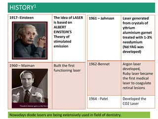


This document provides an overview of lasers in dentistry. It discusses the history and development of lasers, how lasers are designed and how laser light interacts with tissues. It describes common dental lasers like CO2 and argon lasers, and their applications. CO2 lasers are well absorbed in oral tissues and useful for soft tissue procedures. Argon lasers are absorbed by hemoglobin and melanin, making them good for coagulation. The document outlines the benefits of lasers for various dental procedures.


