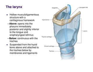
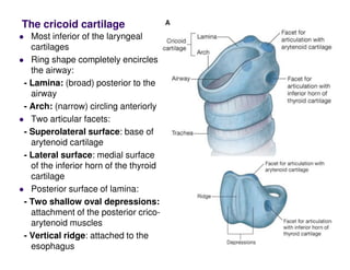
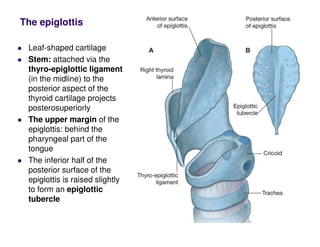
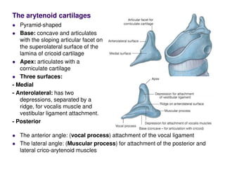
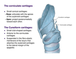
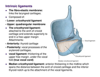
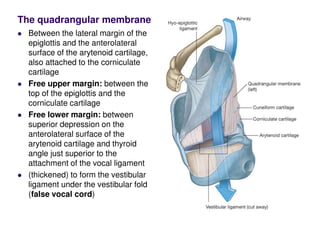
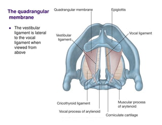
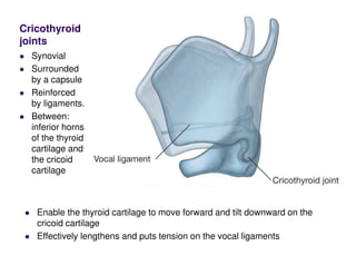
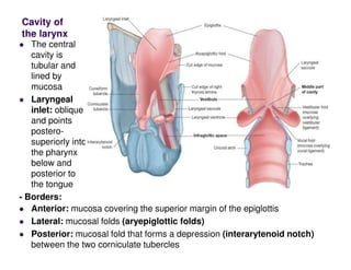
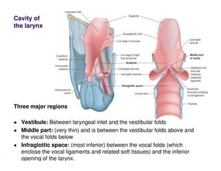
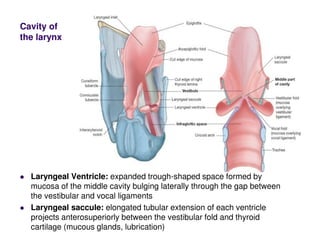
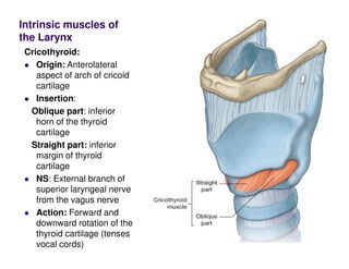
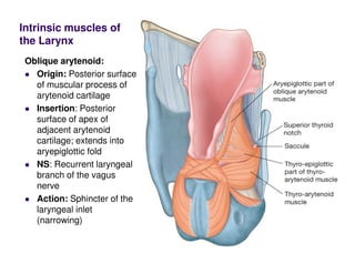
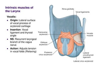
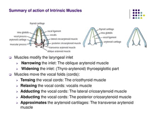
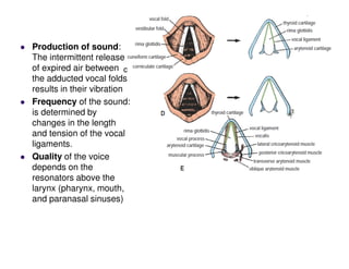
The larynx
Hollow musculoligamentous
structure with a
cartilaginous framework
- Above: opens into the
pharynx immediately
posterior and slightly inferior
to the tongue and
oropharyngeal isthmus
- Below: continuous with the
trachea
Suspended from the hyoid
bone above and attached to
the trachea below by
membranes and ligaments
The cricoid cartilage
Most inferior of the laryngeal
cartilages
Ring shape completely encircles
the airway:
- Lamina: (broad) posterior to the
airway
- Arch: (narrow) circling anteriorly
Two articular facets:
- Superolateral surface: base of
arytenoid cartilage
- Lateral surface: medial surface
of the inferior horn of the thyroid
cartilage
Posterior surface of lamina:
- Two shallow oval depressions:
attachment of the posterior crico-
arytenoid muscles
- Vertical ridge: attached to the
esophagus
The thyroid cartilage
Largest: right and a left
lamina
Posteriorly: Separated
Anteriorly: Joined
Laryngeal prominence:
The most superior point of
the site of fusion between
the two broad flat laminae
Superior thyroid notch:
superior to the laryngeal
prominence,
Inferior thyroid notch:
along the base of the
thyroid cartilage
- The posterior margin of each lamina is elongated to form:
Superior horn: connected to the posterior end of the greater horn of the
hyoid bone by a lateral thyrohyoid ligament.
Inferior horn: articulation with the cricoid cartilage
The epiglottis
Leaf-shaped cartilage
Stem: attached via the
thyro-epiglottic ligament
(in the midline) to the
posterior aspect of the
thyroid cartilage projects
posterosuperiorly
The upper margin of the
epiglottis: behind the
pharyngeal part of the
tongue
The inferior half of the
posterior surface of the
epiglottis is raised slightly
to form an epiglottic
tubercle
The arytenoid cartilages
Pyramid-shaped
Base: concave and articulates
with the sloping articular facet on
the superolateral surface of the
lamina of cricoid cartilage
Apex: articulates with a
corniculate cartilage
Three surfaces:
- Medial
- Anterolateral: has two
depressions, separated by a
ridge, for vocalis muscle and
vestibular ligament attachment.
- Posterior
The anterior angle: (vocal process) attachment of the vocal ligament
The lateral angle: (Muscular process) for attachment of the posterior and
lateral crico-arytenoid muscles
The corniculate cartilages
Small conical cartilages:
- Base: articulate with the apices
of the arytenoid cartilages
- Apex: project posteromedially
toward each other.
The Cuneiform cartilages:
Small club-shaped cartilages
Anterior to the corniculate
cartilages
Suspended in the fibro-elastic
membrane of the larynx that
attaches the arytenoid cartilages
to the lateral margin of the
epiglottis
Extrinsic ligaments
The thyrohyoid membrane:
tough fibro-elastic ligament
between the superior margin of
the thyroid cartilage below and
the hyoid bone above
- Opening for the superior laryngeal
arteries, nerves, and lymphatics
- lateral thyrohyoid ligaments:
thickening in the posterior
borders of the thyrohyoid
membrane
- Median thyrohyoid ligament:
Anterior thickening in the midline
Hyo-epiglottic ligament: from
the midline of the epiglottis to the
body of the hyoid bone.
Cricotracheal ligament
Intrinsic ligaments
The fibro-elastic membrane:
links the laryngeal cartilages.
Composed of:
- Lower: cricothyroid ligament
- Upper: quadrangular membrane
The cricothyroid ligament:
attached to the arch of cricoid
cartilage and extends superiorly to
end in a free upper margin
Attachments:
- Anteriorly: thyroid cartilage
- Posteriorly: vocal processes of the
arytenoid cartilages.
vocal ligament: thickening of the
upper free margin. under the vocal
fold (true vocal cord)
Median cricothyroid ligament: anterior thickening in the midline which
spans the distance between the arch of cricoid cartilage and the inferior
thyroid notch up to the attachment of the vocal ligaments.
The quadrangular membrane
Between the lateral margin of the
epiglottis and the anterolateral
surface of the arytenoid cartilage,
also attached to the corniculate
cartilage
Free upper margin: between the
top of the epiglottis and the
corniculate cartilage
Free lower margin: between
superior depression on the
anterolateral surface of the
arytenoid cartilage and thyroid
angle just superior to the
attachment of the vocal ligament
(thickened) to form the vestibular
ligament under the vestibular fold
(false vocal cord)
The quadrangular
membrane
The vestibular
ligament is lateral
to the vocal
ligament when
viewed from
above
Cricothyroid
joints
Synovial
Surrounded
by a capsule
Reinforced
by ligaments.
Between:
inferior horns
of the thyroid
cartilage and
the cricoid
cartilage
Enable the thyroid cartilage to move forward and tilt downward on the
cricoid cartilage
Effectively lengthens and puts tension on the vocal ligaments
Crico-arytenoid joints
Synovial
Surrounded by a
capsule
Reinforced by
ligaments.
Between articular
facets on the
superolateral
surfaces of the
cricoid cartilage and
the bases of the
arytenoid cartilages
Enable the arytenoid cartilages to slide away or toward each other
and to rotate so that the vocal processes pivot either toward or
away from the midline (abduction and adduction of the vocal
ligaments)
Cavity of
the larynx
The central
cavity is
tubular and
lined by
mucosa
Laryngeal
inlet: oblique
and points
postero-
superiorly into
the pharynx
below and
posterior to
the tongue
- Borders:
Anterior: mucosa covering the superior margin of the epiglottis
Lateral: mucosal folds (aryepiglottic folds)
Posterior: mucosal fold that forms a depression (interarytenoid notch)
between the two corniculate tubercles
Cavity of
the larynx
Inferior opening
Continuous with the lumen of the trachea
Completely encircled by the cricoid cartilage
Horizontal in position
Continuously opened
Cavity of
the larynx
Vestibule: Between laryngeal inlet and the vestibular folds
Middle part: (very thin) and is between the vestibular folds above and
the vocal folds below
Infraglottic space: (most inferior) between the vocal folds (which
enclose the vocal ligaments and related soft tissues) and the inferior
opening of the larynx.
Three major regions
Cavity of
the larynx
Laryngeal Ventricle: expanded trough-shaped space formed by
mucosa of the middle cavity bulging laterally through the gap between
the vestibular and vocal ligaments
Laryngeal saccule: elongated tubular extension of each ventricle
projects anterosuperiorly between the vestibular fold and thyroid
cartilage (mucous glands, lubrication)
Cavity of
the larynx
Rima vestibuli:
triangular opening
between the two
adjacent vestibular
folds at the entrance to
the middle chamber of
the laryngeal cavity.
Rima glottidis:
narrow triangular
opening between the
two adjacent vocal
folds. This opening
separates the middle
chamber above from
the infraglottic cavity
below
Intrinsic muscles of
the Larynx
Cricothyroid:
Origin: Anterolateral
aspect of arch of cricoid
cartilage
Insertion:
Oblique part: inferior
horn of the thyroid
cartilage
Straight part: inferior
margin of thyroid
cartilage
NS: External branch of
superior laryngeal nerve
from the vagus nerve
Action: Forward and
downward rotation of the
thyroid cartilage (tenses
vocal cords)
Intrinsic muscles of
the Larynx
Posterior cricoarytenoid:
Origin: Oval depression
on posterior surface of
lamina of cricoid
cartilage
Insertion: Posterior
surface of muscular
process of arytenoid
cartilage
NS: Recurrent laryngeal
branch of the vagus
nerve
Action: Abduction of the
arytenoid cartilage
Intrinsic muscles of
the Larynx
Lateral cricoarytenoid:
Origin: Superior surface
of arch of cricoid
cartilage
Insertion: Anterior
surface of muscular
process of arytenoid
cartilage
NS: Recurrent laryngeal
branch of the vagus
nerve
Action: Internal rotation
of the arytenoid cartilage
and adduction of vocal
folds
Intrinsic muscles of
the Larynx
Transverse arytenoid:
Origin: Lateral border of
posterior surface of
arytenoid cartilage
Insertion: Lateral border
of posterior surface of
opposite arytenoid
cartilage
NS: Recurrent laryngeal
branch of the vagus
nerve
Action: Adduction of
arytenoid cartilages
Intrinsic muscles of
the Larynx
Oblique arytenoid:
Origin: Posterior surface
of muscular process of
arytenoid cartilage
Insertion: Posterior
surface of apex of
adjacent arytenoid
cartilage; extends into
aryepiglottic fold
NS: Recurrent laryngeal
branch of the vagus
nerve
Action: Sphincter of the
laryngeal inlet
(narrowing)
Intrinsic muscles of
the Larynx
Thyro-arytenoid:
Origin: Thyroid angle
and adjacent cricothyroid
ligament
Insertion: Anterolateral
surface of arytenoid
cartilage; some fibers
continue in aryepiglottic
folds to the lateral
margin of the epiglottis
NS: Recurrent laryngeal
branch of the vagus
nerve
Action: Sphincter of
vestibule and of
laryngeal inlet (widening)
Intrinsic muscles of
the Larynx
Vocalis:
Origin: Lateral surface
of vocal process of
arytenoid cartilage
Insertion: Vocal
ligament and thyroid
angle
NS: Recurrent laryngeal
branch of the vagus
nerve
Action: Adjusts tension
in vocal folds (Relaxing)
Blood supply
of the larynx
Superior
laryngeal
artery :
(from
superior
thyroid
artery)
Inferior
laryngeal
artery:
(from
inferior
thyroid
artery)
Superior laryngeal
nerve:
- Internal laryngeal
nerve: (sensory)
supplies the laryngeal
cavity down to the level
of the vocal folds
- External laryngeal
nerve: (Motor) supplies
the cricothyroid muscle
Recurrent laryngeal
nerve:
- sensory: to the laryngeal
cavity below the level of
the vocal folds
- motor: to all intrinsic
muscles of the larynx
except for the
cricothyroid
Nerve supply of the larynx
Muscles modify the laryngeal inlet
Narrowing the inlet: The oblique arytenoid muscle
Widening the inlet: (Thyro-arytenoid) thyroepiglottic part
Muscles move the vocal folds (cords):
Tensing the vocal cords: The cricothyroid muscle
Relaxing the vocal cords: vocalis muscle
Adducting the vocal cords: The lateral cricoarytenoid muscle
Abducting the vocal cords: The posterior cricoarytenoid muscle
Approximates the arytenoid cartilages: The transverse arytenoid
muscle
Summary of action of Intrinsic Muscles
Quiet inspiration: the
vocal folds are abducted
and the rima glottidis is
triangular in shape
Deep inspiration: the
vocal folds are
maximally abducted
glottis becomes a
diamond shape
because of the maximal
lateral rotation of the
arytenoid cartilages
Expiration: the vocal
folds are adducted,
leaving a small gap
between them
Movements of the
Vocal Folds with
Respiration
Production of sound:
The intermittent release
of expired air between
the adducted vocal folds
results in their vibration
Frequency of the sound:
is determined by
changes in the length
and tension of the vocal
ligaments.
Quality of the voice
depends on the
resonators above the
larynx (pharynx, mouth,
and paranasal sinuses)
Section of the external laryngeal nerve
produces weakness of the voice because the
vocal fold cannot be tensed. (cricothyroid
muscle is paralyzed)
Unilateral complete section of the recurrent
laryngeal nerve: results in the vocal fold on
the affected side assuming the position
midway between abduction and adduction. It
lies just lateral to the midline. Speech is not
greatly affected because the other vocal fold
compensates to some extent and moves
toward the affected vocal fold
Bilateral complete section of the recurrent
laryngeal nerve results in both vocal folds
assuming the position midway between
abduction and adduction. Breathing is
impaired because the rima glottidis is partially
closed, and speech is lost
Unilateral partial section of the recurrent
laryngeal nerve: results in a greater degree
of paralysis of the abductor muscles than of
the adductor muscles
Bilateral partial section of the recurrent
laryngeal nerve: results in bilateral paralysis
of the abductor muscles and the drawing
together of the vocal folds Acute
breathlessness (dyspnea) and stridor follow
cricothyroidotomy or tracheostomy is
necessary

larynx.pdf

  • 1.
    The larynx Hollow musculoligamentous structurewith a cartilaginous framework - Above: opens into the pharynx immediately posterior and slightly inferior to the tongue and oropharyngeal isthmus - Below: continuous with the trachea Suspended from the hyoid bone above and attached to the trachea below by membranes and ligaments
  • 2.
    The cricoid cartilage Mostinferior of the laryngeal cartilages Ring shape completely encircles the airway: - Lamina: (broad) posterior to the airway - Arch: (narrow) circling anteriorly Two articular facets: - Superolateral surface: base of arytenoid cartilage - Lateral surface: medial surface of the inferior horn of the thyroid cartilage Posterior surface of lamina: - Two shallow oval depressions: attachment of the posterior crico- arytenoid muscles - Vertical ridge: attached to the esophagus
  • 3.
    The thyroid cartilage Largest:right and a left lamina Posteriorly: Separated Anteriorly: Joined Laryngeal prominence: The most superior point of the site of fusion between the two broad flat laminae Superior thyroid notch: superior to the laryngeal prominence, Inferior thyroid notch: along the base of the thyroid cartilage - The posterior margin of each lamina is elongated to form: Superior horn: connected to the posterior end of the greater horn of the hyoid bone by a lateral thyrohyoid ligament. Inferior horn: articulation with the cricoid cartilage
  • 4.
    The epiglottis Leaf-shaped cartilage Stem:attached via the thyro-epiglottic ligament (in the midline) to the posterior aspect of the thyroid cartilage projects posterosuperiorly The upper margin of the epiglottis: behind the pharyngeal part of the tongue The inferior half of the posterior surface of the epiglottis is raised slightly to form an epiglottic tubercle
  • 5.
    The arytenoid cartilages Pyramid-shaped Base:concave and articulates with the sloping articular facet on the superolateral surface of the lamina of cricoid cartilage Apex: articulates with a corniculate cartilage Three surfaces: - Medial - Anterolateral: has two depressions, separated by a ridge, for vocalis muscle and vestibular ligament attachment. - Posterior The anterior angle: (vocal process) attachment of the vocal ligament The lateral angle: (Muscular process) for attachment of the posterior and lateral crico-arytenoid muscles
  • 6.
    The corniculate cartilages Smallconical cartilages: - Base: articulate with the apices of the arytenoid cartilages - Apex: project posteromedially toward each other. The Cuneiform cartilages: Small club-shaped cartilages Anterior to the corniculate cartilages Suspended in the fibro-elastic membrane of the larynx that attaches the arytenoid cartilages to the lateral margin of the epiglottis
  • 7.
    Extrinsic ligaments The thyrohyoidmembrane: tough fibro-elastic ligament between the superior margin of the thyroid cartilage below and the hyoid bone above - Opening for the superior laryngeal arteries, nerves, and lymphatics - lateral thyrohyoid ligaments: thickening in the posterior borders of the thyrohyoid membrane - Median thyrohyoid ligament: Anterior thickening in the midline Hyo-epiglottic ligament: from the midline of the epiglottis to the body of the hyoid bone. Cricotracheal ligament
  • 8.
    Intrinsic ligaments The fibro-elasticmembrane: links the laryngeal cartilages. Composed of: - Lower: cricothyroid ligament - Upper: quadrangular membrane The cricothyroid ligament: attached to the arch of cricoid cartilage and extends superiorly to end in a free upper margin Attachments: - Anteriorly: thyroid cartilage - Posteriorly: vocal processes of the arytenoid cartilages. vocal ligament: thickening of the upper free margin. under the vocal fold (true vocal cord) Median cricothyroid ligament: anterior thickening in the midline which spans the distance between the arch of cricoid cartilage and the inferior thyroid notch up to the attachment of the vocal ligaments.
  • 9.
    The quadrangular membrane Betweenthe lateral margin of the epiglottis and the anterolateral surface of the arytenoid cartilage, also attached to the corniculate cartilage Free upper margin: between the top of the epiglottis and the corniculate cartilage Free lower margin: between superior depression on the anterolateral surface of the arytenoid cartilage and thyroid angle just superior to the attachment of the vocal ligament (thickened) to form the vestibular ligament under the vestibular fold (false vocal cord)
  • 10.
    The quadrangular membrane The vestibular ligamentis lateral to the vocal ligament when viewed from above
  • 11.
    Cricothyroid joints Synovial Surrounded by a capsule Reinforced byligaments. Between: inferior horns of the thyroid cartilage and the cricoid cartilage Enable the thyroid cartilage to move forward and tilt downward on the cricoid cartilage Effectively lengthens and puts tension on the vocal ligaments
  • 12.
    Crico-arytenoid joints Synovial Surrounded bya capsule Reinforced by ligaments. Between articular facets on the superolateral surfaces of the cricoid cartilage and the bases of the arytenoid cartilages Enable the arytenoid cartilages to slide away or toward each other and to rotate so that the vocal processes pivot either toward or away from the midline (abduction and adduction of the vocal ligaments)
  • 13.
    Cavity of the larynx Thecentral cavity is tubular and lined by mucosa Laryngeal inlet: oblique and points postero- superiorly into the pharynx below and posterior to the tongue - Borders: Anterior: mucosa covering the superior margin of the epiglottis Lateral: mucosal folds (aryepiglottic folds) Posterior: mucosal fold that forms a depression (interarytenoid notch) between the two corniculate tubercles
  • 14.
    Cavity of the larynx Inferioropening Continuous with the lumen of the trachea Completely encircled by the cricoid cartilage Horizontal in position Continuously opened
  • 15.
    Cavity of the larynx Vestibule:Between laryngeal inlet and the vestibular folds Middle part: (very thin) and is between the vestibular folds above and the vocal folds below Infraglottic space: (most inferior) between the vocal folds (which enclose the vocal ligaments and related soft tissues) and the inferior opening of the larynx. Three major regions
  • 16.
    Cavity of the larynx LaryngealVentricle: expanded trough-shaped space formed by mucosa of the middle cavity bulging laterally through the gap between the vestibular and vocal ligaments Laryngeal saccule: elongated tubular extension of each ventricle projects anterosuperiorly between the vestibular fold and thyroid cartilage (mucous glands, lubrication)
  • 17.
    Cavity of the larynx Rimavestibuli: triangular opening between the two adjacent vestibular folds at the entrance to the middle chamber of the laryngeal cavity. Rima glottidis: narrow triangular opening between the two adjacent vocal folds. This opening separates the middle chamber above from the infraglottic cavity below
  • 18.
    Intrinsic muscles of theLarynx Cricothyroid: Origin: Anterolateral aspect of arch of cricoid cartilage Insertion: Oblique part: inferior horn of the thyroid cartilage Straight part: inferior margin of thyroid cartilage NS: External branch of superior laryngeal nerve from the vagus nerve Action: Forward and downward rotation of the thyroid cartilage (tenses vocal cords)
  • 19.
    Intrinsic muscles of theLarynx Posterior cricoarytenoid: Origin: Oval depression on posterior surface of lamina of cricoid cartilage Insertion: Posterior surface of muscular process of arytenoid cartilage NS: Recurrent laryngeal branch of the vagus nerve Action: Abduction of the arytenoid cartilage
  • 20.
    Intrinsic muscles of theLarynx Lateral cricoarytenoid: Origin: Superior surface of arch of cricoid cartilage Insertion: Anterior surface of muscular process of arytenoid cartilage NS: Recurrent laryngeal branch of the vagus nerve Action: Internal rotation of the arytenoid cartilage and adduction of vocal folds
  • 21.
    Intrinsic muscles of theLarynx Transverse arytenoid: Origin: Lateral border of posterior surface of arytenoid cartilage Insertion: Lateral border of posterior surface of opposite arytenoid cartilage NS: Recurrent laryngeal branch of the vagus nerve Action: Adduction of arytenoid cartilages
  • 22.
    Intrinsic muscles of theLarynx Oblique arytenoid: Origin: Posterior surface of muscular process of arytenoid cartilage Insertion: Posterior surface of apex of adjacent arytenoid cartilage; extends into aryepiglottic fold NS: Recurrent laryngeal branch of the vagus nerve Action: Sphincter of the laryngeal inlet (narrowing)
  • 23.
    Intrinsic muscles of theLarynx Thyro-arytenoid: Origin: Thyroid angle and adjacent cricothyroid ligament Insertion: Anterolateral surface of arytenoid cartilage; some fibers continue in aryepiglottic folds to the lateral margin of the epiglottis NS: Recurrent laryngeal branch of the vagus nerve Action: Sphincter of vestibule and of laryngeal inlet (widening)
  • 24.
    Intrinsic muscles of theLarynx Vocalis: Origin: Lateral surface of vocal process of arytenoid cartilage Insertion: Vocal ligament and thyroid angle NS: Recurrent laryngeal branch of the vagus nerve Action: Adjusts tension in vocal folds (Relaxing)
  • 25.
    Blood supply of thelarynx Superior laryngeal artery : (from superior thyroid artery) Inferior laryngeal artery: (from inferior thyroid artery)
  • 27.
    Superior laryngeal nerve: - Internallaryngeal nerve: (sensory) supplies the laryngeal cavity down to the level of the vocal folds - External laryngeal nerve: (Motor) supplies the cricothyroid muscle Recurrent laryngeal nerve: - sensory: to the laryngeal cavity below the level of the vocal folds - motor: to all intrinsic muscles of the larynx except for the cricothyroid Nerve supply of the larynx
  • 28.
    Muscles modify thelaryngeal inlet Narrowing the inlet: The oblique arytenoid muscle Widening the inlet: (Thyro-arytenoid) thyroepiglottic part Muscles move the vocal folds (cords): Tensing the vocal cords: The cricothyroid muscle Relaxing the vocal cords: vocalis muscle Adducting the vocal cords: The lateral cricoarytenoid muscle Abducting the vocal cords: The posterior cricoarytenoid muscle Approximates the arytenoid cartilages: The transverse arytenoid muscle Summary of action of Intrinsic Muscles
  • 29.
    Quiet inspiration: the vocalfolds are abducted and the rima glottidis is triangular in shape Deep inspiration: the vocal folds are maximally abducted glottis becomes a diamond shape because of the maximal lateral rotation of the arytenoid cartilages Expiration: the vocal folds are adducted, leaving a small gap between them Movements of the Vocal Folds with Respiration
  • 30.
    Production of sound: Theintermittent release of expired air between the adducted vocal folds results in their vibration Frequency of the sound: is determined by changes in the length and tension of the vocal ligaments. Quality of the voice depends on the resonators above the larynx (pharynx, mouth, and paranasal sinuses)
  • 31.
    Section of theexternal laryngeal nerve produces weakness of the voice because the vocal fold cannot be tensed. (cricothyroid muscle is paralyzed) Unilateral complete section of the recurrent laryngeal nerve: results in the vocal fold on the affected side assuming the position midway between abduction and adduction. It lies just lateral to the midline. Speech is not greatly affected because the other vocal fold compensates to some extent and moves toward the affected vocal fold Bilateral complete section of the recurrent laryngeal nerve results in both vocal folds assuming the position midway between abduction and adduction. Breathing is impaired because the rima glottidis is partially closed, and speech is lost
  • 32.
    Unilateral partial sectionof the recurrent laryngeal nerve: results in a greater degree of paralysis of the abductor muscles than of the adductor muscles Bilateral partial section of the recurrent laryngeal nerve: results in bilateral paralysis of the abductor muscles and the drawing together of the vocal folds Acute breathlessness (dyspnea) and stridor follow cricothyroidotomy or tracheostomy is necessary