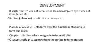
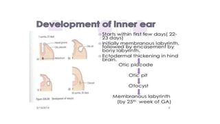
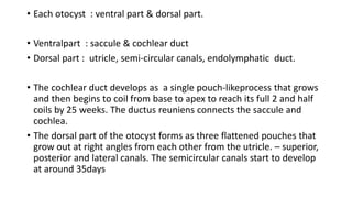
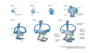
INNER EAR
DEVELOPMENT
• It starts from 3rd week of intrauterine life and complete by 16 week of
intrauterine life.
Otic discs ( placodes) - otic pits – otocysts..
• Placode or otic disc: Ectoderm over the hindbrain, thickens to
form otic discs
• Otic pits : otic discs which invaginate to form oticpits.
• Otocysts: otic pits separate from the surface to form otocysts
• Each otocyst : ventral part & dorsal part.
• Ventralpart : saccule & cochlear duct
• Dorsal part : utricle, semi-circular canals, endolymphatic duct.
• The cochlear duct develops as a single pouch-likeprocess that grows
and then begins to coil from base to apex to reach its full 2 and half
coils by 25 weeks. The ductus reuniens connects the saccule and
cochlea.
• The dorsal part of the otocyst forms as three flattened pouches that
grow out at right angles from each other from the utricle. – superior,
posterior and lateral canals. The semicircular canals start to develop
at around 35days
• The organ of Corti starts developing as a single block of heaped up
ectodermal cells at about 11 weeks.
• Within the membranous labyrinth the sensory cells of the three
cristae, two maculae and the organ of Corti are beginning to develop
from areas of ectodermal specialization.
Anatomy
Bony labrynth
• Has a central part called bony vestibule.
• Anteriorly – connected to cochlea
• Posteriorly –connect to three bony semi-circular canals.
• divided to three parts : a) bony vestibule
• b) semi circular canals
• c) cochlea
Bony vestibule:
• Boundaries: lateral wall : medial wall to middle ear
medial wall : internal acoustic meatus
anterior : cochlea
posterior : three semi circular canals
bean shaped opening present lateral to vestibule : fenestra vestibulae (oval window)
occupied by foot plate of stapes and annular ligament (is a ring of fibrous tissue that
connects the base of the stapes to the oval window of the inner ear).
Three RECESS:
Spherical recess : anteriorly, is a depression on the medial wall ….space for saccule
elliptical recess: psnt posteriorly, is a space for utricle. Both recess are separated by
vestibular crest.
Cochlear recess :The vestibular crest bifurcates below to enclose a small depression.
For cochlear nerve fibers.
Maculae cribrosa media: wall with minute holes for passage of inferior vestibular
nerve filaments.
• Mike’s dot : aka macula cribrosa superior , the elliptical recess perforated by
small holes for the nerves to utricle and ampula of sup and lat semi-circular
canals. ( An imp land mark for trans-labyrinthine approach )
Semi circular canals:
• Three in number.
superior, posterior or vertical & lateral or horizontal
Unequal in length
Occupies two-third of circle
Diameter of 0.8mm
Ampulla : all three canals show dilation at one end.
Crus commune : non-ampulated ends of posterior and
Superior semi-circular canal (4mm length)
Open into medial part of vestibule
• SUPERIOR SEMICIRCULAR CANAL:
• 15 – 20 mm length,
• Lie transverse to the bony axis of petrous portion of temporal bone.
• Ampulated end : open in to upper lateral part of vestibule, non ampulated end : fuses with posterior
canal to form crus commune open in to medial part of vestibule.
• LATERAL SEMICIRCULAR CANAL :
• 12 TO 15mm long
• Lies 30degree angle to horizontal plane
• Ampulated end : open in to upper part of vestibule, non ampulated: in to lower part below the crus
commune.
• POSTERIOR SEMICIRCULAR CANAL:
• 18- 22 mm long
• Lies parallel and close to posterior surface of petrous portion of temporal bone.
• Ampulated end : opens in to lower part of vestibule . Non ampulated: joins crus commune
SOLID ANGLE:
• Angle formed by three semi-circular canals.
• Aka “TRAUTMAN’S TRIANGLE”
Weak point, which infections of
temporal bone may traverse and
affect cerebellum , involving the
posterior cranial fossa
cochlea
• Resemble a common snail
• It is 5mm from base to apex , 9mm around its base , 30mm in length
• It is a hallow tube having 2 and three-fourth turn around a conical central axis- MODIOLUS
• Base is towards the interal auditory meatus and is perforated for cochlear nerve passage.
• Apex lies medial to tensor tympani muscle.
Bony cochlea:
• has three longitudinal compartments :
• 1) scala vestibuli
• 2) scala tympani
• 3) scala media.
• Scala vestibuli communicate with scala tympani at apex of cochlea : HELICOTREMA
• Scala vestibuli : is the continuity with the vestibule at the oval window , closed by stapes
foot plate.
• Scala tympani : is the lower most channel, close by secondary tm of round window.
• Promontory: a bony bulge in medial wall, represent basal coil of cochlea.
• Fenestra cochlea or Round window : lateral wall of internal ear, scala tympani is closed
by secondary TM or RW.
• Helicotrema: the scala tympani and scala vestibuli ,communicate each other at the apex of
cochlea. Filled with perilymph .
• Aqueduct of cochlea : it connect scala tympani to subarachnoid space.
• it regulate perilymph and pressure in bony labyrinth.
• Peripheral tractus spiralis foraminosa: openings in the cochlear area of the
bottom of the internal acoustic meatus through which the fibers of the cochlear
nerve leave the bony labyrinth to enter the cranial cavity
PERILYMPH
• It fills the space between bony and membranous labyrinth.
• It resembles extracellular fluid and rich in “ SODIUM IONS”
• The “Aqueduct of cochlea” provides communication between scala tympani and
subarachnoid space.
• Source:
• csf reaching labyrinth via aqueduct of cochlea
• filterd of blood serum from capillaries of spiral ligament
Membraneous labyrinth:
• Divided in to three parts :
• a) memb vestibular labyrinth
• b) memb semi circular canals
• c) memb cochlear labyrinth
Membranous vestibular labyrinth
• It consists of : saccule, utricle , the endolymphatic duct and sac.
• MACULA: The vestibular receptor organs of saccule and utricle are called macula.
• SACCULE: It lies in a spherical recess in medial wall of bony vestibule.
• it is hook shaped, lies in a vertical position
• area is about 2.4mm.
• saccular macula contains of approximately 18,000 hair cells.
• UTRICLE: it lies in elliptical recess of the bony vestibule. Utricle is bigger than saccule.
• lies in horizontal position , area is about 4.32mm.
• utricular macula contains of approximately 33,000 hair cells.
Endolymphatic duct and sac
• The utricle and saccule unite to form “ UTRICULOSACCULAR DUCT” . It continue
as a “ Endolymphatic duct”
• The terminal part of endolymphatic duct dilate and form endolymphatic sac.
• They both involve in reabsorption and regulation of endolymph.
• The two vestibular receptor organs are:
• . Macula and cristae.
The vestibular receptor composed of :
a)Hair cells or Sensory neuroepithelium
b) supporting cells
c) gelatinous mass.
HAIR CELLS:
Type 1:
flask shaped , surrounded by nerve chalice formed by afferent fiber of
vestibulocochlear nerve.
predominantly seen in the summit of CRISTAE.
resemble inner hair cell of organ of corti.
Type 2 :
cylindrical in shape, no nerve chalice.
predominantly seen in periphery of CRISTAE.
resemble outer hair cell of organ of corti.
type 2 hair cell synapse with the outer membrane of calyces surrounding type 1
hair cells.
• The upper surface of hair cell contain:
• STERCOCILIA and KINOCILLIUM
• Stercocilia : approximately 70 in number
• smaller than kinocilium
• stercocilia in macula are few microns long, whereas in crista : 35um.
• they contain actin filaments,
• Kinocillium : single in number .
• tall and prominent
• not uniformly arranged in macula.
• The upper surface of the cell contains a thicker region called “ CUTICULAR PLATE” into
which the steriocilia extend their rootlets.
• STRIOLA: a curved line divide macula into medial and lateral halves.
supporting cells : “ DEITERS CELLS”
contain dense material , rich in actin , strongly attached to the hair cell
base.
GELATINOUS MASS : composed of mucopolysaccharides secreted by supporting cells.
OTOLITH or STATOCONIA : consist of small cylindrical and hexagonally shaped bodies
with pointed ends made up of a calcium carbonate crystals .
• These are anchored with gelatinous layer forming OTOCONIAL MEMBRANE .
Otoconial layer:
• Consist of gelatinous layer, sub gelatinous space and
otoconia.
• Measure about 3-19 um
• Consist of protein matrix and inorganic calcium
carbonate crystals.
Membranous semicircular canal
• Cristae Ampullaris: lie in the ampullated end of three semi circular duct
• response to angular acceleration .
• Lie the vestibular epithelium along with hair cells.
• CUPULA: it is a gelatinous mass , extend from surface of cristae to ceiling of ampulla.
• it is dome shaped hence called cupula.
• the gelatinous mass made of glycoprotiens and proteoglycans arranged in
filamentous network.
• the haircells project into gelatinous mass.
Membranous cochlea
Cochlear Duct : it is subdivided by two longitudinally running membranes , that separate
into three chambers, the scala tympani, scala media, sclala vestibuli.
Organ of corti : runs in a spiral along the floor of the scala media, situated on acellular
layer called basement membrane.
• Osseous spiral lamina: the basiliar membrane stretches across the cochlear duct
from a bony shelf around central bony modiolus.
• Spiral ganglion : comprised of bipolar neuron , innervate the hair cells and centrally
from cochlear nerve .
SCALA MEDIA:
• Occupies the mid portion of the cochlear canal , triangular in cross section.
• Boundaries:
• Floor: basiliar membrane (BM)
• Roof: reissner’s membrane
• lateral wall: stria vascularis and bony wall of cochlea.
• 1) Basiliar membrane supports the “ORGAN OF CORTI”
• zona arcuata : thin , inner layer of BM.
• zona pectinata : thicker , outer layer of BM.
• Its length increases as it proceeds from basal coil to apical coil.
• High frequency sounds heard at basal , low frequency sound at apical coil .
• 2) Reissner’s membrane: separate scala media from scala vestibuli.
• has two layers of cell : Mesothelial cell , layer facing scala tympani
• Endothelial cell, layer facing scala media. Thick layer cover
by microvilli.
• 3) Stria vascularis : lateral wall, contain vascular epithelium.
• Has a layer of fibrocytes and connective tissue called “SPIRAL LIGAMENT”
• Has three layers of cell : Marginal cells: facing scala media
• Intermediate cells: highly convuluted membrane
• Basal cell
• The cell contain ion pumps, enzymes and proteins, asso with homeostatic mechanism maintain
the composition of fluid of cochlea.
• It secretes “ ENDOLYMPH”
• High in potassium (K) , Low in sodium (Na)
• Absorption : Endolymphatic sac
• THE LONGITUDINAL FLOW THEORY : flow from cochlear duct - saccule - utricle –
endolymphatic duct - endolymphatic sac.
•
ORGAN OF CORTI:
• “ SENSORY ORGAN OF HEARING”
• Spread like a ribbon along the basilar membrane .
• It is 35mm in length .
• COMPONENTS OF ORGAN OF CORTI
• 1) Tunnel of corti
• 2) Hair cells
• 3) Supporting cells
• 4)Tectorial membrane .
a) Tunnel of corti : this tunnel, situated between the inner and out rods,
• contain fluid called “ cortilymph”
• B) Hair cells : Important receptor cells of hearing transdce sound energy in to electrical
energy.
• Two in number : inner and outer
• INNER HAIR CELLS : single in row, flask shaped . Contain 3 or 4 steriocilia.
• this cells do not insert in to tectorial membrane.
• sound induced motion of the basilar membrane stimulate the inner
hair cell bundle via fluid motion of the endolymph between tectorial membrane and hair
cell apex.
• The nerve fibers are afferent cochlear fibers , supply by 95% .
•
• OUTER HAIR CELLS: three or four in rows , cyllindrical in shape.
• the sterocilia insert in to tectorial membrane .
• afferent supply by spiral ganglion of cochlear nerve (5%)
• efferent supply from superior olivary complex bundle.
•
SUPPORTING CELLS
• 1) DEITERS CELLS : situated between outer hair cells, rich in actin
• strongly attached at the base of hair cells and support ohc.
2) CELLS OF HENSEN : elongated columnar shaped cells, rich in lipid droplets.
3) CELLS OF BOETTCHER: polyhedral shape cells, present on basilar membrane .
support the organ of corti
4) CELLS OF CLAUDIUS : single layer of cuboidal cells, cover the basilar membrane.
TECTORIAL MEMBRANE
• The membrane which overlies the organ of corti
• They extend along the longitudinal length of cochlea along basilar membrane.
• Consist of gelatinous matrix and delicate fibers.
• The shearing force between the hair cells and tectorial membrane stimulate hair cells.
Arterial supply of labyrinth
• Internal Auditory or Labyrinthine artery
• 1) ant vestibular art : utricle and lateral and superior scc.
• 2) common cochlear art:
A) Main cochlear artery : supplies cochlea (80%)
B) Vestibulocochlear artery :
1) Posterior vestibular artery: saccule and posterior scc
2) cochlear branch : 20% of cochlea
Venous drainage
• Mainly through :
• Internal Auditory Vein
• Vein of Cochlear aqueduct
• Vein of vestibular aqueduct
• these three veins drains in to :
• Inferior petrosal and sigmoid sinuses .
Nerve supply
• The Vestibular nerve is the main supply.
• With in the Internal acoustic meatus , form a “ vestibular ganglion” aka
“ ganglion of scarpa”
• through ganglion the nerve split In to:
• superior branch supplies : “Macula of utricle & ampulla of
sup and lat scc”
• Inferior branch supplies : “ Macula of saccule”
• posterior branch : through ‘ foramen singulare’ supply the
ampula of post scc.
• Foramen singulare: present at posterio - inferior part of iam .
INTERNAL AUCOSTIC MEATUS
• Canal present with in the pterous part of temporal bone of skull with in the posterior
canal fossa and the inner ear.
• Passage for Facial nerve, Vestibulocochlear nerve and Labrynthine artery.
• It is divided into :
• Ant Superior & Ant inferior
• Post Superior & Post inferior
• BILLS BAR : separate Anterior and posterior part
• FALCIFORM CREST : separate superior and inferior part
THANK YOU

Inner ear embryology and anatomy

  • 1.
  • 2.
    DEVELOPMENT • It startsfrom 3rd week of intrauterine life and complete by 16 week of intrauterine life. Otic discs ( placodes) - otic pits – otocysts.. • Placode or otic disc: Ectoderm over the hindbrain, thickens to form otic discs • Otic pits : otic discs which invaginate to form oticpits. • Otocysts: otic pits separate from the surface to form otocysts
  • 4.
    • Each otocyst: ventral part & dorsal part. • Ventralpart : saccule & cochlear duct • Dorsal part : utricle, semi-circular canals, endolymphatic duct. • The cochlear duct develops as a single pouch-likeprocess that grows and then begins to coil from base to apex to reach its full 2 and half coils by 25 weeks. The ductus reuniens connects the saccule and cochlea. • The dorsal part of the otocyst forms as three flattened pouches that grow out at right angles from each other from the utricle. – superior, posterior and lateral canals. The semicircular canals start to develop at around 35days
  • 6.
    • The organof Corti starts developing as a single block of heaped up ectodermal cells at about 11 weeks. • Within the membranous labyrinth the sensory cells of the three cristae, two maculae and the organ of Corti are beginning to develop from areas of ectodermal specialization.
  • 7.
  • 8.
    Bony labrynth • Hasa central part called bony vestibule. • Anteriorly – connected to cochlea • Posteriorly –connect to three bony semi-circular canals. • divided to three parts : a) bony vestibule • b) semi circular canals • c) cochlea
  • 9.
    Bony vestibule: • Boundaries:lateral wall : medial wall to middle ear medial wall : internal acoustic meatus anterior : cochlea posterior : three semi circular canals bean shaped opening present lateral to vestibule : fenestra vestibulae (oval window) occupied by foot plate of stapes and annular ligament (is a ring of fibrous tissue that connects the base of the stapes to the oval window of the inner ear). Three RECESS: Spherical recess : anteriorly, is a depression on the medial wall ….space for saccule elliptical recess: psnt posteriorly, is a space for utricle. Both recess are separated by vestibular crest. Cochlear recess :The vestibular crest bifurcates below to enclose a small depression. For cochlear nerve fibers. Maculae cribrosa media: wall with minute holes for passage of inferior vestibular nerve filaments.
  • 10.
    • Mike’s dot: aka macula cribrosa superior , the elliptical recess perforated by small holes for the nerves to utricle and ampula of sup and lat semi-circular canals. ( An imp land mark for trans-labyrinthine approach )
  • 12.
    Semi circular canals: •Three in number. superior, posterior or vertical & lateral or horizontal Unequal in length Occupies two-third of circle Diameter of 0.8mm Ampulla : all three canals show dilation at one end. Crus commune : non-ampulated ends of posterior and Superior semi-circular canal (4mm length) Open into medial part of vestibule
  • 13.
    • SUPERIOR SEMICIRCULARCANAL: • 15 – 20 mm length, • Lie transverse to the bony axis of petrous portion of temporal bone. • Ampulated end : open in to upper lateral part of vestibule, non ampulated end : fuses with posterior canal to form crus commune open in to medial part of vestibule. • LATERAL SEMICIRCULAR CANAL : • 12 TO 15mm long • Lies 30degree angle to horizontal plane • Ampulated end : open in to upper part of vestibule, non ampulated: in to lower part below the crus commune. • POSTERIOR SEMICIRCULAR CANAL: • 18- 22 mm long • Lies parallel and close to posterior surface of petrous portion of temporal bone. • Ampulated end : opens in to lower part of vestibule . Non ampulated: joins crus commune
  • 14.
    SOLID ANGLE: • Angleformed by three semi-circular canals. • Aka “TRAUTMAN’S TRIANGLE”
  • 15.
    Weak point, whichinfections of temporal bone may traverse and affect cerebellum , involving the posterior cranial fossa
  • 16.
    cochlea • Resemble acommon snail • It is 5mm from base to apex , 9mm around its base , 30mm in length • It is a hallow tube having 2 and three-fourth turn around a conical central axis- MODIOLUS • Base is towards the interal auditory meatus and is perforated for cochlear nerve passage. • Apex lies medial to tensor tympani muscle.
  • 17.
    Bony cochlea: • hasthree longitudinal compartments : • 1) scala vestibuli • 2) scala tympani • 3) scala media. • Scala vestibuli communicate with scala tympani at apex of cochlea : HELICOTREMA
  • 18.
    • Scala vestibuli: is the continuity with the vestibule at the oval window , closed by stapes foot plate. • Scala tympani : is the lower most channel, close by secondary tm of round window. • Promontory: a bony bulge in medial wall, represent basal coil of cochlea. • Fenestra cochlea or Round window : lateral wall of internal ear, scala tympani is closed by secondary TM or RW. • Helicotrema: the scala tympani and scala vestibuli ,communicate each other at the apex of cochlea. Filled with perilymph . • Aqueduct of cochlea : it connect scala tympani to subarachnoid space. • it regulate perilymph and pressure in bony labyrinth.
  • 20.
    • Peripheral tractusspiralis foraminosa: openings in the cochlear area of the bottom of the internal acoustic meatus through which the fibers of the cochlear nerve leave the bony labyrinth to enter the cranial cavity
  • 21.
    PERILYMPH • It fillsthe space between bony and membranous labyrinth. • It resembles extracellular fluid and rich in “ SODIUM IONS” • The “Aqueduct of cochlea” provides communication between scala tympani and subarachnoid space. • Source: • csf reaching labyrinth via aqueduct of cochlea • filterd of blood serum from capillaries of spiral ligament
  • 23.
    Membraneous labyrinth: • Dividedin to three parts : • a) memb vestibular labyrinth • b) memb semi circular canals • c) memb cochlear labyrinth
  • 24.
    Membranous vestibular labyrinth •It consists of : saccule, utricle , the endolymphatic duct and sac. • MACULA: The vestibular receptor organs of saccule and utricle are called macula. • SACCULE: It lies in a spherical recess in medial wall of bony vestibule. • it is hook shaped, lies in a vertical position • area is about 2.4mm. • saccular macula contains of approximately 18,000 hair cells. • UTRICLE: it lies in elliptical recess of the bony vestibule. Utricle is bigger than saccule. • lies in horizontal position , area is about 4.32mm. • utricular macula contains of approximately 33,000 hair cells.
  • 25.
    Endolymphatic duct andsac • The utricle and saccule unite to form “ UTRICULOSACCULAR DUCT” . It continue as a “ Endolymphatic duct” • The terminal part of endolymphatic duct dilate and form endolymphatic sac. • They both involve in reabsorption and regulation of endolymph.
  • 26.
    • The twovestibular receptor organs are: • . Macula and cristae. The vestibular receptor composed of : a)Hair cells or Sensory neuroepithelium b) supporting cells c) gelatinous mass.
  • 27.
    HAIR CELLS: Type 1: flaskshaped , surrounded by nerve chalice formed by afferent fiber of vestibulocochlear nerve. predominantly seen in the summit of CRISTAE. resemble inner hair cell of organ of corti. Type 2 : cylindrical in shape, no nerve chalice. predominantly seen in periphery of CRISTAE. resemble outer hair cell of organ of corti. type 2 hair cell synapse with the outer membrane of calyces surrounding type 1 hair cells.
  • 29.
    • The uppersurface of hair cell contain: • STERCOCILIA and KINOCILLIUM • Stercocilia : approximately 70 in number • smaller than kinocilium • stercocilia in macula are few microns long, whereas in crista : 35um. • they contain actin filaments, • Kinocillium : single in number . • tall and prominent • not uniformly arranged in macula. • The upper surface of the cell contains a thicker region called “ CUTICULAR PLATE” into which the steriocilia extend their rootlets. • STRIOLA: a curved line divide macula into medial and lateral halves.
  • 31.
    supporting cells :“ DEITERS CELLS” contain dense material , rich in actin , strongly attached to the hair cell base. GELATINOUS MASS : composed of mucopolysaccharides secreted by supporting cells. OTOLITH or STATOCONIA : consist of small cylindrical and hexagonally shaped bodies with pointed ends made up of a calcium carbonate crystals . • These are anchored with gelatinous layer forming OTOCONIAL MEMBRANE .
  • 33.
    Otoconial layer: • Consistof gelatinous layer, sub gelatinous space and otoconia. • Measure about 3-19 um • Consist of protein matrix and inorganic calcium carbonate crystals.
  • 34.
    Membranous semicircular canal •Cristae Ampullaris: lie in the ampullated end of three semi circular duct • response to angular acceleration . • Lie the vestibular epithelium along with hair cells. • CUPULA: it is a gelatinous mass , extend from surface of cristae to ceiling of ampulla. • it is dome shaped hence called cupula. • the gelatinous mass made of glycoprotiens and proteoglycans arranged in filamentous network. • the haircells project into gelatinous mass.
  • 36.
    Membranous cochlea Cochlear Duct: it is subdivided by two longitudinally running membranes , that separate into three chambers, the scala tympani, scala media, sclala vestibuli. Organ of corti : runs in a spiral along the floor of the scala media, situated on acellular layer called basement membrane.
  • 38.
    • Osseous spirallamina: the basiliar membrane stretches across the cochlear duct from a bony shelf around central bony modiolus. • Spiral ganglion : comprised of bipolar neuron , innervate the hair cells and centrally from cochlear nerve .
  • 39.
    SCALA MEDIA: • Occupiesthe mid portion of the cochlear canal , triangular in cross section. • Boundaries: • Floor: basiliar membrane (BM) • Roof: reissner’s membrane • lateral wall: stria vascularis and bony wall of cochlea. • 1) Basiliar membrane supports the “ORGAN OF CORTI” • zona arcuata : thin , inner layer of BM. • zona pectinata : thicker , outer layer of BM. • Its length increases as it proceeds from basal coil to apical coil. • High frequency sounds heard at basal , low frequency sound at apical coil .
  • 40.
    • 2) Reissner’smembrane: separate scala media from scala vestibuli. • has two layers of cell : Mesothelial cell , layer facing scala tympani • Endothelial cell, layer facing scala media. Thick layer cover by microvilli. • 3) Stria vascularis : lateral wall, contain vascular epithelium. • Has a layer of fibrocytes and connective tissue called “SPIRAL LIGAMENT” • Has three layers of cell : Marginal cells: facing scala media • Intermediate cells: highly convuluted membrane • Basal cell • The cell contain ion pumps, enzymes and proteins, asso with homeostatic mechanism maintain the composition of fluid of cochlea. • It secretes “ ENDOLYMPH” • High in potassium (K) , Low in sodium (Na) • Absorption : Endolymphatic sac • THE LONGITUDINAL FLOW THEORY : flow from cochlear duct - saccule - utricle – endolymphatic duct - endolymphatic sac. •
  • 41.
    ORGAN OF CORTI: •“ SENSORY ORGAN OF HEARING” • Spread like a ribbon along the basilar membrane . • It is 35mm in length . • COMPONENTS OF ORGAN OF CORTI • 1) Tunnel of corti • 2) Hair cells • 3) Supporting cells • 4)Tectorial membrane .
  • 43.
    a) Tunnel ofcorti : this tunnel, situated between the inner and out rods, • contain fluid called “ cortilymph” • B) Hair cells : Important receptor cells of hearing transdce sound energy in to electrical energy. • Two in number : inner and outer • INNER HAIR CELLS : single in row, flask shaped . Contain 3 or 4 steriocilia. • this cells do not insert in to tectorial membrane. • sound induced motion of the basilar membrane stimulate the inner hair cell bundle via fluid motion of the endolymph between tectorial membrane and hair cell apex. • The nerve fibers are afferent cochlear fibers , supply by 95% . •
  • 44.
    • OUTER HAIRCELLS: three or four in rows , cyllindrical in shape. • the sterocilia insert in to tectorial membrane . • afferent supply by spiral ganglion of cochlear nerve (5%) • efferent supply from superior olivary complex bundle. •
  • 46.
    SUPPORTING CELLS • 1)DEITERS CELLS : situated between outer hair cells, rich in actin • strongly attached at the base of hair cells and support ohc. 2) CELLS OF HENSEN : elongated columnar shaped cells, rich in lipid droplets. 3) CELLS OF BOETTCHER: polyhedral shape cells, present on basilar membrane . support the organ of corti 4) CELLS OF CLAUDIUS : single layer of cuboidal cells, cover the basilar membrane.
  • 47.
    TECTORIAL MEMBRANE • Themembrane which overlies the organ of corti • They extend along the longitudinal length of cochlea along basilar membrane. • Consist of gelatinous matrix and delicate fibers. • The shearing force between the hair cells and tectorial membrane stimulate hair cells.
  • 48.
    Arterial supply oflabyrinth • Internal Auditory or Labyrinthine artery • 1) ant vestibular art : utricle and lateral and superior scc. • 2) common cochlear art: A) Main cochlear artery : supplies cochlea (80%) B) Vestibulocochlear artery : 1) Posterior vestibular artery: saccule and posterior scc 2) cochlear branch : 20% of cochlea
  • 50.
    Venous drainage • Mainlythrough : • Internal Auditory Vein • Vein of Cochlear aqueduct • Vein of vestibular aqueduct • these three veins drains in to : • Inferior petrosal and sigmoid sinuses .
  • 51.
    Nerve supply • TheVestibular nerve is the main supply. • With in the Internal acoustic meatus , form a “ vestibular ganglion” aka “ ganglion of scarpa” • through ganglion the nerve split In to: • superior branch supplies : “Macula of utricle & ampulla of sup and lat scc” • Inferior branch supplies : “ Macula of saccule” • posterior branch : through ‘ foramen singulare’ supply the ampula of post scc. • Foramen singulare: present at posterio - inferior part of iam .
  • 53.
    INTERNAL AUCOSTIC MEATUS •Canal present with in the pterous part of temporal bone of skull with in the posterior canal fossa and the inner ear. • Passage for Facial nerve, Vestibulocochlear nerve and Labrynthine artery. • It is divided into : • Ant Superior & Ant inferior • Post Superior & Post inferior • BILLS BAR : separate Anterior and posterior part • FALCIFORM CREST : separate superior and inferior part
  • 55.