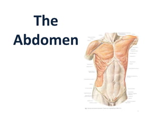
The abdomen is the region between the thorax and pelvis bounded superiorly by the diaphragm and inferiorly by the pelvis. It contains most of the digestive organs and some reproductive organs. The abdominal walls consist of skin, superficial fascia, muscles, and peritoneum. The abdomen can be divided into quadrants or nine regions to describe organ locations. The inguinal canal transmits structures between the abdomen and lower limbs and is the site of inguinal hernias.
rectus sheath, the sheath covering rectus muscle of anterior abdominal wall, formation of the sheath, the muscles involved in ts formation, and the contents the sheath is covering
Abdomen MCQs with Answers Key (below)
(Anterior Abdominal Wall)
NOTE: For each of the following multiple choice questions select the one most appropriate answer:
1. Rectus Abdominus Muscle is divided in bellies by tendinous intersections. What is by far the most common configuration of the muscle bellies of the rectus Abdominus.
(A) 2 Bellies and symmetric
(B) 2 Bellies and asymmetric
(C) 4 Bellies and asymmetric
(D) 6 Bellies and symmetric
(E) 8 Bellies and symmetric
2. A person was stung by a bee in the left lumbar region. The nerves supplying the region accompany the branches of
(A) Musculophrenic Artery
(B) Anterior Intercostal Arteries
(C) Posterior Intercostal Arteries
(D) Superior Epigastric Artery
(E) Inferior Epigastric Artery
3. A patient comes to your clinic whom you operated for obstructed irreducible indirect Left Inguinal hernia one month ago. He says “It has been over four weeks from the surgery and I still have much discomfort. Inside of my thigh is numb, burns or simply hurts when touched. When I move in certain ways I get a stabbing pain in that area accompanied with a sensation of being bit by a bunch of wasps (Bees).” Which nerve is most likely damaged?
(A) Genital branch of Genitofemoral nerve
(B) Illioinguinal Nerve
(C) Cremasteric Nerve
(D) Illiohypogastric Nerve
(E) Subcoastal Nerve
4. This patient has more chances of developing which type of hernia in future
(A) Right Direct Inguinal Hernia
(B) Left Direct Inguinal Hernia
(C) Right Indirect Inguinal Hernia
(D) Left Indirect Inguinal Hernia
(E) Umbilical Hernia
5. During Laproscopic repair of Direct inguinal Hernia, the site of hernia will be located in
(A) Median Umbilical fold
(B) Medial Umbilical fold
(C) Medial Inguinal Fossa
(D) Lateral Inguinal Fossa
(E) Lateral umbilical Fold
6. Median Umbilical Fold
(A) Is a remnant of Urachus
(B) Is a remnant of Umbilical Artery
(C) Contains Inferior Epigastric Artery
(D) Is a remnant Umbilical Vein
(E) Contains Ductus Venosus
7. While operating for Indirect Inguinal Hernia there started an unusual rapid oozing of blood, which filled the site with blood. The Surgeon had to stop to control the bleed. Which artery is most likely injured?
(A) Inferior Epigastric
(B) Cremasteric
(C) Testicular
(D) External Illiac
(E) Internal iliac
8. You are examining a patient for Hernia during exam. The examiner asks you to differentiate between inguinal and Femoral Hernia. Your best response will be
(A) Femoral Hernia is above and medial to Pubic tubercle
(B) Femoral Hernia is below and medial to Pubic tubercle
(C) Femoral Hernia is above and Lateral to Pubic tubercle
(D) Femoral Hernia is below and Lateral to Pubic tubercle
(E) None of Above
9. A patient was diagnosed with Testicular Carcinoma (Seminoma). He comes to you and asks what stage is his cancer i
Abdominal anatomical and symptoms and symptoms and Marasmus of the fetus first and symptoms to the signs on a verification dsujŝkkkllllllllljnvvvhĵjbvvghhjjĵkķkkkkkkkkkkkllķ
Anterior abdominal wall , Rectus sheath and Inguinal.pptxJudeChinecherem
In this detailed lecture note, we embark on a comprehensive journey through the complex and crucial anatomy of the abdominal wall. The abdominal wall is not just a physical barrier; it is a dynamic structure with multiple layers, muscles, and intricate structures that play a fundamental role in protecting our internal organs, providing support, and enabling various bodily functions.
We will delve deep into the layers of the abdominal wall, understanding the significance of each component - from the outermost skin to the innermost peritoneum. Through detailed illustrations, diagrams, and explanations, you will gain a profound insight into the anatomical intricacies of this region.
Moreover, this lecture note provides valuable insights into the clinical relevance of the abdominal wall. Learn about common medical conditions and surgical procedures related to the abdominal wall, including hernias, trauma, and abdominal wall reconstruction. Whether you are a medical student, healthcare professional, or simply intrigued by the wonders of the human body, this resource will enrich your knowledge and understanding of this vital anatomical structure.
Join us on this educational journey as we unravel the mysteries of the abdominal wall, exploring its anatomy, functions, and clinical significance. Whether you're studying medicine, pursuing a career in healthcare, or just eager to expand your knowledge, this lecture note is a valuable resource for anyone interested in the fascinating world of human anatomy."
Muscles Of Anterolateral Abdominal Wall.pptxaqsaaroob1
I described about the whole anatomy of anterolateral abdominal wall. Muscles, ligaments attach directly to anterolateral abdominal wall. Also add the topic of inguinal canal complete.
Unit 8 - Information and Communication Technology (Paper I).pdfThiyagu K
This slides describes the basic concepts of ICT, basics of Email, Emerging Technology and Digital Initiatives in Education. This presentations aligns with the UGC Paper I syllabus.
Instructions for Submissions thorugh G- Classroom.pptxJheel Barad
This presentation provides a briefing on how to upload submissions and documents in Google Classroom. It was prepared as part of an orientation for new Sainik School in-service teacher trainees. As a training officer, my goal is to ensure that you are comfortable and proficient with this essential tool for managing assignments and fostering student engagement.
How to Make a Field invisible in Odoo 17Celine George
It is possible to hide or invisible some fields in odoo. Commonly using “invisible” attribute in the field definition to invisible the fields. This slide will show how to make a field invisible in odoo 17.
The Indian economy is classified into different sectors to simplify the analysis and understanding of economic activities. For Class 10, it's essential to grasp the sectors of the Indian economy, understand their characteristics, and recognize their importance. This guide will provide detailed notes on the Sectors of the Indian Economy Class 10, using specific long-tail keywords to enhance comprehension.
For more information, visit-www.vavaclasses.com
The French Revolution, which began in 1789, was a period of radical social and political upheaval in France. It marked the decline of absolute monarchies, the rise of secular and democratic republics, and the eventual rise of Napoleon Bonaparte. This revolutionary period is crucial in understanding the transition from feudalism to modernity in Europe.
For more information, visit-www.vavaclasses.com
How to Create Map Views in the Odoo 17 ERPCeline George
The map views are useful for providing a geographical representation of data. They allow users to visualize and analyze the data in a more intuitive manner.
Read| The latest issue of The Challenger is here! We are thrilled to announce that our school paper has qualified for the NATIONAL SCHOOLS PRESS CONFERENCE (NSPC) 2024. Thank you for your unwavering support and trust. Dive into the stories that made us stand out!
Synthetic Fiber Construction in lab .pptxPavel ( NSTU)
Synthetic fiber production is a fascinating and complex field that blends chemistry, engineering, and environmental science. By understanding these aspects, students can gain a comprehensive view of synthetic fiber production, its impact on society and the environment, and the potential for future innovations. Synthetic fibers play a crucial role in modern society, impacting various aspects of daily life, industry, and the environment. ynthetic fibers are integral to modern life, offering a range of benefits from cost-effectiveness and versatility to innovative applications and performance characteristics. While they pose environmental challenges, ongoing research and development aim to create more sustainable and eco-friendly alternatives. Understanding the importance of synthetic fibers helps in appreciating their role in the economy, industry, and daily life, while also emphasizing the need for sustainable practices and innovation.
Ethnobotany and Ethnopharmacology:
Ethnobotany in herbal drug evaluation,
Impact of Ethnobotany in traditional medicine,
New development in herbals,
Bio-prospecting tools for drug discovery,
Role of Ethnopharmacology in drug evaluation,
Reverse Pharmacology.
2. Abdomen
• The abdomen is the part of the
trunk between the thorax and
the pelvis.
• It is a flexible, dynamic container,
housing most of the organs of
the alimentary system and part
of the urogenital system.
The abdomen consists of:
• abdominal walls
• abdominal cavity
• abdominal viscera
3. Anterior Abdominal Wall
Boundaries
• Superior:
• xiphoid process
• costal cartilages of the 7th-10th ribs
• Inferior:
• iliac crest
• inguinal fold
• pubic symphysis
• Lateral:
• posterior axillary line
4. Surface landmarks and regions of the
anterior abdominal wall
Topographical divisions of the abdomen are used to
describe the location of abdominal organs and the
pain associated with abdominal problems.
The two schemes most often used are:
1.A four-quadrant pattern
2.A nine-region organizational description.
5. Four-quadrant pattern
• Transverse Transumbilical
plane, passing through the
umbilicus (and the
intervertebral [IV] disc between
the L3 and L4 vertebrae)
• Vertical median plane passing
longitudinally through the
body, dividing it into right and
left halves to form four
quadrants-the right upper, left
upper, right lower, and left
lower quadrants
6. Using abdominal quadrants to locate
major viscera
• Liver and gallbladder
are in the right upper
quadrant.
• Stomach and spleen are
in the left upper
quadrant
• Cecum and appendix are
in the right lower
quadrant
• Descending colon and
sigmoid colon are in the
left lower quadrant.
7. Nine-region organizational pattern
• The nine regions are delineated by
four planes
two sagittal (vertical) and two transverse
(horizontal) planes.
1. Midclavicular planes that pass from the
midpoint of the clavicles (approximately 9 cm
from the midline) to the midinguinal points.
2. Subcostal plane is immediately inferior to the
costal margins, which places it at the lower
border of the costal cartilage of rib X and
passes posteriorly through the body of
vertebra LIII.
3. Intertubercular plane connects the tubercles
of the iliac crests, which are palpable
structures 5 cm posterior to the anterior
superior iliac spines, and passes through the
upper part of the body of vertebra LV.
8. The right and left midclavicular
lines subdivide it into:
Epigastrium:
• Epigastric region
• Right hypochondric region
• Left hypochondric region
Mesogastrium:
• Umbilical region
• Regio lateralis dex.
• Regio lateralis sin.
Hypogastrium:
• Pubic region
• Right inguinal region
• Left inguinal region
10. 3. Superficial fascia
• Below the umbilicus,
it forms two layers: a
superficial fatty layer
and a deeper
membranous layer.
11. Superficial fatty layer of superficial fascia
(Camper's fascia)
• Contains fat and varies in
thickness.
• It is continuous over the inguinal
ligament with the superficial
fascia of the thigh and with a
similar layer in the perineum.
• Continues over the penis and,
after losing its fat and fusing with
the deeper layer of superficial
fascia.
• Continues into the scrotum
where it forms a specialized
fascial layer containing smooth
muscle fibers (the dartos fascia)
12. Deeper membranous layer of superficial
fascia (Scarpa's fascia)
• Is thin and membranous, and
contains little or no fat.
• Inferiorly, it fuses with the
deep fascia of the thigh (the
fascia lata).
• It continues into the anterior
part of the perineum where
it is referred to as the
superficial perineal fascia
(Colles fascia).
13. 4.Muscles
There are five (bilaterally paired) in the
anterolateral abdominal wall:
three flat muscles -
• external oblique,
• internal oblique, and
•transversus abdominis
two vertical muscles –
• rectus abdominis and
• pyramidalis
22. Functions and actions of anterolateral
abdominal muscles
•Move the trunk and help to maintain posture
(resisting lumbar lordosis).
• The rectus abdominis is a powerful flexor
•Support the abdominal viscera and protect them
from most injuries.
•Compress the abdominal contents to maintain or
increase the intraabdominal pressure
•Produce the force required for defecation (discharge
of feces), micturition (urination), vomiting, and
parturition (childbirth).
23. Rectus sheath
• The rectus abdominis
and pyramidalis
muscles are enclosed in
an aponeurotic
tendinous sheath (the
rectus sheath) formed
by a unique layering of
the aponeuroses of the
external and internal
oblique, and
transversus abdominis
muscles
24. Organization of the rectus sheath
A. Transverse section
through the upper ¾ of
the rectus sheath
B. Transverse section
through the lower ¼ of
the rectus sheath
25. Upper ¾ of the rectus sheath
The anterior wall of the rectus sheath consists of
• the aponeurosis of the external oblique
• & half of the aponeurosis of the internal oblique
26. Upper ¾ of the rectus sheath Cont’d
The posterior wall of the rectus sheath consists of
• The other half of the aponeurosis of the internal oblique
• & aponeurosis of the transversus abdominis
27. Lower 1/4 of the rectus sheath
The anterior wall of the sheath consists of the
aponeuroses of the external oblique, internal oblique,
and transversus abdominis m.
There is no posterior wall at the lower ¼ of the rectus
sheath
28. Arterial supply and venous drainage
Superficially:
• The superior part of the
wall is supplied by branches
from the musculophrenic
artery, a terminal branch of
the internal thoracic artery.
• The inferior part of the wall
is supplied by the medially
placed superficial epigastric
artery and the laterally
placed superficial
circumflex iliac artery, both
branches of the femoral
artery.
29. Arterial supply and venous drainage
At a deeper level:
• The superior part
of the wall is
supplied by the
superior epigastric
artery, a terminal
branch of the
internal thoracic
artery.
• The lateral part of
the wall is supplied
by branches of the
tenth and
eleventh
intercostal
arteries and the
subcostal artery.
30. Arterial supply and venous drainage
At a deeper level:
• The inferior part of
the wall is supplied
by the medially
placed inferior
epigastric artery
and the laterally
placed deep
circumflex iliac
artery, both
branches of the
external iliac artery.
The superior and inferior epigastric arteries both enter the
rectus sheath. They are posterior to the rectus abdominis
muscle throughout their course, and anastomose with each
other
31. Veins:
In the upper abdomen:
• Thoracoepigastric v.
In the lower abdomen:
• Superficial epigastric v.
• Superficial circumflex
iliac v.
• External pudendal v.
Around the umbilicus:
• Parumbilical veins
Deep veins:
• Intercostal vv.
• Superior epigastric v.
• Inferior epigastric
32. Lymphatic drainage
From the upper
abdominal half to:
• Axillary lymph nodes
From the lower abdominal
half to :
•Superficial inguinal
lymph nodes
33. Innervation
• Intercostal nn. Th7 –
Th11
• Subcostal n.Th12
•Branches of lumbal
plexus Th12 – L4:
- Iliohypogastric n.
- Ilioinguinal n.
- Genitofemoral n
34. INGUINAL CANAL
• Surgically an important canal because it is the site of
inguinal hernias
• obliquely located;tubelike
• 3-4cm. in length.
• Has two openings :
• Superficial inguinal ring
external oblique apon.
-medial
• Deep ingunal ring:
transversalis fascia
- Lateral
36. INGUINAL CANAL
Anterior wall
The anterior wall of the inguinal
canal is formed along its entire
length by the aponeurosis of
the external oblique muscle
Posterior wall
The posterior wall of the inguinal
canal is formed along its entire
length by the transversalis
fascia
Superior wall
The roof (superior wall) of the
inguinal canal is formed by the
arching fibers of the
transversus abdominis and
internal oblique muscles
Inferior wall
The floor (inferior wall) of the
inguinal canal is formed by the
medial one-half of the inguinal
ligament
37. Contents
• The contents of the inguinal canal are:
• images the spermatic cord in men,
• images the round ligament of the uterus, and
• images genital branch of the genitofemoral nerve in
women.
• These structures enter the inguinal canal through the
deep inguinal ring and exit it through the superficial
inguinal ring.
38. Boundaries of abdomen
• Superiorly: The inferior
thoracic aperture forms the
superior opening to the
abdomen, and is closed by the
diaphragm.
• Inferiorly: the deep abdominal
wall is continuous with the
pelvic wall at the pelvic inlet.
39. Boundaries of abdomen
• Anteriorly: anteriorly, a
segmented muscle (the
rectus abdominis) on each
side spans the distance
between the inferior
thoracic wall and the pelvis
• Laterally: lateral parts of the
abdominal wall are
predominantly formed by
three layers of muscles
• Posteriorly: vertebral
column, the quadratus
lumborum, psoas major,
and iliacus muscles