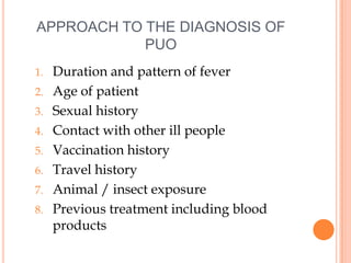
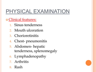
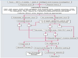
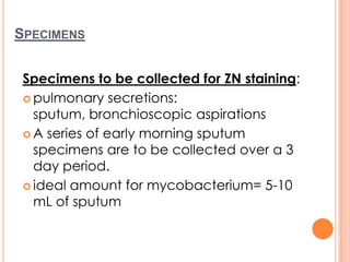
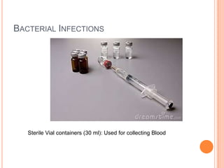
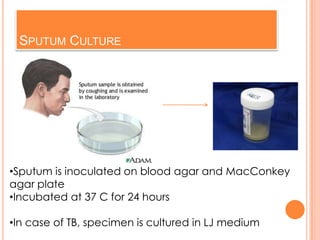
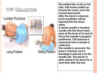
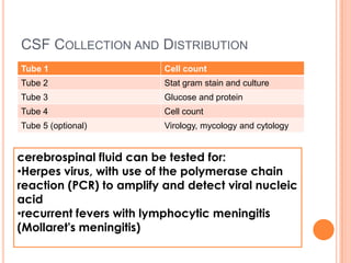
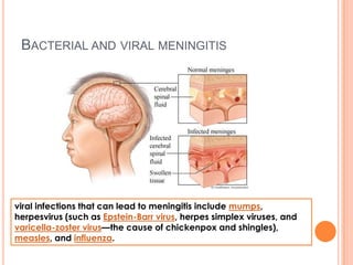
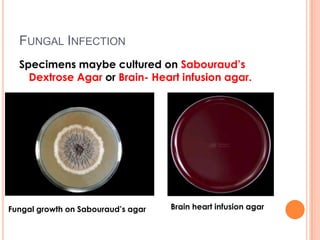
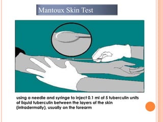
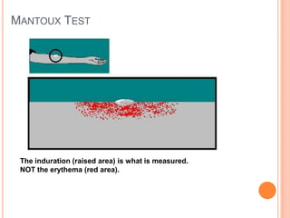
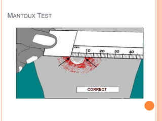
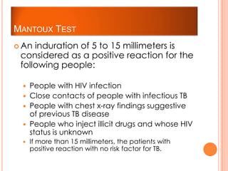
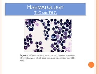
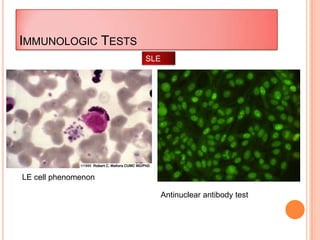
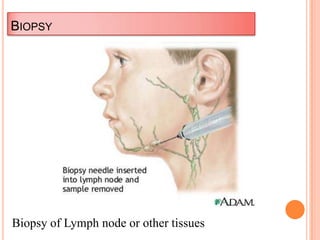
LABORATORY DIAGNOSIS OF
PYREXIA OF UNKNOWN
APPROACH TO THE DIAGNOSIS OF
PUO
1. Duration and pattern of fever
2. Age of patient
3. Sexual history
4. Contact with other ill people
5. Vaccination history
6. Travel history
7. Animal / insect exposure
8. Previous treatment including blood
products
PHYSICAL EXAMINATION
 Clinical features:
1. Sinus tenderness
2. Mouth ulceration
3. Chorioretinitis
4. Chest- pneumonitis
5. Abdomen- hepatic
tenderness, splenomegaly
6. Lymphadenopathy
7. Arthritis
8. Rash
BACTERIAL INFECTIONS
Specimens:
 Blood: for blood
cultures, peripheral blood
smear, haematology, serology
and other tests
 Urine analysis: for Urinary Tract
Infections
 Sputum: in cases of lung
infections
 Pus: in localised abscesses
SPECIMENS
Specimens to be collected for ZN staining:
 pulmonary secretions:
sputum, bronchioscopic aspirations
 A series of early morning sputum
specimens are to be collected over a 3
day period.
 ideal amount for mycobacterium= 5-10
mL of sputum
OTHER SPECIMENS
 fecal specimens
 tissue and body fluids:
pleural, pericardial and peritoneal fluids)
 CSF
 bone marrow aspirates
 *Note: Blood and stool specimens are
usually cultured from AIDS patient
BACTERIAL INFECTIONS
 Collection
 All the specimens should be collected
preferably prior to antibiotic therapy.
 These specimens are to be collected in a
sterile containers under aseptic conditions
 Blood is collected in blood culture bottles
for culture and in a sterile vial for serology.
 Mid stream urine specimen should be
collected in a sterile universal container.
BACTERIAL INFECTIONS
Sterile Vial containers (30 ml): Used for collecting Blood
BACTERIAL INFECTIONS
Sterile Universal Container: Used for collecting Urine
CULTURING
BACTERIAL INFECTIONS
BLOOD CULTURE:
Procedure:
 Take 5 ml of blood in each bottle of 50
ml of glucose broth + 50 ml
taurocholate broth
 Incubate these broths at 37ºC for 24
hours
 Subcultures are made on Blood agar
from (glucose broth) and MacConkey
agar (from taurocholate broth)
Blood Agar MacConkey Agar
Blood agar and MacConkey agar are
incubated at 37ºC for 24 hours
BLOOD CULTURE
 Febrile agglutinins
 refers to serologic studies for salmonellosis,
brucellosis, and rickettsial diseases.
 These studies are useful, having low sensitivity
and variable specificity.
 Multiple blood samples (no fewer than three and
rarely more than six, including samples for
anaerobic culture) should be cultured in the
laboratory for at least 2 weeks to ensure that any
HACEK group organisms that may be present
have time to grow
BLOOD CULTURE
 Lysis-centrifugation blood culture techniques should
be employed in cases where prior antimicrobial
therapy or fungal or atypical mycobacterial infection
is suspected.
 Blood culture media should be supplemented with
L-cysteine or pyridoxal to assist in the isolation of
nutritionally variant streptococci.
 It should be noted that sequential cultures positive
for multiple organisms may reflect self-injection of
contaminated substances.
URINE CULTURE
 A calibrated volume of midstream urine
specimen is inoculated on:
 blood agar
 MacConkey agar
•Incubated at 37ºC for 24 hours
*In renal
tuberculosis, culture should
be performed in
Lowenstein Jensen
Urine cultures, including cultures
for mycobacteria, fungi, and
CMV, are indicated.
SPUTUM CULTURE
•Sputum is inoculated on blood agar and MacConkey
agar plate
•Incubated at 37 C for 24 hours
•In case of TB, specimen is cultured in LJ medium
CSF COLLECTION
The patient lies on his or her
side, with knees pulled up
toward the chest, and chin
tucked downward.
After the back is cleaned,
local anesthetic will be
injected into the lower
spine.
A spinal needle is inserted,
usually into the lower back
area at the level of L3 and L4
Once the needle is properly
positioned, CSF pressure is
measured and a sample is
collected.
The needle is removed, the
area is cleaned, and a
bandage is placed over the
needle site. The person is
often asked to lie down for a
short time after the test.
CSF COLLECTION AND DISTRIBUTION
Tube 1 Cell count
Tube 2 Stat gram stain and culture
Tube 3 Glucose and protein
Tube 4 Cell count
Tube 5 (optional) Virology, mycology and cytology
cerebrospinal fluid can be tested for:
•Herpes virus, with use of the polymerase chain
reaction (PCR) to amplify and detect viral nucleic
acid
•recurrent fevers with lymphocytic meningitis
(Mollaret's meningitis)
BACTERIAL AND VIRAL MENINGITIS
viral infections that can lead to meningitis include mumps,
herpesvirus (such as Epstein-Barr virus, herpes simplex viruses, and
varicella-zoster virus—the cause of chickenpox and shingles),
measles, and influenza.
PUS CULTURE
 Pus is inoculated in glucose broth, blood
agar and MacConkey agar.
 Incubate at 37°C for 24 hours
 For M. tuberculosis, pus should be
cultured on LJ medium.
 If suspecting anaerobic organism,
culture of pus should be performed
under anaerobic conditions
IDENTIFICATION
 On the basis of:
 Colony morphology
 Gram Staining
 Ziehl-Neelsen Staining- For M.
tuberculosis
 Biochemical reactions
 Agglutination
ZIEHL-NEELSEN STAINING
 The ZN stain is mostly used to identify acid-fast
mycobacteria, the most important of which is
Mycobacterium Tuberculosis, the organism
responsible for tuberculosis (TB).
 the tubercle bacilli having a lipid-rich cell wall
that takes up phenol-dye solutions (eg. carbol
fuchsin, the main dye used in the ZN stain) and
after subsequent differentiation, retains the
phenol-dye.
ZIEHL-NEELSEN STAINING
Solutions
Concentrated carbol fuchsin
20% Sulphuric acid
2% methylene blue
ZIEHL-NEELSEN STAINING- PROCEDURE
1. Flood the slide with concentrated carbol fuchsin
and heat slide from below intermittently until
steam arises (Strains should not boil or
evaporate). Heating  better penetration of strain
into the cell.
2. Decolorize the smear with 20% Sulphuric acid
and wash with water. Repeat this step till the
red/pink colour stops coming out
3. The smear is counterstained with 2% methylene
blue for 1-2 minutes
4. Wash with water and air dry.
ZIEHL- NEELSEN STAINING
Identification of Mycobacteria spp. by qualified clinical
laboratories entails several of the following:
 Confirmation that the isolate recovered in broth or on
solid media is an acid-fast organism.
 Categorize (presumptively) the isolate by phenotypic
characteristics, such as colony morphology,
photoreactivity, growth rate, and optimum growth
temperature.
 Identification through tests based on enzyme systems of
the organism, metabolic by-products, and inhibition of
growth by exposure to selected biochemicals.
 Chromatographic detection of mycolic acid.
 Identification by DNA hybridization (e.g., Gen-Probe-San
Diego, Calif.)
 Identification by PCR (polymerse chain reaction) tests.
BIOCHEMICAL TESTS
The biochemical tests most often utilized are:
 niacin accumulation
 nitrate reduction
 TCH (inhibition of growth when exposed to
thiophene-2-carboxylic acid hydrazide)
 growth in 5% NaCl, tellurite reduction
 growth on MacConkey agar
 Catalase
 hydrolysis of Tween 80
 iron uptake
 tests for the enzymes aryl-sulfatase, urease, and
pyrazinamidase.
SEROLOGY
 Useful in:
 Infectious mononucleosis- Paul-Bunnell
test
 Enteric fever
 Hepatitis A, B infections
 CMV infections
 Amoebiasis
PARASITIC INFECTION
Stained peripheral blood smears (thick and thin) will
help in diagnosis of:
 Malaria
 Leishmaniasis
 Filariasis
 Toxoplasmosis
 Tripanoma
 Entamoeba histolytica
Wet blood film may show microfilaria in filarsis
VIRAL INFECTION
Paul- Bunnell test is useful in infectious
mononucleosis
CMV
Epstein barr virus
Hep A and B viruses
Arboviruses
Enteroviruses
Adenovirsues
Myoxviruses
Human immunodeficiency virus
Haemorrhagic fever viruses
FUNGAL CAUSES
 Candida albicans
 Streptococcus neoformans
 Histoplasm
 Aspergillous spp
 Coccidioides innipil neocystis carinii
FUNGAL INFECTION
Specimens maybe cultured on Sabouraud’s
Dextrose Agar or Brain- Heart infusion agar.
Fungal growth on Sabouraud’s agar Brain heart infusion agar
OTHER TESTS
1. Skin tests
2. Haematology
3. Immunologic tests
4. Biopsy
Mantoux Skin Test
using a needle and syringe to inject 0.1 ml of 5 tuberculin units
of liquid tuberculin between the layers of the skin
(intradermally), usually on the forearm
MANTOUX TEST
The induration (raised area) is what is measured.
NOT the erythema (red area).
MANTOUX TEST
MANTOUX TEST
 An induration of 5 to 15 millimeters is
considered as a positive reaction for the
following people:
 People with HIV infection
 Close contacts of people with infectious TB
 People with chest x-ray findings suggestive
of previous TB disease
 People who inject illicit drugs and whose HIV
status is unknown
 If more than 15 millimeters, the patients with
positive reaction with no risk factor for TB.
SKIN TESTS FOR HISTOPLASMOSIS
HAEMATOLOGY
TLC AND DLC
IMMUNOLOGIC TESTS
LE cell phenomenon
Antinuclear antibody test
SLE
BIOPSY
Biopsy of Lymph node or other tissues
TREATMENT
 vital-sign instability or neutropenia is an indication
for empirical therapy with a fluoroquinolone plus
piperacillin
 If the PPD skin test is positive or if granulomatous
hepatitis or other granulomatous disease is present,
then a therapeutic trial with isoniazid and rifampin,
with treatment usually continued for up to 6 weeks.
BIBLIOGRAPHY
 Dr. Fauci and Dr. Longo. “Harrison’s Internal
Medicine”. America: The McGraw-Hill
Companies, Inc 2008.
 Skin Pathonline. “Ziehl Neelsen Stain for AFB”. 9
May. 2011
<http://skinpathonline.wordpress.com/2011/05/09/zi
ehl%E2%80%93neelsen-stain-for-acid-fast-
organisms-method-and-tips/>.
 http://www.enotes.com/nursing-encyclopedia/acid-
fast-culture
 http://www.virusesinmay.com/docs/2006/VIM06%20
Kesson%20PUO.pdf
Laboratory diagnosis of PUO

Laboratory diagnosis of PUO

  • 1.
  • 2.
    APPROACH TO THEDIAGNOSIS OF PUO 1. Duration and pattern of fever 2. Age of patient 3. Sexual history 4. Contact with other ill people 5. Vaccination history 6. Travel history 7. Animal / insect exposure 8. Previous treatment including blood products
  • 3.
    PHYSICAL EXAMINATION  Clinicalfeatures: 1. Sinus tenderness 2. Mouth ulceration 3. Chorioretinitis 4. Chest- pneumonitis 5. Abdomen- hepatic tenderness, splenomegaly 6. Lymphadenopathy 7. Arthritis 8. Rash
  • 5.
  • 6.
    BACTERIAL INFECTIONS Specimens:  Blood:for blood cultures, peripheral blood smear, haematology, serology and other tests  Urine analysis: for Urinary Tract Infections  Sputum: in cases of lung infections  Pus: in localised abscesses
  • 7.
    SPECIMENS Specimens to becollected for ZN staining:  pulmonary secretions: sputum, bronchioscopic aspirations  A series of early morning sputum specimens are to be collected over a 3 day period.  ideal amount for mycobacterium= 5-10 mL of sputum
  • 8.
    OTHER SPECIMENS  fecalspecimens  tissue and body fluids: pleural, pericardial and peritoneal fluids)  CSF  bone marrow aspirates  *Note: Blood and stool specimens are usually cultured from AIDS patient
  • 9.
    BACTERIAL INFECTIONS  Collection All the specimens should be collected preferably prior to antibiotic therapy.  These specimens are to be collected in a sterile containers under aseptic conditions  Blood is collected in blood culture bottles for culture and in a sterile vial for serology.  Mid stream urine specimen should be collected in a sterile universal container.
  • 10.
    BACTERIAL INFECTIONS Sterile Vialcontainers (30 ml): Used for collecting Blood
  • 11.
    BACTERIAL INFECTIONS Sterile UniversalContainer: Used for collecting Urine
  • 12.
  • 13.
    BLOOD CULTURE: Procedure:  Take5 ml of blood in each bottle of 50 ml of glucose broth + 50 ml taurocholate broth  Incubate these broths at 37ºC for 24 hours  Subcultures are made on Blood agar from (glucose broth) and MacConkey agar (from taurocholate broth)
  • 14.
    Blood Agar MacConkeyAgar Blood agar and MacConkey agar are incubated at 37ºC for 24 hours
  • 15.
    BLOOD CULTURE  Febrileagglutinins  refers to serologic studies for salmonellosis, brucellosis, and rickettsial diseases.  These studies are useful, having low sensitivity and variable specificity.  Multiple blood samples (no fewer than three and rarely more than six, including samples for anaerobic culture) should be cultured in the laboratory for at least 2 weeks to ensure that any HACEK group organisms that may be present have time to grow
  • 16.
    BLOOD CULTURE  Lysis-centrifugationblood culture techniques should be employed in cases where prior antimicrobial therapy or fungal or atypical mycobacterial infection is suspected.  Blood culture media should be supplemented with L-cysteine or pyridoxal to assist in the isolation of nutritionally variant streptococci.  It should be noted that sequential cultures positive for multiple organisms may reflect self-injection of contaminated substances.
  • 17.
    URINE CULTURE  Acalibrated volume of midstream urine specimen is inoculated on:  blood agar  MacConkey agar •Incubated at 37ºC for 24 hours *In renal tuberculosis, culture should be performed in Lowenstein Jensen Urine cultures, including cultures for mycobacteria, fungi, and CMV, are indicated.
  • 18.
    SPUTUM CULTURE •Sputum isinoculated on blood agar and MacConkey agar plate •Incubated at 37 C for 24 hours •In case of TB, specimen is cultured in LJ medium
  • 19.
    CSF COLLECTION The patientlies on his or her side, with knees pulled up toward the chest, and chin tucked downward. After the back is cleaned, local anesthetic will be injected into the lower spine. A spinal needle is inserted, usually into the lower back area at the level of L3 and L4 Once the needle is properly positioned, CSF pressure is measured and a sample is collected. The needle is removed, the area is cleaned, and a bandage is placed over the needle site. The person is often asked to lie down for a short time after the test.
  • 20.
    CSF COLLECTION ANDDISTRIBUTION Tube 1 Cell count Tube 2 Stat gram stain and culture Tube 3 Glucose and protein Tube 4 Cell count Tube 5 (optional) Virology, mycology and cytology cerebrospinal fluid can be tested for: •Herpes virus, with use of the polymerase chain reaction (PCR) to amplify and detect viral nucleic acid •recurrent fevers with lymphocytic meningitis (Mollaret's meningitis)
  • 21.
    BACTERIAL AND VIRALMENINGITIS viral infections that can lead to meningitis include mumps, herpesvirus (such as Epstein-Barr virus, herpes simplex viruses, and varicella-zoster virus—the cause of chickenpox and shingles), measles, and influenza.
  • 22.
    PUS CULTURE  Pusis inoculated in glucose broth, blood agar and MacConkey agar.  Incubate at 37°C for 24 hours  For M. tuberculosis, pus should be cultured on LJ medium.  If suspecting anaerobic organism, culture of pus should be performed under anaerobic conditions
  • 23.
    IDENTIFICATION  On thebasis of:  Colony morphology  Gram Staining  Ziehl-Neelsen Staining- For M. tuberculosis  Biochemical reactions  Agglutination
  • 24.
    ZIEHL-NEELSEN STAINING  TheZN stain is mostly used to identify acid-fast mycobacteria, the most important of which is Mycobacterium Tuberculosis, the organism responsible for tuberculosis (TB).  the tubercle bacilli having a lipid-rich cell wall that takes up phenol-dye solutions (eg. carbol fuchsin, the main dye used in the ZN stain) and after subsequent differentiation, retains the phenol-dye.
  • 25.
    ZIEHL-NEELSEN STAINING Solutions Concentrated carbolfuchsin 20% Sulphuric acid 2% methylene blue
  • 26.
    ZIEHL-NEELSEN STAINING- PROCEDURE 1.Flood the slide with concentrated carbol fuchsin and heat slide from below intermittently until steam arises (Strains should not boil or evaporate). Heating  better penetration of strain into the cell. 2. Decolorize the smear with 20% Sulphuric acid and wash with water. Repeat this step till the red/pink colour stops coming out 3. The smear is counterstained with 2% methylene blue for 1-2 minutes 4. Wash with water and air dry.
  • 27.
    ZIEHL- NEELSEN STAINING Identificationof Mycobacteria spp. by qualified clinical laboratories entails several of the following:  Confirmation that the isolate recovered in broth or on solid media is an acid-fast organism.  Categorize (presumptively) the isolate by phenotypic characteristics, such as colony morphology, photoreactivity, growth rate, and optimum growth temperature.  Identification through tests based on enzyme systems of the organism, metabolic by-products, and inhibition of growth by exposure to selected biochemicals.  Chromatographic detection of mycolic acid.  Identification by DNA hybridization (e.g., Gen-Probe-San Diego, Calif.)  Identification by PCR (polymerse chain reaction) tests.
  • 28.
    BIOCHEMICAL TESTS The biochemicaltests most often utilized are:  niacin accumulation  nitrate reduction  TCH (inhibition of growth when exposed to thiophene-2-carboxylic acid hydrazide)  growth in 5% NaCl, tellurite reduction  growth on MacConkey agar  Catalase  hydrolysis of Tween 80  iron uptake  tests for the enzymes aryl-sulfatase, urease, and pyrazinamidase.
  • 29.
    SEROLOGY  Useful in: Infectious mononucleosis- Paul-Bunnell test  Enteric fever  Hepatitis A, B infections  CMV infections  Amoebiasis
  • 30.
    PARASITIC INFECTION Stained peripheralblood smears (thick and thin) will help in diagnosis of:  Malaria  Leishmaniasis  Filariasis  Toxoplasmosis  Tripanoma  Entamoeba histolytica Wet blood film may show microfilaria in filarsis
  • 31.
    VIRAL INFECTION Paul- Bunnelltest is useful in infectious mononucleosis CMV Epstein barr virus Hep A and B viruses Arboviruses Enteroviruses Adenovirsues Myoxviruses Human immunodeficiency virus Haemorrhagic fever viruses
  • 32.
    FUNGAL CAUSES  Candidaalbicans  Streptococcus neoformans  Histoplasm  Aspergillous spp  Coccidioides innipil neocystis carinii
  • 33.
    FUNGAL INFECTION Specimens maybecultured on Sabouraud’s Dextrose Agar or Brain- Heart infusion agar. Fungal growth on Sabouraud’s agar Brain heart infusion agar
  • 34.
    OTHER TESTS 1. Skintests 2. Haematology 3. Immunologic tests 4. Biopsy
  • 35.
    Mantoux Skin Test usinga needle and syringe to inject 0.1 ml of 5 tuberculin units of liquid tuberculin between the layers of the skin (intradermally), usually on the forearm
  • 36.
    MANTOUX TEST The induration(raised area) is what is measured. NOT the erythema (red area).
  • 37.
  • 38.
    MANTOUX TEST  Aninduration of 5 to 15 millimeters is considered as a positive reaction for the following people:  People with HIV infection  Close contacts of people with infectious TB  People with chest x-ray findings suggestive of previous TB disease  People who inject illicit drugs and whose HIV status is unknown  If more than 15 millimeters, the patients with positive reaction with no risk factor for TB.
  • 39.
    SKIN TESTS FORHISTOPLASMOSIS
  • 40.
  • 41.
    IMMUNOLOGIC TESTS LE cellphenomenon Antinuclear antibody test SLE
  • 42.
    BIOPSY Biopsy of Lymphnode or other tissues
  • 43.
    TREATMENT  vital-sign instabilityor neutropenia is an indication for empirical therapy with a fluoroquinolone plus piperacillin  If the PPD skin test is positive or if granulomatous hepatitis or other granulomatous disease is present, then a therapeutic trial with isoniazid and rifampin, with treatment usually continued for up to 6 weeks.
  • 44.
    BIBLIOGRAPHY  Dr. Fauciand Dr. Longo. “Harrison’s Internal Medicine”. America: The McGraw-Hill Companies, Inc 2008.  Skin Pathonline. “Ziehl Neelsen Stain for AFB”. 9 May. 2011 <http://skinpathonline.wordpress.com/2011/05/09/zi ehl%E2%80%93neelsen-stain-for-acid-fast- organisms-method-and-tips/>.  http://www.enotes.com/nursing-encyclopedia/acid- fast-culture  http://www.virusesinmay.com/docs/2006/VIM06%20 Kesson%20PUO.pdf