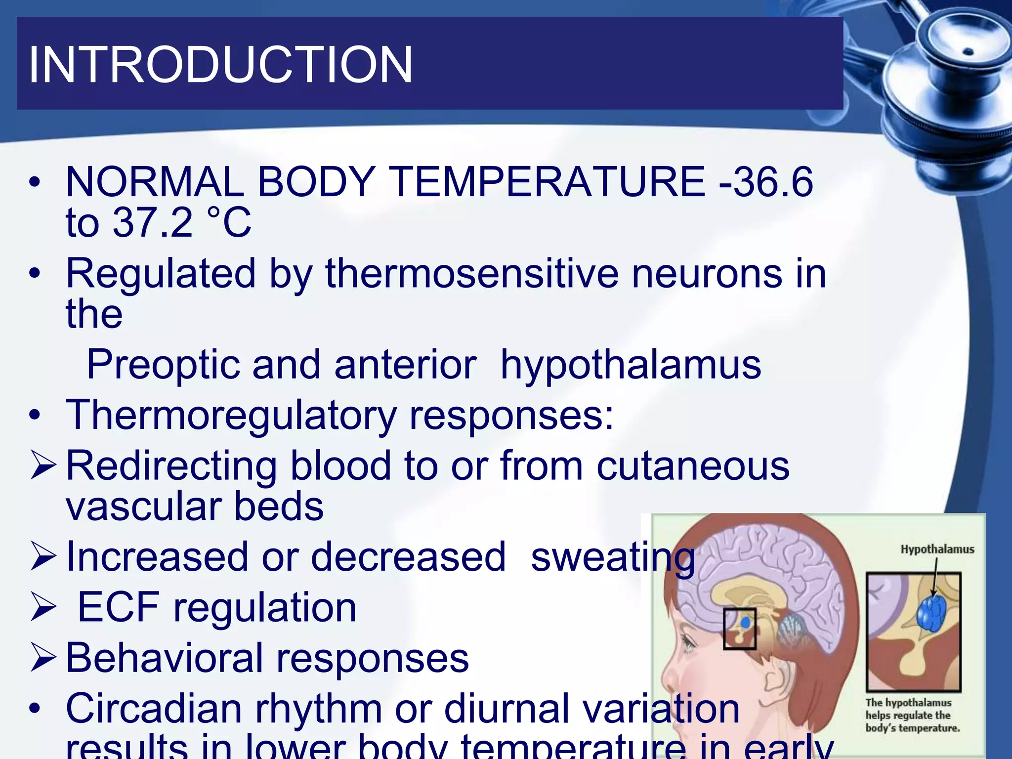
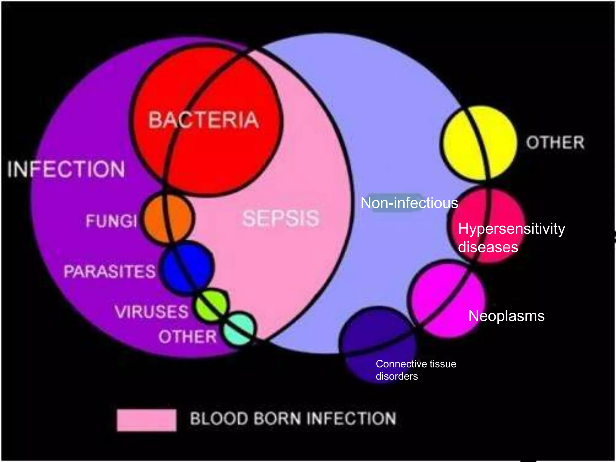
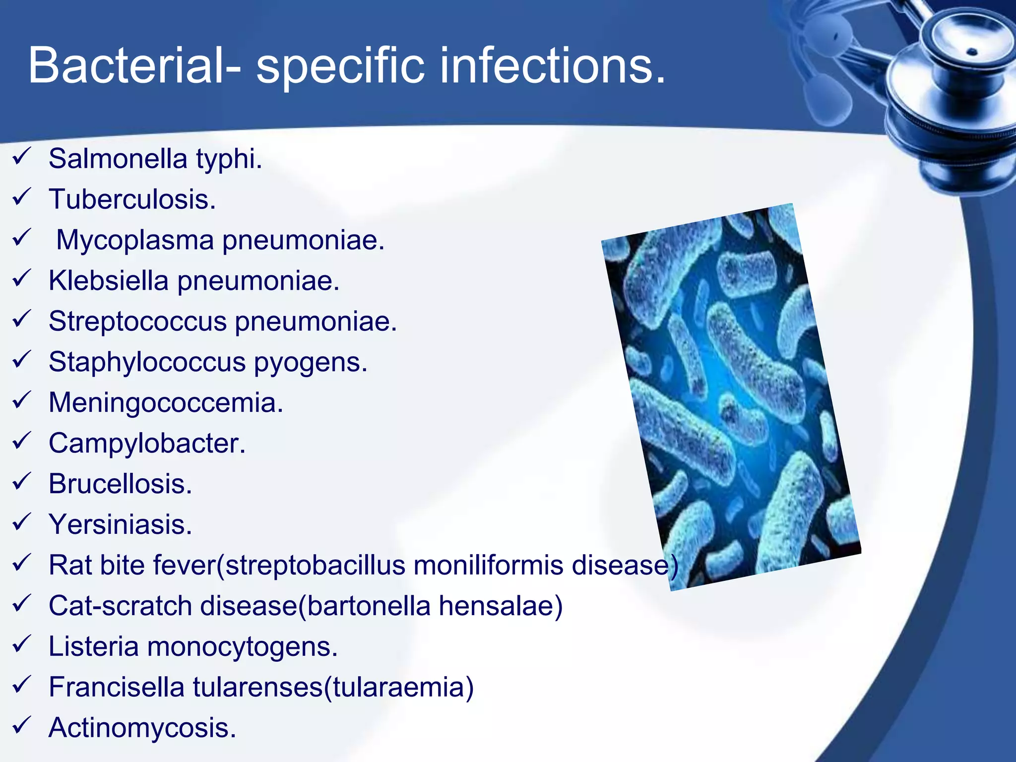
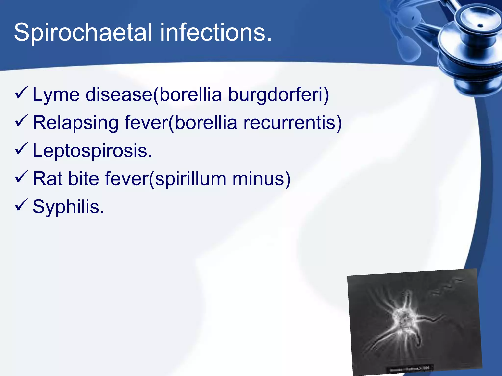
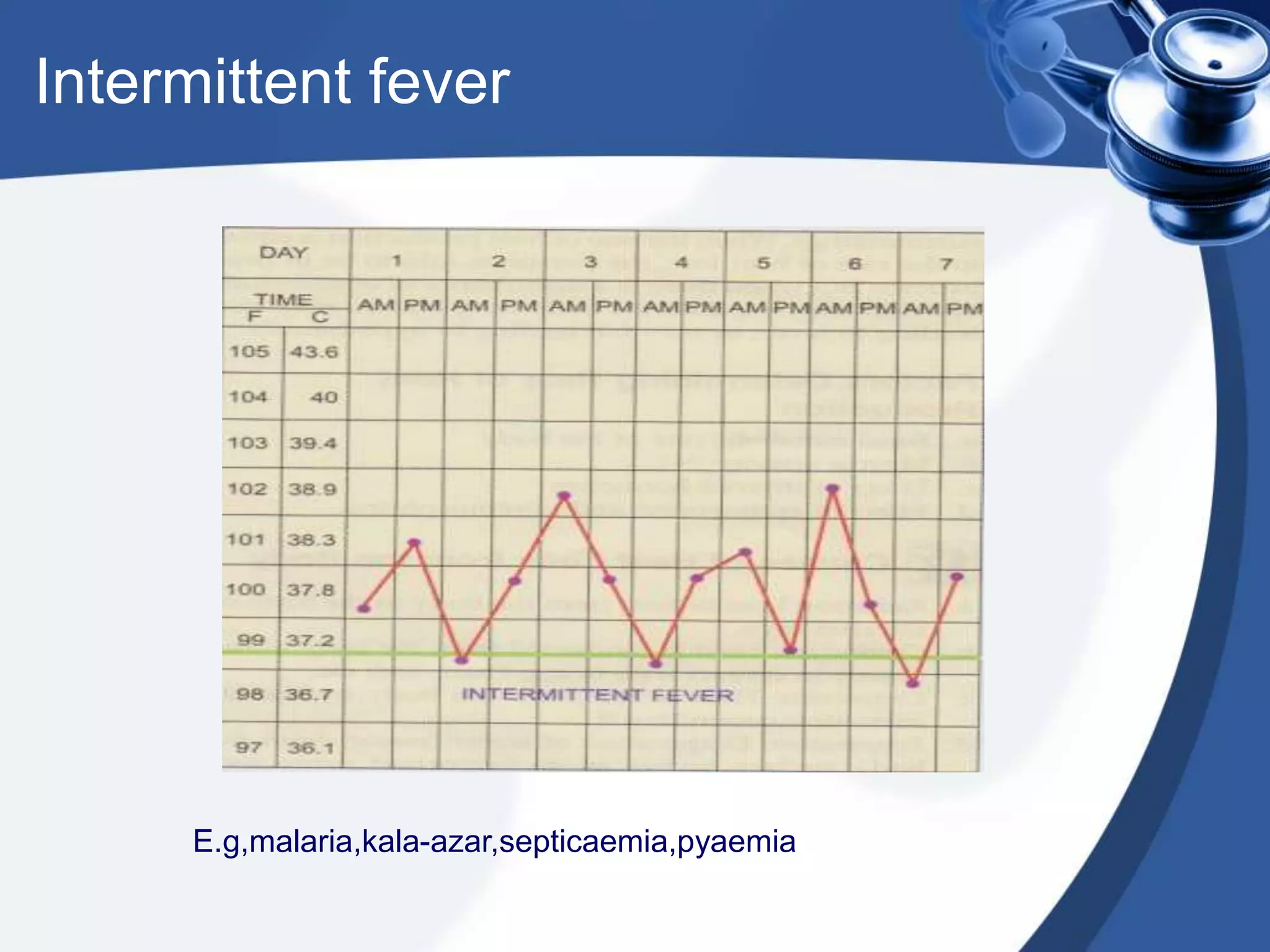
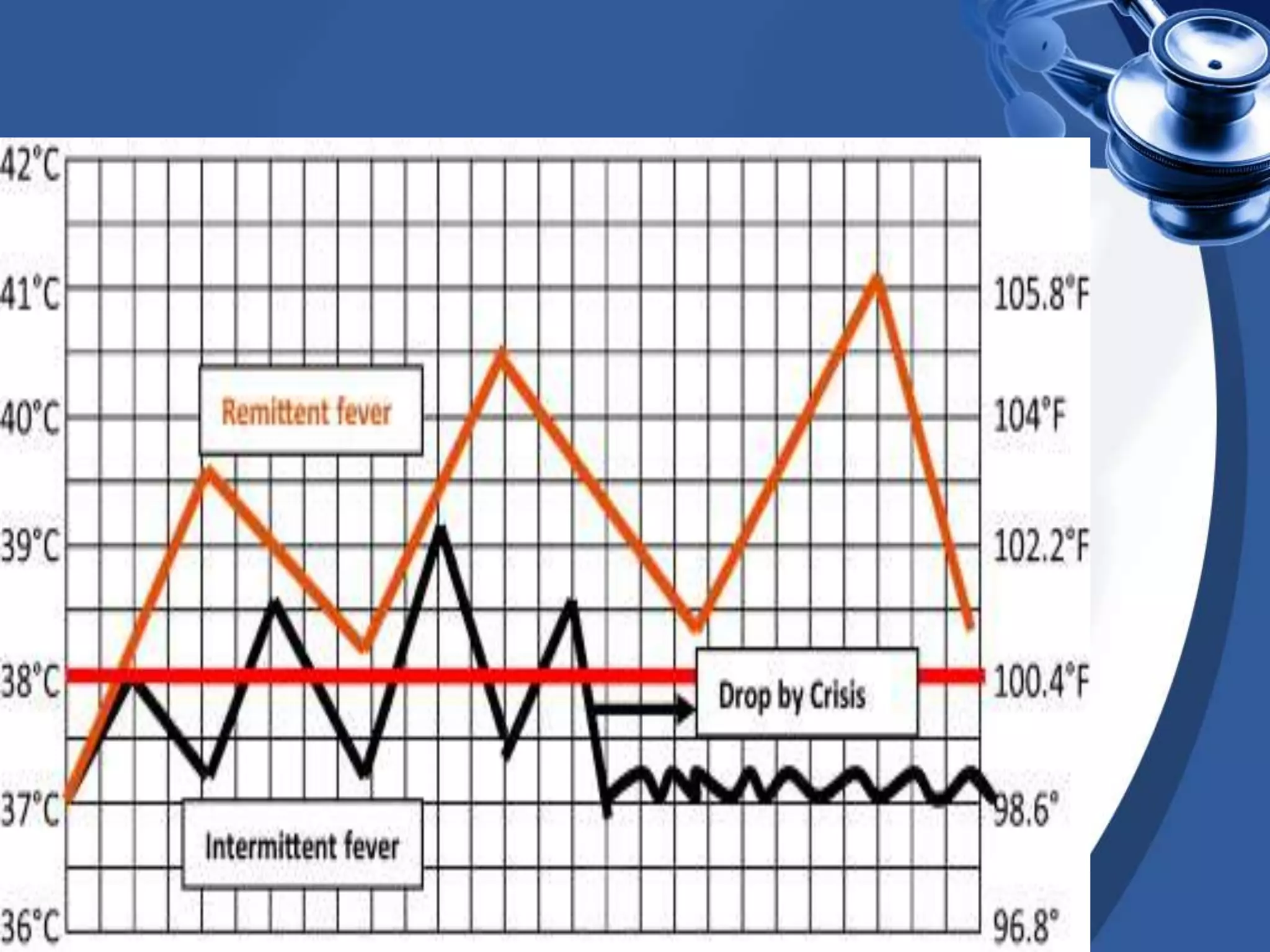
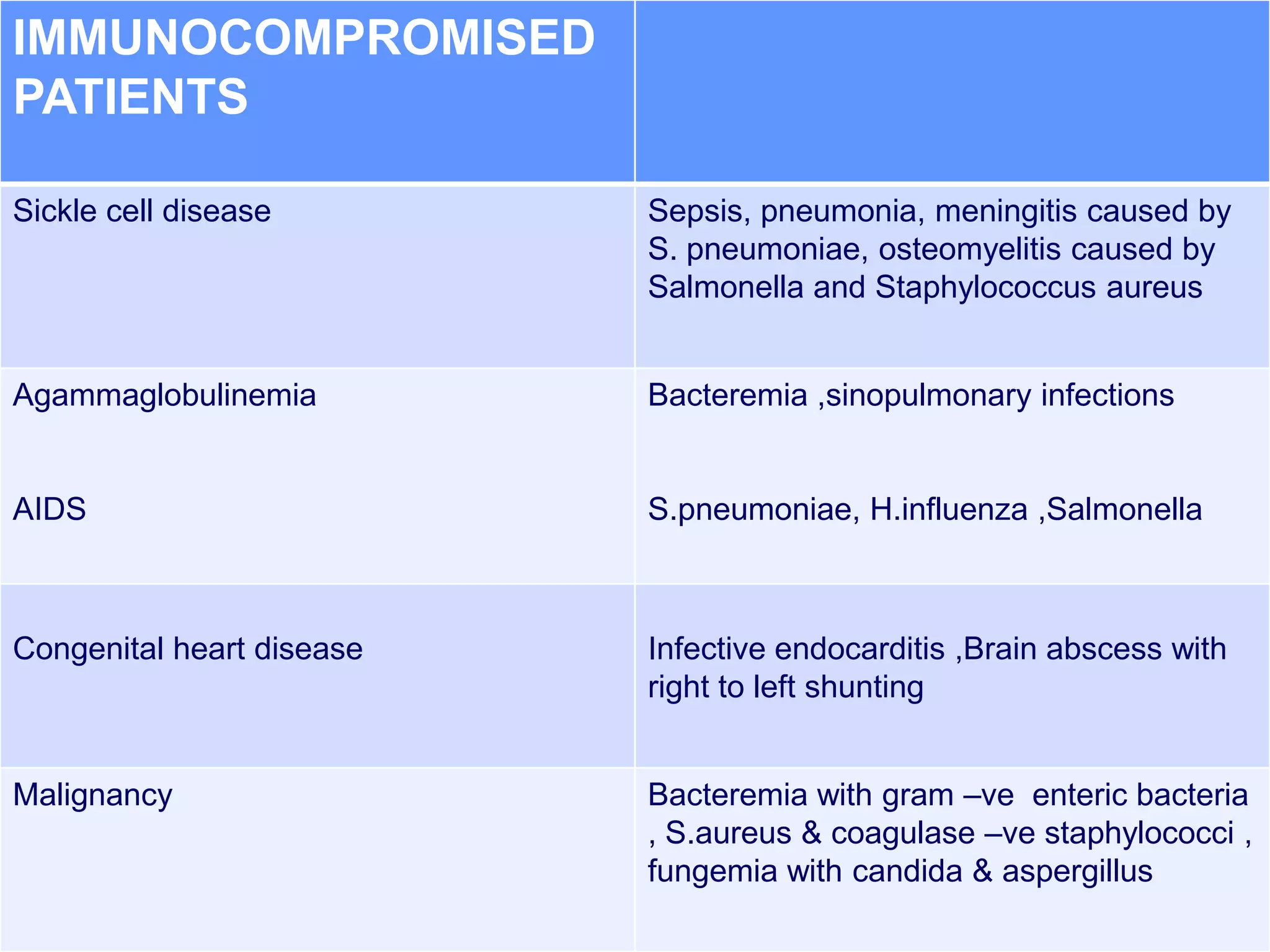
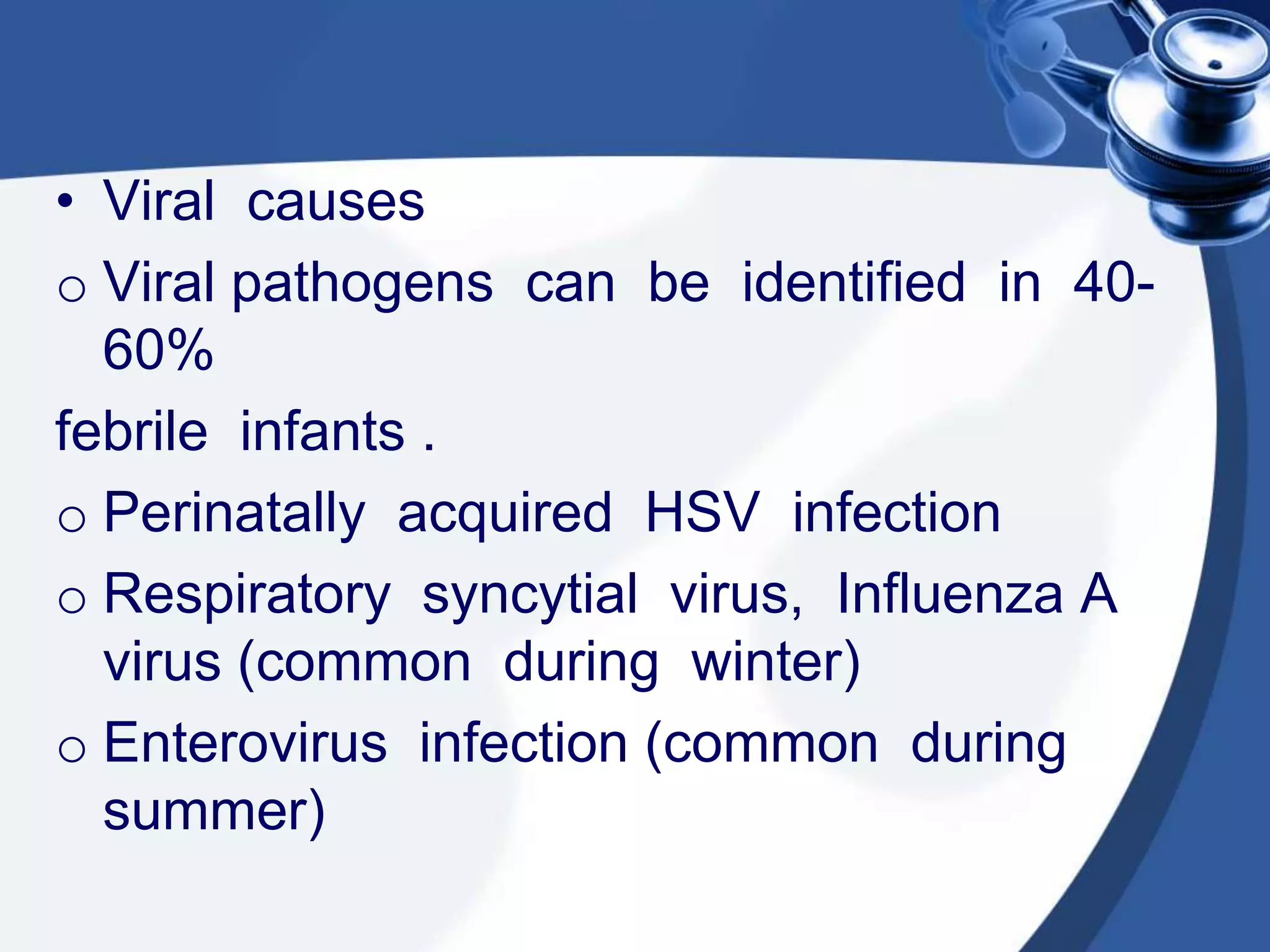
Tutorial on,

Fever of Unknown Origin.
Tutorial on,


Fever of Unknown Origin.
FEVER.
INTRODUCTION

• NORMAL BODY TEMPERATURE -36.6
  to 37.2 °C
• Regulated by thermosensitive neurons in
  the
   Preoptic and anterior hypothalamus
• Thermoregulatory responses:
 Redirecting blood to or from cutaneous
  vascular beds
 Increased or decreased sweating
 ECF regulation
 Behavioral responses
• Circadian rhythm or diurnal variation
• Skin temperature is 0.4 °C(0.7° F)
  lower than oral temperature
• Rectal or eardrum temperature is 0.4
  °C (0.7° F) higher than oral temperature
• Oral temperature is the reference and
  fever is diagnosed when it exceeds
  (37.8° C).
DEFINITION OF FEVER

• Fever is a controlled increase in body
  temperature above the normal
  hypothalamic set point
• Rectal temperature >38 °C
• Hyperpyrexia is > 40° C
PATHOGENESIS OF FEVER
Causes for fever?????
Infectious causes

Non infectious causes
Non-infectious
                      Hypersensitivity
                      diseases


                       Neoplasms


  Connective tissue
  disorders
Bacterial- specific infections.
   Salmonella typhi.
   Tuberculosis.
   Mycoplasma pneumoniae.
   Klebsiella pneumoniae.
   Streptococcus pneumoniae.
   Staphylococcus pyogens.
   Meningococcemia.
   Campylobacter.
   Brucellosis.
   Yersiniasis.
   Rat bite fever(streptobacillus moniliformis disease)
   Cat-scratch disease(bartonella hensalae)
   Listeria monocytogens.
   Francisella tularenses(tularaemia)
   Actinomycosis.
Bacterial-localised infection.

 Abscesses:
  Abdominal,brain,dental,hepatic,pelvic,perinephric,rec
  tal,subphrenic.
 Cholangitis.
 Infective endocarditis.
 Mastoiditis.
 Osteomyelitis.
 Pyelonephritis.
 Sinusitis.
Spirochaetal infections.

 Lyme disease(borellia burgdorferi)
 Relapsing fever(borellia recurrentis)
 Leptospirosis.
 Rat bite fever(spirillum minus)
 Syphilis.
Fungal infections

 Blastomycosis.
 Coccidiomycosis.
 Histoplasmosis.
 Aspergillosis.
Chlamydial infections.

LGV
Psittacosis.
Ricketssial infections.

 Q fever.
 Rocky mountain spotted fever.
 Ehrlichia canis.
 Tick borne typhus.
Viral infections.

 Varicella-zooster.
 Mumps,measles,rubella.
 Influenza virus
 HIV.
 Herpes simplex virus.
 Infectious mononucleosis
 Yellow fever(flavi virus fabricus)
 Cytomegalo-virus.
Parasitic.

 Malaria.
 Amoebiosis.
 Babesiosis.
 Giardiasis.
 Toxoplasmosis.
 Trichinosis.
 Trypanosomiosis.
 Visceral larva migrans(toxocara)
 Shchitosomiasis.
Connective tissue diseases.

 Juvenile rheumatoid arthritis.
 Systemic lupus erythematosus.
 Juvenile dermato-myositis.
 Bechet’s disease.
 Polyarteritis nodosa
 Giant cell arteritis
Hypersensitivity diseases.

 Hypersensitivity pneumonia.
 Drug fever.
 Serum sickness.
 Pancreatitis.
Neoplasms.

 Lymphoma
 Hodgkin’s lymphoma
 Leukaemia
 Neuroblastoma
 Wilm’s tumour
 Atrial myxoma
Granulomatous diseases like :

Sarcoidosis.
Granulomatous hepatitis.
Familial heriditary diseases:

 Familial mediterrenian fever.
 Anhidrotic ectodermal dysplasia.
 Hyper triglyceridaemia
 Sickle cell crisis.
 Familial dysautonomia.
Miscellaneous conditions:
 Chronic active hepatitis.
 Diabetes insipidus.
 Inflammatory bowel disease.
 Pulmonary embolism
 Thromboembolism
 Hypothalamic-central fever
 Factious fever
 Aplastic anaemia.
 Agranulocytosis.
Patterns of fever.
Continuous fever.


• Eg. Lobar pneumonia, infective endocarditis,enteric fever.
Remittent fever.
Intermittent fever


      E.g,malaria,kala-azar,septicaemia,pyaemia
Quotidian fever


        Eg,plasmodium falciparum
Relapsing fever.


• Tertian fever.        Saddle back           Periodic fever.
•                       fever.

•      Quartan fever.       Biphasic fever.
•
•           Pel-Ebstien fever
Tertian fever.


• Eg.plasmodium vivax,ovale,falciparum.
Quartan fever.


Eg. Plasmodium malariae.
 Pel-Ebstien fever-         lasting for 3-10 days followed by an
  afebrile period of 3-10 days,e.g.hodgkins and other lymphomas.


 Saddle back fever- fever lasts for 2-3 days
  followed by a remission for 2-3 days and
  reappearance after 2-3 days,e.g.Dengue fever.


 Biphasic fever- single illness two distinct
  periods of fever over one or more weeks(camel back
  pattern),e.g. poliomyeletis,leptospirosis,dengue fever,
  yellow fever,colarado tick fever,spirillary bat
  fever,african haemorrhagic fever.
Periodic fever- fever syndromes with regular
  periodicity,e.g. cyclic neutropenia,PFAPA,familial
  Mediterranean fever.
Classification of fever


Fever with focus.

Fever without focus.
FEVER WITHOUT A FOCUS

• Fever without a focus refers to a
  rectal temperature of 38 degree
  centigrade or higher as the sole
  presenting feature.

• It has two subcategories :-
   -fever without localizing signs
   -fever of unknown origin.
Fever of unknown origin

 Best reserved for children with a fever ;
 lasted for 3 or more weeks
 with temperature >38 C
 for which cause could not be identified after 3 wks of
  evaluation as an outpatient or after 1wk of evaluation in
  hospital .

   Fever without localizing signs

  Patients with fever not meeting above criteria , and specifically
  those admitted to hospital with neither an apparent site of
  infection nor a noninfectious diagnosis .
Fever without localizing signs

• Fever of acute onset , with duration of <1 wk and without
  localizing signs is a common diagnostic dilemma in
  children < 36 months of age .


• Etiology and evaluation of this type depends upon age
  of the child , 3 age groups are considered :


I.    Neonates or infants to 1 month of age .
II.   Infants > 1 month to 3 months of age .
III. children > 3 months to 3 yrs of age .
Febrile patients at increased risk for serious
 bacterial infections
RISK GROUP                       DIAGNOSTIC
                                 CONSIDERATIONS

Immunocompetent
patients
Neonates(<28 days)               Sepsis and meningitis- group B
                                 streptococcus, E. coli, listeria, HSV,
                                 enteroviruses

Infants 1-3 months               Serious bacterial disease 10-15%
                                 ,bacteremia 5% UTI


Infants & children 3-36 months   Occult bacteremia 0.5% of children
                                 immunized with Hib pneumococcal
                                 conjugate vaccine ,UTI
IMMUNOCOMPROMISED
PATIENTS

Sickle cell disease        Sepsis, pneumonia, meningitis caused by
                           S. pneumoniae, osteomyelitis caused by
                           Salmonella and Staphylococcus aureus


Agammaglobulinemia         Bacteremia ,sinopulmonary infections


AIDS                       S.pneumoniae, H.influenza ,Salmonella


Congenital heart disease   Infective endocarditis ,Brain abscess with
                           right to left shunting


Malignancy                 Bacteremia with gram –ve enteric bacteria
                           , S.aureus & coagulase –ve staphylococci ,
                           fungemia with candida & aspergillus
Fever without localizing signs in
   NEONATES
• Neonates having fever without focus display limited
  signs of infection making difficult to clinically
  distinguish between a serious bacterial infection & self
  limited viral illness .

• SERIOUS BACTERIAL INFECTION-7% neonates ,
  includes sepsis,meningitis,UTI,enteritis,osteomyelitis
  ,suppurative osteoarthritis .

• Organisms responsible - group B streptococcus &
  Listeria(Late onset neonatal sepsis & meningitis) ,
  community acquired pathogens (Salmonella, E.coli,
  Haemophillus influenza type B, Streptococcus
  pneumoniae, staphylococcus aureus )
• Viral causes
o Viral pathogens can be identified in 40-
  60%
febrile infants .
o Perinatally acquired HSV infection
o Respiratory syncytial virus, Influenza A
  virus (common during winter)
o Enterovirus infection (common during
  summer)
Guidelines for investigations

• If fever is recorded at home by reliable
  parent , then treat as febrile neonate .
• In false cases excess covering should be
  removed & temp. retaken in 15-30min .
• Blood ,urine ,CSF should be cultured .
• CSF study includes cell counts, glucose,
  protein levels, gram stain & culture.
• HSV & Enteroviruse polymerase chain
  reaction .
• Stool culture ,chest radiograph .
Treatment

 For ill appearing febrile infants ceftriaxone
  (50mg/kg every 24 hours – normal CSF finding,
  80mg/kg every 24 hours – CSF pleocytosis) or
  cefotaxime(50 mg/kg every 6 hr) ,plus
  Ampicillin(50mg/kg every 6 hr) to cover
  L.monocytogenes & Enterococcus .
Meningitis – vancomycin (15 mg/kg every 6 hr) for
  penicillin resistant S. pneumoniae .
Acyclovir for HSV infection .
 For infants with fever who appear generally well
  & have a total WBC count of 5000-15000 cells/ml
  & normal urinalysis results are unlikely to have
  serious bacterial infection .
1 Month to 3 Months:
• Majority of the cases are of viral origin
• Viral diseases have a distinct seasonal pattern
            Eg - 1.Respiratory syncytial virus and
                  influenza A in winter season
                 2.Entero virus in summer
• Although viral infection are common one should
  suspect serious bacterial infections.
• Common bacteria :
                  Group B streptococci
                  Salmonella enteritis
                  Streptococcus pneumoniae
                  H influenza etc…
• Common conditions:
             > Pyelonephritis
               >Otitis media
             >Pneumonia
             >Skin and soft tissue infections
• These infants require prompt
  hospitalization and immediate
  antimicrobial therapy
• Based on culture of blood ,urine ,CSF the
  infants are classified in to low and high
  risk group
Low risk criteria in 1-3months old with
fever

                         BOSTON          ROCHESTER
                        CRITERIA:         CRITERIA:


                        CBC: <20,000       CBC : 5000-
  Infants are at low     WBC /mm3            15000
  risk if they appear                       WBC/mm3
  well , have normal
        physical
   examination and
  laboratory test as      Urine : -ve       Urine :<10
         follows          leucocyte        WBC/HPF at
                           esterase            40x


                        CSF: leucocyte     Stool :<5 WBC
                            count         /HPF if diarrhea
                         <10x106/litre
PHILADELPHIA               PITTSBURGH
 PROTOCOL:                 GUIDELINES:

      CBC :<15000              CBC:5000-15000
       WBC/mm3                   WBC/mm3


  Urine :<10 WBC/HPF,
                             Urine :9 WBC/mm3,no
  no bacteria on Gram
                             bacteria on gram stain
          stain


                           CSF:5WBC/mm3,-ve gram
  CSF:<8 WBC   /mm3,no      stain ,if bloody tap then
  bacteria on Gram stain     WBC :RBC </= 1:500


  Chest radiograph :no
        infiltrate            Chest radiograph: no
                                    infiltrate


  Stool : no RBC,few to
          no WBC             Stool :5 WBC/HPF with
                                     diarrhea
Management :


 Low risk :                       High risk:
 • with out antibiotic under      • Intensive parenteral
   close observation                antimicrobial therapy
 • Empirical antibiotic therapy   • Ampicillin plus either
                                    ceftriaxone / cefotaxime
                                  • If CSF shows abnormal
                                    findings vancomycin
                                    included against penicillin
                                    resistant S.Pneumoniae
3 Month to 36 Months of Age

 • 30% of these infants with fever have no
   localizing signs of infection
 • Majority are viral
 • But serious bacterial infection do occur
 • Pathogens are same as 1 to 3 months of age
 • S.pneumoniae, N.meningitidis and
   Solmonella acount for the most of occult
   bacteremia.
 • Hib was important cause in young children in
   the pre immunization era.
• Risk actors indicating occult bacteremia
                 1.temperature >39° c
                 2.WBC count >15000/micro litre
                 3.elevated
                        a.ANC
                        b.CRP
                        c.ESR
• Occult bacteremia caused by S. pneumoniae,
  Hib is the common condition
• It may resolve spontaneously without sequelae
  or can lead to localized infection like meningitis,
  pneumonia, cellulitis ,pericarditis etc..
MANAGEMENT:

               Immunization against
              Hib and S.pneumoniae
              with conjugate vaccine.


               Hospitalization and
               prompt antimicrobial
                    therapy


                Based on the blood,
                 urine ,CSF culture
                antibiotics are given.
Fever of Unknown Origin
• Definition :
   It is a term best reserved for children with a
  fever documented by a health care provider &
  fever :
 has lasted for 3 or more weeks .
 with temperature > 38 degree C on most days .
 & for which cause could not be identified after
  3 weeks of evaluation as an outpatient or after
  1 week of evaluation in hospital .
Classification :

• Fever of unknown origin is classified
  into following 4 categories :
1. Classic FUO
2. Health care associated FUO
3. Immune defficient FUO
4. HIV –related FUO
1. CLASSIC FUO
 Definition: fever of > 38 degree C , lasted for > 3 wks , &
  cause could not be identified after 3 wks of evaluation as
  outpatient or after 1 wk in hospital .
 Patient location : community , clinic , or hospital .
 Leading causes : cancer , infections , inflammatory conditions ,
 undiagnosed , habitual hyperthermia .
 History emphasis : H/O travel , contacts , animal & insect
  exposure , medications , immunization , family history , cardiac
  valve disorder .
 Examination emphasis : Fundi ,oropharynx , temporal artery ,
 abdomen , lymph nodes , spleen , joints , skin , nails ,
 genitalia , lower limb deep veins .
,


 Investigation emphasis : Imaging , biopsies ,
    erythrocyte sedimentation rate , skin test .


 Management : Observation , outpatient
    temperature chart , investigations , avoidance of
    empirical drug treatment .


 Time course of disease : For months .
2. HEALTH CARE ASSOCIATED FUO

 Definition :Fever of > 38 degree C , lasted for > 1 week ,
  not present or incubating on admission .


 Patient location :   Acute care hospital .


 Leading causes :   Hospital acquired infections , post-
  operative complication , drug fever .


 History emphasis : Operation & procedures , devices    used ,
  anatomic considerations , drug treatment .
 Examination emphasis :     Wounds , drains , devices , sinuses
  , urine .
 Investigation emphasis :   Imaging , bacterial cultures
  & other microbiological investigations .


 Management :    Depends upon situation .


 Time course of disease :    Lasts for weeks .
3. IMMUNE DEFICIENT FUO

 Definition : Fever   of > 38 degree C , lasted for > 1 wk , &
  negative culture after 48 hrs .


 Patient location : Hospital   or clinic .


 Leading causes : Majority are due to infections but
  cause has been documented in only 40-60% .


 History emphasis :    Stage of chemotherapy , drugs
  administered , underlying immunosuppressive disorder .
 Examination emphasis :    Skin folds , IV sites , lungs.


 Investgation emphasis :   Chest radiograph , bacterial
  cultures ,


 Management : Antimicrobial   treatment .


 Time course of disease :    Lasts for days .
4. HIV – RELATED FUO

 Definition :Fever of >38 degree C , >3 wks for outpatients ,
  >1 wk for inpatients & HIV infection confirmed .


 Patient location :   Community , clinic or hospital .


 Leading causes : HIV (primary infection) , typical & atypical
  mycobacteria , CMV , toxoplasmosis , cryptococcosis ,
  lymphomas , immune reconstitution inflammatory syndrome
  (IRIS) .
 Examination emphasis : Mouth , sinuses , skin , lymph   nodes
  , eyes .
 Investigation emphasis :    Blood & lymphocyte count ,
  serologic tests , chest X-ray , stool examination, biopsies
  of lung , bone marrow & liver for cultures and cytologic
  tests , brain imaging .


 Management :     Antiviral & antimicrobial protocols ,
  vaccines , revision of treatment regimen , good nutrition .


 Time course of disease :    Lasts for weeks to months .
ETIOLOGY OF PUO
CAUSES OF PUO

 • Infectious causes
 • Non infectious causes
1. Infectious causes

• Bacterial cause
- salmonella
-brucellosis
-meningococcal
-mycoplasma pneumonia
-TB
-actinomycosis
• Sphirochaetal
   -B burgdorferi
   -leptospirosis
     -relapsing fever
    -syphillis
• Parasitic
-amoebiasis
-giardiasis
-toxoplasmosis
-babesios
-malaria
• Fungal
-blastomycosis
-histoplasmosis
-coccidiodomycosis
• Chlamidial
 -lym venerium
  -psittacosisl
Ricketsial
-Q fever
-tick borne typhus
-rocky mountain spotted fever
• Viruses
  -CMV
   -HIV
   -hepatitis
• Local septic infection
-dental abscess
-subphrenic abscess
-sinusitis
-Tonsillitis
-hepatic abscess
-bronchiectasis
-mastoiditis
• Local infection without pus formation
 -UTI
 -Ulcerative colitis
 -Diverticulitis
 -phlebitis
 -regional enteritis
• 2 Non infectious cause
 -Collagen vascular disorder-
 -JRA
 -SLE
 -bechets disease
 -juvenile dermatomyosis
• Neoplastic
 -leukaemia
 -lymphoma
 -neuroblastoma
 -wilms tumour
• Metabolic
 -gout
 -porphyria
• Endocrine
 -thyrotoxicosis
 -Addisons disease
• HS reaction
• -serum sickness
• Misc
 -liver cirrhosis
 -familiai mediterannean fever
 -poisoning
 -sarcaidosis
 -whipples disease
 -Fictious fever malingering
APPROACH TO PUO


 HISTORY

   PHYSICAL
   EXAMINATION

     LABORATORY
     INESTIGATIONS
GENERAL INFO
• History should be taken from the child or
  reliable informant.

• Significance of age-
           1-5 yrs - common causes are RTI,UTI
             diarrhoea and osteomyelitis .
           5-10 yrs-measles, mumps,chicken
  pox,Typhoid
            >10yrs- TB, Typhoid , Rheumatic
  fever
GENDER.

• Females-urinary tract infections ,pelvic
  infections , .
• Males-allergic fever(hay fever), typhoid ,,
  tuberculosis.
       malaria.


ADDRESS
• ENDEMIC- e.g,mandya for malaria and
  japanese encephalitis.
• Epidemics , out breaks in that area
CHIEF COMPLAINTS

• History of Fever and other symptoms Should
  be taken in chronological order.
• They give clue towards system involved
   eg:-
      fever , cough, rhinitis , sore throat -RTI
      fever, vomiting ,diarrhoea ,pain abdomen -
  GIT
  infection
      fever ,dysuria ,loin pain -UTI
      fever ,drowsiness ,convulsions -
                         meningitis, encephalitis
HOPI (FEVER)

      1. Onset


    Acute     Insidious
  1.Measles     1.Typhoid
  2.Mumps       2.Malignancies
  3.Acute
    sinusitis
Duration

       short         long
    1.malaria        1.Brucellosis
    2.Dengue         2.malignancies
    3.measles         3. Rheumatic fever
    4.varicella

.
PROGRESSION

• Viral fever-peaks in 2 days and
  declines.

• Bacterial fever-worsens day by day
  without treatment.

• Parasite fever like malaria-shows cyclic
  cold,hot and sweating stages.
TYPE

• Continuous-Pneumonia , Urinary tract
  infection.

• Remittent-Viral, collagen vascular
  disease

• Intermittent –Malaria , Brucellosis.

• Step ladder fever-Typhoid.
SEVERITY

          Low grade          High grade
    1.TB                       1.Dengue
    2.HIV                      2.Malaria
    3.Sinusitis                3.typhoid
    4.Diptheria.
 Relapses??-Malaria , louse & tick borne ,
pel-ebstein fever
Associated with-

• Chills and rigors-?????
              1.malaria          3.Brucellosis
              2.otitis media
• Myalgia-?????
             1.Brucellosis   3.Dengue
             2.Bartonellosis
• Sweating-?????
              1.Meningitis    3.TB
              2.Bacteraemia    4.Malaria
HISTORY OF

• travel?-endemic areas?,how long?any
  precaution?. Epidemics in resident area ?
• Pets,pica-toxoplasmosis,visceral larva migrans.
• Contact with animals? –leptospirosis,brucellosis.
• Tick bites-relapsing fever, Q fever.
• Blood transfusion-malaria,hepatitis-B virus.

• Migrating joint pains-Rheumatic fever
• Loss of weight-malignancies!
• History of recurrent fever , oral
  thrush(immunocompromised)
• Joint pain,rash,photosensitivity(autoimmune)
•Past history-

•Surgeries(occult infection)
• Family history-
             similar complaints suggest
 infectious disease.
 genetic background-familial
 dysautonomia(recurrent hyperpyrexia)

• Personal history –
  diet –unpasteurized milk(brucellosis,TB)
• Raw egg -salmonella species infection

• loss of appetite- malignancies
 ,TB, appendicitis.
• Breast feeding- Children who are not
  breast fed are susceptible for infection

• Immunization history – vaccination
  induced fever.e.g,DPT,measles
• Treatment history-penicillin ,
  antihistamines.(drug induced fever)
PHYSICAL EXAMINATION


       A careful and complete physical
     examination is essential to find clue
        to the underlying diagnosis


    Repetitive examination ,preferable
    daily examination is important to
    pick up subtle or new signs
 Look for the child’s general appearance , whether,
sick and toxic or looks well in spite of fever.
     including sweating during fever


Continues absence of sweat , in the presence of
 elevated or changing temperature           because


                         suggest
 Dehydration due to vomiting, diarrhea, central or
 nephrogenic diabetic insipidus.

 It also suggests exposure of child to atropine
 Look for temperature pattern during hospitalization

 Pulse rate –relative bradycardia –typhoid, meningitis
                                 dengue,weil’s disease.

 Skin – look for – rashes , petechiae, splinter
  hemorrhages, subctaneous nodules – vasculitis ,
  endocarditis.


 Built and nourishment –by anthropometric
  measurment .
Head to toe examination

Ophthalmic examination
 Anemia- malaria, kala azar , ALL , SABE
 Icterus – infectious hepatitis, malaria, weil’s disease ,
  liver abscess, infectious mononucleosis
 Proptosis –orbital tumor , thyrotoxicosis, orbital infection
  , wegener granulomatosis , metastasis(neuroblastoma)
 Petechial conjunctival hemorrhage , roth’s spots –
  infective endocarditis
 Uveitis –sarcoidosis, SLE, kawasaki disease , vasculitis
 Chorioretinitis – CMV, toxoplasmosis ,
  syphilis

 Palpebral conjunctivitis in a febrile patient -
  measles coxsackievirus infection,
  tuberculosis , infectious mononucleosis.

     Bulbar conjunctivitis – kawasaki disease ,
    leptospirosis
• In hypothalamic dysfunction – failure of
  pupillary constriction –due to absence of
  sphinctr constrictor muscle
• Choroid tubercles in fundus – tuberculosis
• Tenderness to tapping over sinus – sinusitis
• Oral cavity
• Hyperemia of pharynx – with or with out
  exudate – infectious mononucleosis , CMV
  infection ,
• Tender tooth – periapical abscess
• Ulceration – disseminated histoplasmosis
• Recurrent oral candidiasis – disorder of
  immune system
• Smooth tongue with absence of fungiform
  papillae – familial dysautonomia
• Fever blister – seen in - pneumococcal ,
  streptococcal ,malaria, rickettsial infection
• Neck
• Enlargment or tenderness of thyroid gland –
  thyroiditis
• Heart- Murmur – infective endocarditis
• Abdomen –splenomegaly – infective endocarditis,
  malaria, kala azar , CML,
• abdominal tenderness-pelvic abccess.
• Loin tenderness-pyelonephritis.
• Hepatomegaly- liver abscess , primary or
  metastatic malignancy
• Muscle and bone should be palpated

• Point tenderness- occult osteomyelitis or bone marrow
  invasion from neoplasm

• Painful and swollen joints – arthritis – rheumatic fever ,
  SABE , leukemia,

• Tenderness over trapezius muscle –clue to -
  subdiaphragmatic abscess

• Generalised muscle tenderness – arboviral infection,
  dermatomyositis , trichinosis ,
• Rectal examination – perirectal
  lymphadenopathy or tenderness – deep
  pelvic abscess , iliac adenitis , pelvic
  osteomyelites
• Genitalia
• Testicular nodule- periarteritis nodosa
• Epididymal nodules- disseminated
  granulomatosis
• Reflexes
• Hyperactive deep tendon reflex –
  thyrotoxicosis
LABORATORY EVALUATION
CASE BY CASE BASIS


                   CASE
                     1


                 LABORATORY
               INVESTIGATIONS


        CASE                    CASE
          2                       3
HOW TO GO ABOUT LABORATORY
INVESTIGATIONS


                                 DETAILED
                                 HISTORY
  LABORATORY     PRESENTATION
INVESTIGATIONS    OF ILLNESS
                                  CLINICAL
                                EXAMINATION
HOW TO GO ABOUT LABORATORY
INVESTIGATIONS

                       HISTORY

  SUDDEN IN                          CHRONIC
    ONSET                          PRESENTATION

                                     OUT PATIENT
                                       BASIS
HOSPITALIZATION   INVESTIGATIONS


                                    REGULAR CHECK
                                         UP
WHAT IF HISTORY & PHYSICAL
EXAMINATIONS ARE NOT SIGNIFICANT…!!!


                  NO CLUES


            CONTINUED SURVEILLANCE


               RE-EVALUATION


             NEW CLINICAL FINDINGS
LIST OF INVESTIGATIONS WHICH HELP
US ARRIVE AT THE DIAGNOSIS
•   Blood cell count
•   Urine analysis
•   Blood smear
•   Erythrocyte sedimentation rate
•   Blood and urine culture
•   Radiological diagnosis
•   Bone marrow examination
•   Serologic tests
•   Radionuclide scans
•   CT and MRI
BLOOD CELL COUNT

Differential WBC count is a must….

    ABSOLUTE            <5000/MICRO LITER       INDOLENT BACTERIAL
NEUTROPHIL COUNT                                    INFECTION


   PMN
                   >10000/MICRO
                       LITER

                                            SEVERE BACTERIAL
                                               INFECTIONS
    NON            >500/MICROLITRE
 SEGMENTED
    PMN
BLOOD SMEAR…


                                   MALARIA


               GIEMSA/WRIGHT
 BLOOD SMEAR                      RELAPSING FEVER
                   STAIN


                               TRYPANOSOMIASIS
ERYTHROCYTE SEDIMENTATION RATE


                                     FURTHER
          >30mm/hr   INFLAMATION
                                    EVALUATION


 ESR                 TUBERCULOSIS


         >100mm/hr   MALIGNANCY


                     AUTO IMMUNE
BLOOD CULTURES

Normally aerobic culture is done as anaerobic
 culture gives low yield.

Repeated culture done in case of infective
 endocarditis and osteomyelitis.

Poly microbial infection suggests GI infection.
RADIOLOGICAL EXAMINATIONS


               RADIOLOGICAL
               EXAMINATION


                        PHYSICAL
          HISTORY
                        FINDINGS


                SINUSES &
  CHEST                        GI TRACT
                 MASTOID
BONE MARROW EXAMINATION


                BONE MARROW
                 EXAMINATION

             BIOPSY                      ASPIRATION

METASTATIC            LEUKEMIA
NEOPLASM                                    CULTURE


                                 FUNGI
               FUNGAL
             &PARASITIC
             INFECTIONS                  BACTERIAL
SEROLOGIC TESTS

• SPECIFICITY & SENSITIVITY are the two
  important aspects which should be checked
  before ordering serologic examinations.

                                 INFECTIVE
 CMV                           MONONUCLEOSIS

                 SEROLOGY


 BRUCELLOSIS                     TOXOPLASMOSIS
RADIONUCLEIDE SCANS

 • These are mainly helpful in detecting abdominal
   abscess & osteomyelitis, especially in multifocal
   disease.
 • GALLIUM CITRATE- mainly localizes in
   inflammatory tissues.
 • IODINATED IgG- helpful in detecting localized
   pyogenic abscess.
ECHOCARDIOGRAPHY
&ULTRASONOGRAPHY


• ECHOCARDIOGRAMS detect
  vegetation's on valve leaflets in
  INFECTIVE ENDOCARDITIS.
• ULTRASONOGRAPHY detects intra-
  abdominal abscesses of LIVER &
  SPLEEN.
CT SCAN AND MRI

• These permit detection of neoplasms with out
  the use of surgical exploration.
• CT scan guided aspiration & biopsy has
  reduced the need for surgical exploration.
• MRI is mainly useful in detecting
  osteomyelitis .
SUMMARY
  ETIOLOGY SHOULD BE KEPT IN MIND


    COMPLETE HISTORY IS TAKEN


COMPLETE PHYSICAL EXAMINATION DONE


   REQUIRED INVESTIGATIONS DONE


    WE ARRIVE AT THE DIAGNOSIS
REFERENCES.


Nelson text book of paediatrics 19th
 edition( pg-839-846)

IAP text book of paediatrics(pg-295-302)

O.P Ghai textbook of paediatrics(pg)

Meharban singh textbook of paediatrics
Thank you...

fever of unknown origin

  • 1.
    Tutorial on, Fever ofUnknown Origin.
  • 2.
    Tutorial on, Fever ofUnknown Origin.
  • 3.
  • 4.
    INTRODUCTION • NORMAL BODYTEMPERATURE -36.6 to 37.2 °C • Regulated by thermosensitive neurons in the Preoptic and anterior hypothalamus • Thermoregulatory responses:  Redirecting blood to or from cutaneous vascular beds  Increased or decreased sweating  ECF regulation  Behavioral responses • Circadian rhythm or diurnal variation
  • 5.
    • Skin temperatureis 0.4 °C(0.7° F) lower than oral temperature • Rectal or eardrum temperature is 0.4 °C (0.7° F) higher than oral temperature • Oral temperature is the reference and fever is diagnosed when it exceeds (37.8° C).
  • 6.
    DEFINITION OF FEVER •Fever is a controlled increase in body temperature above the normal hypothalamic set point • Rectal temperature >38 °C • Hyperpyrexia is > 40° C
  • 7.
  • 8.
  • 9.
  • 10.
    Non-infectious Hypersensitivity diseases Neoplasms Connective tissue disorders
  • 11.
    Bacterial- specific infections.  Salmonella typhi.  Tuberculosis.  Mycoplasma pneumoniae.  Klebsiella pneumoniae.  Streptococcus pneumoniae.  Staphylococcus pyogens.  Meningococcemia.  Campylobacter.  Brucellosis.  Yersiniasis.  Rat bite fever(streptobacillus moniliformis disease)  Cat-scratch disease(bartonella hensalae)  Listeria monocytogens.  Francisella tularenses(tularaemia)  Actinomycosis.
  • 12.
    Bacterial-localised infection.  Abscesses: Abdominal,brain,dental,hepatic,pelvic,perinephric,rec tal,subphrenic.  Cholangitis.  Infective endocarditis.  Mastoiditis.  Osteomyelitis.  Pyelonephritis.  Sinusitis.
  • 13.
    Spirochaetal infections.  Lymedisease(borellia burgdorferi)  Relapsing fever(borellia recurrentis)  Leptospirosis.  Rat bite fever(spirillum minus)  Syphilis.
  • 14.
    Fungal infections  Blastomycosis. Coccidiomycosis.  Histoplasmosis.  Aspergillosis.
  • 15.
  • 16.
    Ricketssial infections.  Qfever.  Rocky mountain spotted fever.  Ehrlichia canis.  Tick borne typhus.
  • 17.
    Viral infections.  Varicella-zooster. Mumps,measles,rubella.  Influenza virus  HIV.  Herpes simplex virus.  Infectious mononucleosis  Yellow fever(flavi virus fabricus)  Cytomegalo-virus.
  • 18.
    Parasitic.  Malaria.  Amoebiosis. Babesiosis.  Giardiasis.  Toxoplasmosis.  Trichinosis.  Trypanosomiosis.  Visceral larva migrans(toxocara)  Shchitosomiasis.
  • 19.
    Connective tissue diseases. Juvenile rheumatoid arthritis.  Systemic lupus erythematosus.  Juvenile dermato-myositis.  Bechet’s disease.  Polyarteritis nodosa  Giant cell arteritis
  • 20.
    Hypersensitivity diseases.  Hypersensitivitypneumonia.  Drug fever.  Serum sickness.  Pancreatitis.
  • 21.
    Neoplasms.  Lymphoma  Hodgkin’slymphoma  Leukaemia  Neuroblastoma  Wilm’s tumour  Atrial myxoma
  • 22.
    Granulomatous diseases like: Sarcoidosis. Granulomatous hepatitis.
  • 23.
    Familial heriditary diseases: Familial mediterrenian fever.  Anhidrotic ectodermal dysplasia.  Hyper triglyceridaemia  Sickle cell crisis.  Familial dysautonomia.
  • 24.
    Miscellaneous conditions:  Chronicactive hepatitis.  Diabetes insipidus.  Inflammatory bowel disease.  Pulmonary embolism  Thromboembolism  Hypothalamic-central fever  Factious fever  Aplastic anaemia.  Agranulocytosis.
  • 25.
  • 26.
    Continuous fever. • Eg.Lobar pneumonia, infective endocarditis,enteric fever.
  • 28.
  • 29.
    Intermittent fever E.g,malaria,kala-azar,septicaemia,pyaemia
  • 30.
    Quotidian fever Eg,plasmodium falciparum
  • 32.
    Relapsing fever. • Tertianfever. Saddle back Periodic fever. • fever. • Quartan fever. Biphasic fever. • • Pel-Ebstien fever
  • 33.
    Tertian fever. • Eg.plasmodiumvivax,ovale,falciparum.
  • 34.
  • 35.
     Pel-Ebstien fever- lasting for 3-10 days followed by an afebrile period of 3-10 days,e.g.hodgkins and other lymphomas.  Saddle back fever- fever lasts for 2-3 days followed by a remission for 2-3 days and reappearance after 2-3 days,e.g.Dengue fever.  Biphasic fever- single illness two distinct periods of fever over one or more weeks(camel back pattern),e.g. poliomyeletis,leptospirosis,dengue fever, yellow fever,colarado tick fever,spirillary bat fever,african haemorrhagic fever.
  • 36.
    Periodic fever- feversyndromes with regular periodicity,e.g. cyclic neutropenia,PFAPA,familial Mediterranean fever.
  • 37.
    Classification of fever Feverwith focus. Fever without focus.
  • 38.
  • 39.
    FEVER WITHOUT AFOCUS • Fever without a focus refers to a rectal temperature of 38 degree centigrade or higher as the sole presenting feature. • It has two subcategories :- -fever without localizing signs -fever of unknown origin.
  • 40.
    Fever of unknownorigin Best reserved for children with a fever ;  lasted for 3 or more weeks  with temperature >38 C  for which cause could not be identified after 3 wks of evaluation as an outpatient or after 1wk of evaluation in hospital . Fever without localizing signs Patients with fever not meeting above criteria , and specifically those admitted to hospital with neither an apparent site of infection nor a noninfectious diagnosis .
  • 41.
    Fever without localizingsigns • Fever of acute onset , with duration of <1 wk and without localizing signs is a common diagnostic dilemma in children < 36 months of age . • Etiology and evaluation of this type depends upon age of the child , 3 age groups are considered : I. Neonates or infants to 1 month of age . II. Infants > 1 month to 3 months of age . III. children > 3 months to 3 yrs of age .
  • 42.
    Febrile patients atincreased risk for serious bacterial infections RISK GROUP DIAGNOSTIC CONSIDERATIONS Immunocompetent patients Neonates(<28 days) Sepsis and meningitis- group B streptococcus, E. coli, listeria, HSV, enteroviruses Infants 1-3 months Serious bacterial disease 10-15% ,bacteremia 5% UTI Infants & children 3-36 months Occult bacteremia 0.5% of children immunized with Hib pneumococcal conjugate vaccine ,UTI
  • 43.
    IMMUNOCOMPROMISED PATIENTS Sickle cell disease Sepsis, pneumonia, meningitis caused by S. pneumoniae, osteomyelitis caused by Salmonella and Staphylococcus aureus Agammaglobulinemia Bacteremia ,sinopulmonary infections AIDS S.pneumoniae, H.influenza ,Salmonella Congenital heart disease Infective endocarditis ,Brain abscess with right to left shunting Malignancy Bacteremia with gram –ve enteric bacteria , S.aureus & coagulase –ve staphylococci , fungemia with candida & aspergillus
  • 44.
    Fever without localizingsigns in NEONATES • Neonates having fever without focus display limited signs of infection making difficult to clinically distinguish between a serious bacterial infection & self limited viral illness . • SERIOUS BACTERIAL INFECTION-7% neonates , includes sepsis,meningitis,UTI,enteritis,osteomyelitis ,suppurative osteoarthritis . • Organisms responsible - group B streptococcus & Listeria(Late onset neonatal sepsis & meningitis) , community acquired pathogens (Salmonella, E.coli, Haemophillus influenza type B, Streptococcus pneumoniae, staphylococcus aureus )
  • 45.
    • Viral causes oViral pathogens can be identified in 40- 60% febrile infants . o Perinatally acquired HSV infection o Respiratory syncytial virus, Influenza A virus (common during winter) o Enterovirus infection (common during summer)
  • 46.
    Guidelines for investigations •If fever is recorded at home by reliable parent , then treat as febrile neonate . • In false cases excess covering should be removed & temp. retaken in 15-30min . • Blood ,urine ,CSF should be cultured . • CSF study includes cell counts, glucose, protein levels, gram stain & culture. • HSV & Enteroviruse polymerase chain reaction . • Stool culture ,chest radiograph .
  • 47.
    Treatment  For illappearing febrile infants ceftriaxone (50mg/kg every 24 hours – normal CSF finding, 80mg/kg every 24 hours – CSF pleocytosis) or cefotaxime(50 mg/kg every 6 hr) ,plus Ampicillin(50mg/kg every 6 hr) to cover L.monocytogenes & Enterococcus . Meningitis – vancomycin (15 mg/kg every 6 hr) for penicillin resistant S. pneumoniae . Acyclovir for HSV infection .  For infants with fever who appear generally well & have a total WBC count of 5000-15000 cells/ml & normal urinalysis results are unlikely to have serious bacterial infection .
  • 48.
    1 Month to3 Months: • Majority of the cases are of viral origin • Viral diseases have a distinct seasonal pattern Eg - 1.Respiratory syncytial virus and influenza A in winter season 2.Entero virus in summer • Although viral infection are common one should suspect serious bacterial infections. • Common bacteria : Group B streptococci Salmonella enteritis Streptococcus pneumoniae H influenza etc…
  • 49.
    • Common conditions: > Pyelonephritis >Otitis media >Pneumonia >Skin and soft tissue infections • These infants require prompt hospitalization and immediate antimicrobial therapy • Based on culture of blood ,urine ,CSF the infants are classified in to low and high risk group
  • 50.
    Low risk criteriain 1-3months old with fever BOSTON ROCHESTER CRITERIA: CRITERIA: CBC: <20,000 CBC : 5000- Infants are at low WBC /mm3 15000 risk if they appear WBC/mm3 well , have normal physical examination and laboratory test as Urine : -ve Urine :<10 follows leucocyte WBC/HPF at esterase 40x CSF: leucocyte Stool :<5 WBC count /HPF if diarrhea <10x106/litre
  • 51.
    PHILADELPHIA PITTSBURGH PROTOCOL: GUIDELINES: CBC :<15000 CBC:5000-15000 WBC/mm3 WBC/mm3 Urine :<10 WBC/HPF, Urine :9 WBC/mm3,no no bacteria on Gram bacteria on gram stain stain CSF:5WBC/mm3,-ve gram CSF:<8 WBC /mm3,no stain ,if bloody tap then bacteria on Gram stain WBC :RBC </= 1:500 Chest radiograph :no infiltrate Chest radiograph: no infiltrate Stool : no RBC,few to no WBC Stool :5 WBC/HPF with diarrhea
  • 52.
    Management : Lowrisk : High risk: • with out antibiotic under • Intensive parenteral close observation antimicrobial therapy • Empirical antibiotic therapy • Ampicillin plus either ceftriaxone / cefotaxime • If CSF shows abnormal findings vancomycin included against penicillin resistant S.Pneumoniae
  • 53.
    3 Month to36 Months of Age • 30% of these infants with fever have no localizing signs of infection • Majority are viral • But serious bacterial infection do occur • Pathogens are same as 1 to 3 months of age • S.pneumoniae, N.meningitidis and Solmonella acount for the most of occult bacteremia. • Hib was important cause in young children in the pre immunization era.
  • 54.
    • Risk actorsindicating occult bacteremia 1.temperature >39° c 2.WBC count >15000/micro litre 3.elevated a.ANC b.CRP c.ESR • Occult bacteremia caused by S. pneumoniae, Hib is the common condition • It may resolve spontaneously without sequelae or can lead to localized infection like meningitis, pneumonia, cellulitis ,pericarditis etc..
  • 55.
    MANAGEMENT: Immunization against Hib and S.pneumoniae with conjugate vaccine. Hospitalization and prompt antimicrobial therapy Based on the blood, urine ,CSF culture antibiotics are given.
  • 56.
    Fever of UnknownOrigin • Definition : It is a term best reserved for children with a fever documented by a health care provider & fever :  has lasted for 3 or more weeks .  with temperature > 38 degree C on most days .  & for which cause could not be identified after 3 weeks of evaluation as an outpatient or after 1 week of evaluation in hospital .
  • 57.
    Classification : • Feverof unknown origin is classified into following 4 categories : 1. Classic FUO 2. Health care associated FUO 3. Immune defficient FUO 4. HIV –related FUO
  • 58.
    1. CLASSIC FUO Definition: fever of > 38 degree C , lasted for > 3 wks , & cause could not be identified after 3 wks of evaluation as outpatient or after 1 wk in hospital .  Patient location : community , clinic , or hospital .  Leading causes : cancer , infections , inflammatory conditions , undiagnosed , habitual hyperthermia .  History emphasis : H/O travel , contacts , animal & insect exposure , medications , immunization , family history , cardiac valve disorder .  Examination emphasis : Fundi ,oropharynx , temporal artery , abdomen , lymph nodes , spleen , joints , skin , nails , genitalia , lower limb deep veins .
  • 59.
    ,  Investigation emphasis: Imaging , biopsies , erythrocyte sedimentation rate , skin test .  Management : Observation , outpatient temperature chart , investigations , avoidance of empirical drug treatment .  Time course of disease : For months .
  • 60.
    2. HEALTH CAREASSOCIATED FUO  Definition :Fever of > 38 degree C , lasted for > 1 week , not present or incubating on admission .  Patient location : Acute care hospital .  Leading causes : Hospital acquired infections , post- operative complication , drug fever .  History emphasis : Operation & procedures , devices used , anatomic considerations , drug treatment .  Examination emphasis : Wounds , drains , devices , sinuses , urine .
  • 61.
     Investigation emphasis: Imaging , bacterial cultures & other microbiological investigations .  Management : Depends upon situation .  Time course of disease : Lasts for weeks .
  • 62.
    3. IMMUNE DEFICIENTFUO  Definition : Fever of > 38 degree C , lasted for > 1 wk , & negative culture after 48 hrs .  Patient location : Hospital or clinic .  Leading causes : Majority are due to infections but cause has been documented in only 40-60% .  History emphasis : Stage of chemotherapy , drugs administered , underlying immunosuppressive disorder .
  • 63.
     Examination emphasis: Skin folds , IV sites , lungs.  Investgation emphasis : Chest radiograph , bacterial cultures ,  Management : Antimicrobial treatment .  Time course of disease : Lasts for days .
  • 64.
    4. HIV –RELATED FUO  Definition :Fever of >38 degree C , >3 wks for outpatients , >1 wk for inpatients & HIV infection confirmed .  Patient location : Community , clinic or hospital .  Leading causes : HIV (primary infection) , typical & atypical mycobacteria , CMV , toxoplasmosis , cryptococcosis , lymphomas , immune reconstitution inflammatory syndrome (IRIS) .  Examination emphasis : Mouth , sinuses , skin , lymph nodes , eyes .
  • 65.
     Investigation emphasis: Blood & lymphocyte count , serologic tests , chest X-ray , stool examination, biopsies of lung , bone marrow & liver for cultures and cytologic tests , brain imaging .  Management : Antiviral & antimicrobial protocols , vaccines , revision of treatment regimen , good nutrition .  Time course of disease : Lasts for weeks to months .
  • 66.
  • 67.
    CAUSES OF PUO • Infectious causes • Non infectious causes
  • 68.
    1. Infectious causes •Bacterial cause - salmonella -brucellosis -meningococcal -mycoplasma pneumonia -TB -actinomycosis
  • 69.
    • Sphirochaetal -B burgdorferi -leptospirosis -relapsing fever -syphillis
  • 70.
  • 71.
  • 72.
    • Chlamidial -lymvenerium -psittacosisl
  • 73.
    Ricketsial -Q fever -tick bornetyphus -rocky mountain spotted fever
  • 74.
    • Viruses -CMV -HIV -hepatitis
  • 75.
    • Local septicinfection -dental abscess -subphrenic abscess -sinusitis -Tonsillitis -hepatic abscess -bronchiectasis -mastoiditis
  • 76.
    • Local infectionwithout pus formation -UTI -Ulcerative colitis -Diverticulitis -phlebitis -regional enteritis
  • 77.
    • 2 Noninfectious cause -Collagen vascular disorder- -JRA -SLE -bechets disease -juvenile dermatomyosis
  • 78.
    • Neoplastic -leukaemia -lymphoma -neuroblastoma -wilms tumour
  • 79.
  • 80.
  • 81.
    • HS reaction •-serum sickness
  • 82.
    • Misc -livercirrhosis -familiai mediterannean fever -poisoning -sarcaidosis -whipples disease -Fictious fever malingering
  • 83.
    APPROACH TO PUO HISTORY PHYSICAL EXAMINATION LABORATORY INESTIGATIONS
  • 84.
    GENERAL INFO • Historyshould be taken from the child or reliable informant. • Significance of age- 1-5 yrs - common causes are RTI,UTI diarrhoea and osteomyelitis . 5-10 yrs-measles, mumps,chicken pox,Typhoid >10yrs- TB, Typhoid , Rheumatic fever
  • 85.
    GENDER. • Females-urinary tractinfections ,pelvic infections , . • Males-allergic fever(hay fever), typhoid ,, tuberculosis. malaria. ADDRESS • ENDEMIC- e.g,mandya for malaria and japanese encephalitis. • Epidemics , out breaks in that area
  • 86.
    CHIEF COMPLAINTS • Historyof Fever and other symptoms Should be taken in chronological order. • They give clue towards system involved eg:- fever , cough, rhinitis , sore throat -RTI fever, vomiting ,diarrhoea ,pain abdomen - GIT infection fever ,dysuria ,loin pain -UTI fever ,drowsiness ,convulsions - meningitis, encephalitis
  • 87.
    HOPI (FEVER) 1. Onset Acute Insidious 1.Measles 1.Typhoid 2.Mumps 2.Malignancies 3.Acute sinusitis
  • 88.
    Duration short long 1.malaria 1.Brucellosis 2.Dengue 2.malignancies 3.measles 3. Rheumatic fever 4.varicella .
  • 89.
    PROGRESSION • Viral fever-peaksin 2 days and declines. • Bacterial fever-worsens day by day without treatment. • Parasite fever like malaria-shows cyclic cold,hot and sweating stages.
  • 90.
    TYPE • Continuous-Pneumonia ,Urinary tract infection. • Remittent-Viral, collagen vascular disease • Intermittent –Malaria , Brucellosis. • Step ladder fever-Typhoid.
  • 91.
    SEVERITY Low grade High grade 1.TB 1.Dengue 2.HIV 2.Malaria 3.Sinusitis 3.typhoid 4.Diptheria. Relapses??-Malaria , louse & tick borne , pel-ebstein fever
  • 92.
    Associated with- • Chillsand rigors-????? 1.malaria 3.Brucellosis 2.otitis media • Myalgia-????? 1.Brucellosis 3.Dengue 2.Bartonellosis • Sweating-????? 1.Meningitis 3.TB 2.Bacteraemia 4.Malaria
  • 93.
    HISTORY OF • travel?-endemicareas?,how long?any precaution?. Epidemics in resident area ? • Pets,pica-toxoplasmosis,visceral larva migrans. • Contact with animals? –leptospirosis,brucellosis. • Tick bites-relapsing fever, Q fever. • Blood transfusion-malaria,hepatitis-B virus. • Migrating joint pains-Rheumatic fever • Loss of weight-malignancies! • History of recurrent fever , oral thrush(immunocompromised) • Joint pain,rash,photosensitivity(autoimmune)
  • 94.
    •Past history- •Surgeries(occult infection) •Family history- similar complaints suggest infectious disease. genetic background-familial dysautonomia(recurrent hyperpyrexia) • Personal history – diet –unpasteurized milk(brucellosis,TB)
  • 95.
    • Raw egg-salmonella species infection • loss of appetite- malignancies ,TB, appendicitis.
  • 96.
    • Breast feeding-Children who are not breast fed are susceptible for infection • Immunization history – vaccination induced fever.e.g,DPT,measles • Treatment history-penicillin , antihistamines.(drug induced fever)
  • 97.
    PHYSICAL EXAMINATION  A careful and complete physical examination is essential to find clue to the underlying diagnosis Repetitive examination ,preferable daily examination is important to pick up subtle or new signs
  • 98.
     Look forthe child’s general appearance , whether, sick and toxic or looks well in spite of fever. including sweating during fever Continues absence of sweat , in the presence of elevated or changing temperature because suggest Dehydration due to vomiting, diarrhea, central or nephrogenic diabetic insipidus.  It also suggests exposure of child to atropine
  • 99.
     Look fortemperature pattern during hospitalization  Pulse rate –relative bradycardia –typhoid, meningitis dengue,weil’s disease.  Skin – look for – rashes , petechiae, splinter hemorrhages, subctaneous nodules – vasculitis , endocarditis.  Built and nourishment –by anthropometric measurment .
  • 100.
    Head to toeexamination Ophthalmic examination  Anemia- malaria, kala azar , ALL , SABE  Icterus – infectious hepatitis, malaria, weil’s disease , liver abscess, infectious mononucleosis  Proptosis –orbital tumor , thyrotoxicosis, orbital infection , wegener granulomatosis , metastasis(neuroblastoma)  Petechial conjunctival hemorrhage , roth’s spots – infective endocarditis  Uveitis –sarcoidosis, SLE, kawasaki disease , vasculitis
  • 102.
     Chorioretinitis –CMV, toxoplasmosis , syphilis  Palpebral conjunctivitis in a febrile patient - measles coxsackievirus infection, tuberculosis , infectious mononucleosis.  Bulbar conjunctivitis – kawasaki disease , leptospirosis
  • 103.
    • In hypothalamicdysfunction – failure of pupillary constriction –due to absence of sphinctr constrictor muscle • Choroid tubercles in fundus – tuberculosis
  • 104.
    • Tenderness totapping over sinus – sinusitis • Oral cavity • Hyperemia of pharynx – with or with out exudate – infectious mononucleosis , CMV infection , • Tender tooth – periapical abscess • Ulceration – disseminated histoplasmosis • Recurrent oral candidiasis – disorder of immune system • Smooth tongue with absence of fungiform papillae – familial dysautonomia
  • 105.
    • Fever blister– seen in - pneumococcal , streptococcal ,malaria, rickettsial infection • Neck • Enlargment or tenderness of thyroid gland – thyroiditis • Heart- Murmur – infective endocarditis • Abdomen –splenomegaly – infective endocarditis, malaria, kala azar , CML, • abdominal tenderness-pelvic abccess. • Loin tenderness-pyelonephritis. • Hepatomegaly- liver abscess , primary or metastatic malignancy
  • 106.
    • Muscle andbone should be palpated • Point tenderness- occult osteomyelitis or bone marrow invasion from neoplasm • Painful and swollen joints – arthritis – rheumatic fever , SABE , leukemia, • Tenderness over trapezius muscle –clue to - subdiaphragmatic abscess • Generalised muscle tenderness – arboviral infection, dermatomyositis , trichinosis ,
  • 107.
    • Rectal examination– perirectal lymphadenopathy or tenderness – deep pelvic abscess , iliac adenitis , pelvic osteomyelites • Genitalia • Testicular nodule- periarteritis nodosa • Epididymal nodules- disseminated granulomatosis • Reflexes • Hyperactive deep tendon reflex – thyrotoxicosis
  • 108.
  • 109.
    CASE BY CASEBASIS CASE 1 LABORATORY INVESTIGATIONS CASE CASE 2 3
  • 110.
    HOW TO GOABOUT LABORATORY INVESTIGATIONS DETAILED HISTORY LABORATORY PRESENTATION INVESTIGATIONS OF ILLNESS CLINICAL EXAMINATION
  • 111.
    HOW TO GOABOUT LABORATORY INVESTIGATIONS HISTORY SUDDEN IN CHRONIC ONSET PRESENTATION OUT PATIENT BASIS HOSPITALIZATION INVESTIGATIONS REGULAR CHECK UP
  • 112.
    WHAT IF HISTORY& PHYSICAL EXAMINATIONS ARE NOT SIGNIFICANT…!!! NO CLUES CONTINUED SURVEILLANCE RE-EVALUATION NEW CLINICAL FINDINGS
  • 113.
    LIST OF INVESTIGATIONSWHICH HELP US ARRIVE AT THE DIAGNOSIS • Blood cell count • Urine analysis • Blood smear • Erythrocyte sedimentation rate • Blood and urine culture • Radiological diagnosis • Bone marrow examination • Serologic tests • Radionuclide scans • CT and MRI
  • 114.
    BLOOD CELL COUNT DifferentialWBC count is a must…. ABSOLUTE <5000/MICRO LITER INDOLENT BACTERIAL NEUTROPHIL COUNT INFECTION PMN >10000/MICRO LITER SEVERE BACTERIAL INFECTIONS NON >500/MICROLITRE SEGMENTED PMN
  • 115.
    BLOOD SMEAR… MALARIA GIEMSA/WRIGHT BLOOD SMEAR RELAPSING FEVER STAIN TRYPANOSOMIASIS
  • 116.
    ERYTHROCYTE SEDIMENTATION RATE FURTHER >30mm/hr INFLAMATION EVALUATION ESR TUBERCULOSIS >100mm/hr MALIGNANCY AUTO IMMUNE
  • 117.
    BLOOD CULTURES Normally aerobicculture is done as anaerobic culture gives low yield. Repeated culture done in case of infective endocarditis and osteomyelitis. Poly microbial infection suggests GI infection.
  • 118.
    RADIOLOGICAL EXAMINATIONS RADIOLOGICAL EXAMINATION PHYSICAL HISTORY FINDINGS SINUSES & CHEST GI TRACT MASTOID
  • 119.
    BONE MARROW EXAMINATION BONE MARROW EXAMINATION BIOPSY ASPIRATION METASTATIC LEUKEMIA NEOPLASM CULTURE FUNGI FUNGAL &PARASITIC INFECTIONS BACTERIAL
  • 120.
    SEROLOGIC TESTS • SPECIFICITY& SENSITIVITY are the two important aspects which should be checked before ordering serologic examinations. INFECTIVE CMV MONONUCLEOSIS SEROLOGY BRUCELLOSIS TOXOPLASMOSIS
  • 121.
    RADIONUCLEIDE SCANS •These are mainly helpful in detecting abdominal abscess & osteomyelitis, especially in multifocal disease. • GALLIUM CITRATE- mainly localizes in inflammatory tissues. • IODINATED IgG- helpful in detecting localized pyogenic abscess.
  • 122.
    ECHOCARDIOGRAPHY &ULTRASONOGRAPHY • ECHOCARDIOGRAMS detect vegetation's on valve leaflets in INFECTIVE ENDOCARDITIS. • ULTRASONOGRAPHY detects intra- abdominal abscesses of LIVER & SPLEEN.
  • 123.
    CT SCAN ANDMRI • These permit detection of neoplasms with out the use of surgical exploration. • CT scan guided aspiration & biopsy has reduced the need for surgical exploration. • MRI is mainly useful in detecting osteomyelitis .
  • 125.
    SUMMARY ETIOLOGYSHOULD BE KEPT IN MIND COMPLETE HISTORY IS TAKEN COMPLETE PHYSICAL EXAMINATION DONE REQUIRED INVESTIGATIONS DONE WE ARRIVE AT THE DIAGNOSIS
  • 126.
    REFERENCES. Nelson text bookof paediatrics 19th edition( pg-839-846) IAP text book of paediatrics(pg-295-302) O.P Ghai textbook of paediatrics(pg) Meharban singh textbook of paediatrics
  • 127.