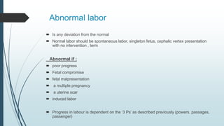
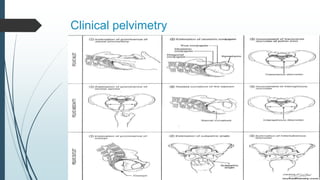
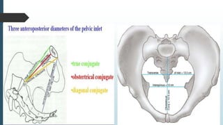
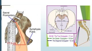
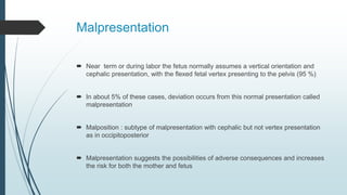
Abnormal labor
Abnormal labor
 Is any deviation from the normal
 Normal labor should be spontaneous labor, singleton fetus, cephalic vertex presentation
with no intervention , term
Abnormal if :
 poor progress
 Fetal compromise
 fetal malpresentation
 a multiple pregnancy
 a uterine scar
 induced labor
 Progress in labour is dependent on the ‘3 Ps’ as described previously (powers, passages,
passenger)
abnormal progress in labour
Prolonged latent phase
 Prolonged latent phase occurs when the latent phase is longer than the arbitrary
time limits of the normal
 more common in primiparous women
 best managed away from the labour suite with simple analgesics,
mobilization and reassurance
 intervention in the form of artificial rupture of membranes (ARM) or oxytocin
infusion will increase the likelihood of poor progress later in the labour and the
need for caesarean birth
Primary arrest
 poor progress in the active first stage of labour (<2 cm cervical dilatation/4 hours)
 More in primiparous
Causes :
 inefficient uterine (most common )
 CPD
 Malspresentation
Secondary arrest
 progress in the active first stage is initially good but then slows or stops
 Same causes of primary arrest
Arrest of second stage of labor
 when delivery is not imminent after the usual interval of pushing in the second
stage of labour
Causes :
 inefficient uterine activity
 malposition
 malpresentation
 CPD
 resistant perineum
Poor progress in the first stage of
labour
 defined as cervical dilatation of less than 2 cm in 4 hours,
usually associated with failure of descent and rotation of the fetal
head
 Management depends on the cause …3p’s
Dysfunctional uterine activity (‘powers’)
Cephalopelvic disproportion (‘passages’ and ‘passenger’)
Malpresentation (the ‘passenger’)
Portogram to assess labor progress
Should be used starting from the
active phase of first stage of labor;
if to be used
Dysfunctional uterine activity (‘powers’)
 weak, irregular and infrequent contractions
 A frequency of four to five contractions per 10 minutes is usually considered
ideal
 the most common cause of poor progress in labour
 more common in primigravidae and in older women
 Management : if confirmed delayed after 4 hours; ARM should be done first and
vaginal examination 2 hours later , if still delayed; consider augmentation of
contractions by oxytocin infusion
 If progress fails to occur despite 4–6 hours of augmentation with oxytocin, a
caesarean section will usually be recommended
Augmentation should be used with caution in multiparous women*
Risk of rupture uterus , PPH and other causes to be involved in the delay as
malpresentation
Augmentation with oxytocin is contraindicated if there are concerns regarding the
condition of the fetus
Use continuous EFM if augmentation started
Cephalopelvic disproportion
 CPD implies anatomical disproportion between the fetal head and maternal pelvis.
 Causes : small pelvis in relation to fetal size
large fetus
fetal neck masses , thyroid goiter
macrocephaly , hydrocephaly
malposition (relative CPD )
Retrospective diagnosis (failure of vaginal delivery )
Findings suggestive of CPD
 Fetal head is not engaged.
 Progress is slow or arrests despite efficient uterine contractions.
 Vaginal examination shows severe moulding and caput formation.
 Head is poorly applied to the cervix.
 Haematuria.
Clinical pelvimetry
 Clinical pelvimetry Has less role in assessing for CPD than in the past
 X ray had a role (radiological pelvimetry )
 CPD is considered a retrospective diagnosis after a failure in trial of vaginal
delivery
Clinical pelvimetry
Malpresentation
 Near term or during labor the fetus normally assumes a vertical orientation and
cephalic presentation, with the flexed fetal vertex presenting to the pelvis (95 %)
 In about 5% of these cases, deviation occurs from this normal presentation called
malpresentation
 Malposition : subtype of malpresentation with cephalic but not vertex presentation
as in occipitoposterior
 Malpresentation suggests the possibilities of adverse consequences and increases
the risk for both the mother and fetus
Causes and risk factors for
malpresentation
 Diminished vertical polarity of the uterine cavity
 Increased or decreased fetal mobility
 Obstructed pelvic inlet, cephalo-pelvic disproportion
 Polyhydramnios
 Oligohydramnios
 Fetal malformation
 Abnormal placentation
 Uterine myoma and uterine synechiae
 Multiparous
 Idiopathic
Abnormal Lie
 Lie: orientation of the fetal spine relative to the
mother’s spine
 Transverse
 Oblique
 Usually transient but if persistent; transverse lie
needs cesarean section
 Occur in 0.3 %
 Risk factors as : grand-multiparity ,Prematurity,
Contraction and deformity of maternal pelvis
,Abnormal placentation
Face presentation
 Is longitudinal lie, full extension of the fetal head on the neck and the occiput against the
upper back
 Anencephaly is found in one third of cases
 Fetal goiter and neck soft tissue tumors also cause deflection
 Contracted pelvis or CPD is found in 10-40% of cases
Face presentation
 Dominator is mentum
 Submento bragmatic is the engagement diameter
 Mentoanterior : vaginal deliveryand Delivers by flexion (mento-anterior)
 Mento posterior : CS
Brow presentation
 Is longitudinal axis with partially deflexed cephalic attitude, midway between full flexion
and full extension
 The frontal bones are the point of designation
 The engagement diameter is the mento-vertical 13.5 cm
 the incidence is 1 to 1500 deliverers
 It is more detected in early labor before flexion occurs to normal attitude. Less
frequently, further extension results in face presentation
 Transient; most convert spontaneously by flexion or extension to either vertex or face
presentation and are managed accordingly
 If persist : CS
Compound presentation
 an extremity is found prolapsed beside the main presenting part
 The combination of an upper extremity and vertex presentation is the most
common
 Prematurity is the most consistent clinical finding
 fetal risk is associated with birth trauma and cord prolapse
 the prolapsed extremity should not be manipulated, as it may retract as the
major presenting part descends
 75% of vertex/upper extremity combinations deliver spontaneously; occult
or undetected cord prolapse is possible, therefore continuous CTG is
recommended
Persistent compound presentation with parts other than vertex and hand in
combination in a term-sized infant has poor prognosis for safe vaginal
delivery and cesarean section is necessary
Breech presentation
 When the buttocks of the fetus enter the pelvis before the head the presentation is
breech
 Its more common in preterm fetuses because the bulk of each pole is more or less
similar
 Types :
1)Frank breech:
the lower extremities are flexed at the hips and extended at the knees.
2)complete breech:
the lower extremities are flexed at the hips and one or both knees are flexed.
3)incomplete breech:
one or both hips are not flexed and one or both feet are below the breech(footling)
Risk factors for breech
 Gestational age
 Hydramnios
 Uterine relaxation
 Multiple fetuses
 Oligohydramnios
 Hydrocephaly, anencephaly
 Previous breech delivery
 Uterine anomalies and pelvic tumor
Breech delivery
Cesarean delivery is commonly but not exclusively used in the following
circumstances
 A large fetus
 Any degree of contraction or unfavorable shape of the pelvis
 Hyperextended head
 When delivery is indicated in the absence of spontaneous labor
Methods of delivery ; CS , assisted vaginal delivery , spontaneous vaginal
delivery and complete breech extraction in CS or second twin
Poor progress in the second stage of
labour
 Delay is diagnosed if delivery is not imminent after 2 hours of pushing in a
nulliparous labour and 1 hour for a parous woman
 Secondary dysfunctional uterine activity (‘powers’) is a common cause of second
stage delay
 Epidural analgesia may delay second stage progress for one hour
 Operative vaginal delivery maybe done if suitable
 Oxytocin shouldn’t be started in this stage
Vaginal delivery with previous uterine scar
VBAC
 Risk of rupture in previous lower segment cs 1:200
 Risk of rupture in previous upper segment cs 2-3 % (classical )
 Success rate : 70-80 % and more if previously delivered vaginally
 Continuous CTG with close observation for signs of rupture uterus
Relative contraindications to VBAC
 Two or more previous caesarean section scars.
 Need for induction of labour (IOL).
 Previous labour outcome suggestive of CPD.
 Previous classical caesarean section is an absolute contraindication.

L32 Abnormal labor

  • 1.
  • 2.
    Abnormal labor  Isany deviation from the normal  Normal labor should be spontaneous labor, singleton fetus, cephalic vertex presentation with no intervention , term Abnormal if :  poor progress  Fetal compromise  fetal malpresentation  a multiple pregnancy  a uterine scar  induced labor  Progress in labour is dependent on the ‘3 Ps’ as described previously (powers, passages, passenger)
  • 3.
  • 4.
    Prolonged latent phase Prolonged latent phase occurs when the latent phase is longer than the arbitrary time limits of the normal  more common in primiparous women  best managed away from the labour suite with simple analgesics, mobilization and reassurance  intervention in the form of artificial rupture of membranes (ARM) or oxytocin infusion will increase the likelihood of poor progress later in the labour and the need for caesarean birth
  • 5.
    Primary arrest  poorprogress in the active first stage of labour (<2 cm cervical dilatation/4 hours)  More in primiparous Causes :  inefficient uterine (most common )  CPD  Malspresentation
  • 6.
    Secondary arrest  progressin the active first stage is initially good but then slows or stops  Same causes of primary arrest
  • 7.
    Arrest of secondstage of labor  when delivery is not imminent after the usual interval of pushing in the second stage of labour Causes :  inefficient uterine activity  malposition  malpresentation  CPD  resistant perineum
  • 8.
    Poor progress inthe first stage of labour  defined as cervical dilatation of less than 2 cm in 4 hours, usually associated with failure of descent and rotation of the fetal head  Management depends on the cause …3p’s Dysfunctional uterine activity (‘powers’) Cephalopelvic disproportion (‘passages’ and ‘passenger’) Malpresentation (the ‘passenger’)
  • 9.
    Portogram to assesslabor progress Should be used starting from the active phase of first stage of labor; if to be used
  • 10.
    Dysfunctional uterine activity(‘powers’)  weak, irregular and infrequent contractions  A frequency of four to five contractions per 10 minutes is usually considered ideal  the most common cause of poor progress in labour  more common in primigravidae and in older women  Management : if confirmed delayed after 4 hours; ARM should be done first and vaginal examination 2 hours later , if still delayed; consider augmentation of contractions by oxytocin infusion  If progress fails to occur despite 4–6 hours of augmentation with oxytocin, a caesarean section will usually be recommended
  • 11.
    Augmentation should beused with caution in multiparous women* Risk of rupture uterus , PPH and other causes to be involved in the delay as malpresentation Augmentation with oxytocin is contraindicated if there are concerns regarding the condition of the fetus Use continuous EFM if augmentation started
  • 12.
    Cephalopelvic disproportion  CPDimplies anatomical disproportion between the fetal head and maternal pelvis.  Causes : small pelvis in relation to fetal size large fetus fetal neck masses , thyroid goiter macrocephaly , hydrocephaly malposition (relative CPD ) Retrospective diagnosis (failure of vaginal delivery )
  • 13.
    Findings suggestive ofCPD  Fetal head is not engaged.  Progress is slow or arrests despite efficient uterine contractions.  Vaginal examination shows severe moulding and caput formation.  Head is poorly applied to the cervix.  Haematuria.
  • 14.
    Clinical pelvimetry  Clinicalpelvimetry Has less role in assessing for CPD than in the past  X ray had a role (radiological pelvimetry )  CPD is considered a retrospective diagnosis after a failure in trial of vaginal delivery
  • 15.
  • 18.
    Malpresentation  Near termor during labor the fetus normally assumes a vertical orientation and cephalic presentation, with the flexed fetal vertex presenting to the pelvis (95 %)  In about 5% of these cases, deviation occurs from this normal presentation called malpresentation  Malposition : subtype of malpresentation with cephalic but not vertex presentation as in occipitoposterior  Malpresentation suggests the possibilities of adverse consequences and increases the risk for both the mother and fetus
  • 19.
    Causes and riskfactors for malpresentation  Diminished vertical polarity of the uterine cavity  Increased or decreased fetal mobility  Obstructed pelvic inlet, cephalo-pelvic disproportion  Polyhydramnios  Oligohydramnios  Fetal malformation  Abnormal placentation  Uterine myoma and uterine synechiae  Multiparous  Idiopathic
  • 20.
    Abnormal Lie  Lie:orientation of the fetal spine relative to the mother’s spine  Transverse  Oblique  Usually transient but if persistent; transverse lie needs cesarean section  Occur in 0.3 %  Risk factors as : grand-multiparity ,Prematurity, Contraction and deformity of maternal pelvis ,Abnormal placentation
  • 21.
    Face presentation  Islongitudinal lie, full extension of the fetal head on the neck and the occiput against the upper back  Anencephaly is found in one third of cases  Fetal goiter and neck soft tissue tumors also cause deflection  Contracted pelvis or CPD is found in 10-40% of cases
  • 22.
    Face presentation  Dominatoris mentum  Submento bragmatic is the engagement diameter  Mentoanterior : vaginal deliveryand Delivers by flexion (mento-anterior)  Mento posterior : CS
  • 23.
    Brow presentation  Islongitudinal axis with partially deflexed cephalic attitude, midway between full flexion and full extension  The frontal bones are the point of designation  The engagement diameter is the mento-vertical 13.5 cm  the incidence is 1 to 1500 deliverers  It is more detected in early labor before flexion occurs to normal attitude. Less frequently, further extension results in face presentation  Transient; most convert spontaneously by flexion or extension to either vertex or face presentation and are managed accordingly  If persist : CS
  • 25.
    Compound presentation  anextremity is found prolapsed beside the main presenting part  The combination of an upper extremity and vertex presentation is the most common  Prematurity is the most consistent clinical finding  fetal risk is associated with birth trauma and cord prolapse
  • 26.
     the prolapsedextremity should not be manipulated, as it may retract as the major presenting part descends  75% of vertex/upper extremity combinations deliver spontaneously; occult or undetected cord prolapse is possible, therefore continuous CTG is recommended Persistent compound presentation with parts other than vertex and hand in combination in a term-sized infant has poor prognosis for safe vaginal delivery and cesarean section is necessary
  • 27.
    Breech presentation  Whenthe buttocks of the fetus enter the pelvis before the head the presentation is breech  Its more common in preterm fetuses because the bulk of each pole is more or less similar  Types : 1)Frank breech: the lower extremities are flexed at the hips and extended at the knees. 2)complete breech: the lower extremities are flexed at the hips and one or both knees are flexed. 3)incomplete breech: one or both hips are not flexed and one or both feet are below the breech(footling)
  • 29.
    Risk factors forbreech  Gestational age  Hydramnios  Uterine relaxation  Multiple fetuses  Oligohydramnios  Hydrocephaly, anencephaly  Previous breech delivery  Uterine anomalies and pelvic tumor
  • 30.
    Breech delivery Cesarean deliveryis commonly but not exclusively used in the following circumstances  A large fetus  Any degree of contraction or unfavorable shape of the pelvis  Hyperextended head  When delivery is indicated in the absence of spontaneous labor Methods of delivery ; CS , assisted vaginal delivery , spontaneous vaginal delivery and complete breech extraction in CS or second twin
  • 31.
    Poor progress inthe second stage of labour  Delay is diagnosed if delivery is not imminent after 2 hours of pushing in a nulliparous labour and 1 hour for a parous woman  Secondary dysfunctional uterine activity (‘powers’) is a common cause of second stage delay  Epidural analgesia may delay second stage progress for one hour  Operative vaginal delivery maybe done if suitable  Oxytocin shouldn’t be started in this stage
  • 32.
    Vaginal delivery withprevious uterine scar VBAC  Risk of rupture in previous lower segment cs 1:200  Risk of rupture in previous upper segment cs 2-3 % (classical )  Success rate : 70-80 % and more if previously delivered vaginally  Continuous CTG with close observation for signs of rupture uterus Relative contraindications to VBAC  Two or more previous caesarean section scars.  Need for induction of labour (IOL).  Previous labour outcome suggestive of CPD.  Previous classical caesarean section is an absolute contraindication.