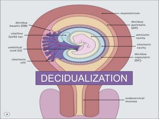
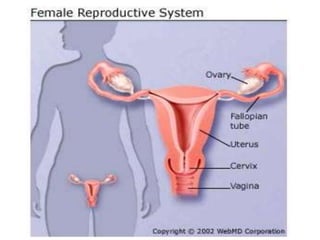
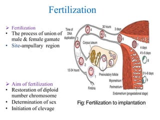
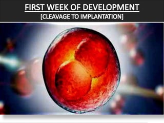
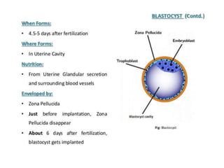
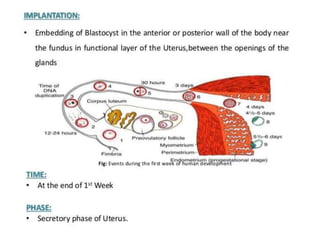
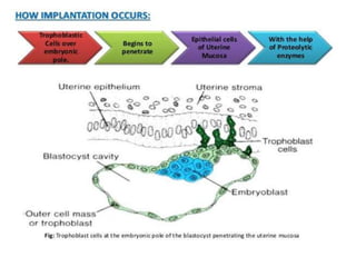
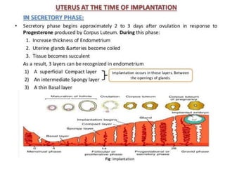
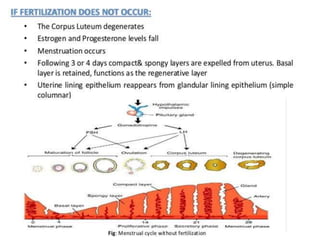
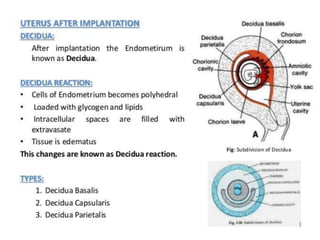
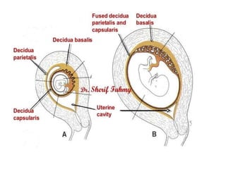
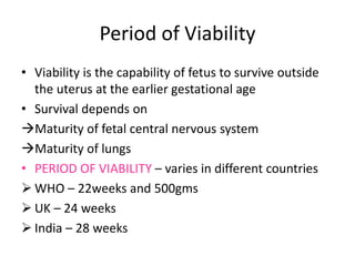
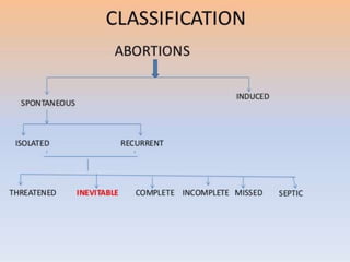
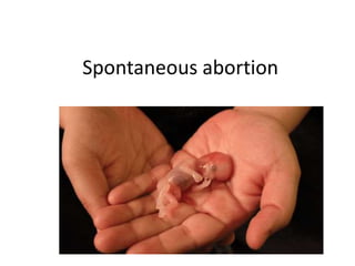
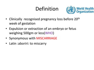
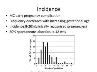
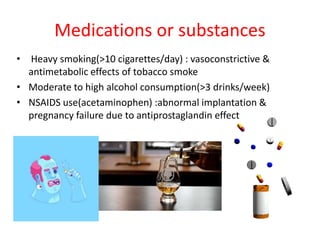
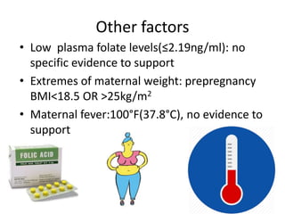
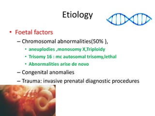
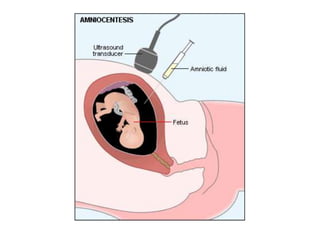
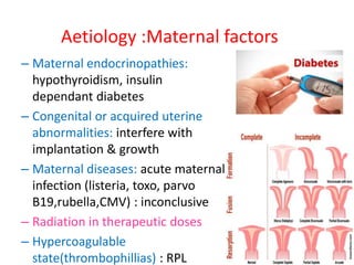
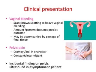
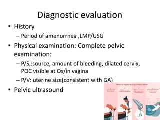
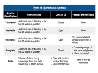
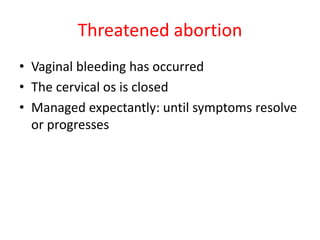
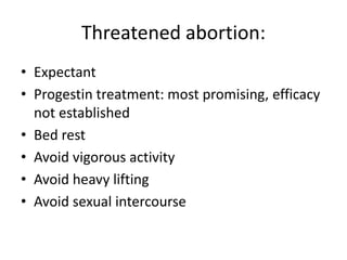
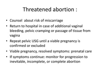
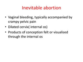
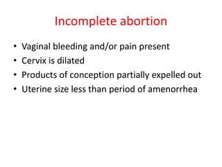
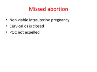
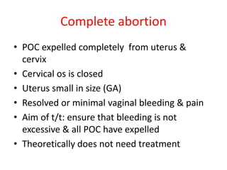
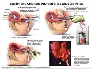
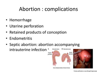
AMNIOINFUSION--
definition-An amnioinfusion is a technique of instilling an isotonic fluid {such as a normal saline or lactated ringer’s solution} into the amniotic cavity during labor to relieve umbilical cord compression and alleviate fetal distress from severe prolonged variable decelerations in the presence of oligohydramnios.
INDICATIONS
Fetal heart rate abnormalities
APGAR scores for those with low scores
Asphyxia during time of birth
Decreasing the rates of cesarean birth related with FHR problem
PROCEDURE
The amnio infusion procedure involves the use of an intrauterine pressure catheter (IUPC), or a single or double lumen type of IUPC.
The IUPC has been designed to attain an accurate monitoring of uterine contractions among women in the intrapartum period.
It has a special port from which the saline fluid or lactated ringer’s solution is being injected, passing through the tubing and going its way into the uterus.
An IUPC is inserted through standard technique once the membranes ruptures, and then it is attached to intravenous extension tubing. If IUPC is not available, a pediatric nasogastric tube can be used instead.
Lactated ringer’s solution without dextrose is infused into the amniotic cavity; normal saline can be an acceptable fluid alternative
Assisting Physician with Amnioinfusion
Explain the procedure to the patient.
Assist in dorsal recumbent position. Assist with draping and exposing vaginal area.
Connect IUPC tubing to IV fluid, flush
Connect the catheter to the monitor cable
Assist physician with insertion of double lumen IUPC and connect IV tubing to the amnioport to begin amnioinfusion.
Uterine Rupture
Deepa Mishra
Assistant Professor (OBG)
Introduction
Uterine rupture is when the muscular wall of the uterus tears during pregnancy or childbirth
Symptoms while classically including increased pain, vaginal bleeding, or a change in contractions are not always present.
Disability or death of the mother or baby may result.
Definition
Uterine rupture is giving way of gravid uterus or dissolution in the continuity of uterine wall anytime after 28 weeks of gestation with or without expulsion of the fetus.
Incidence
Rates of uterine rupture during vaginal birth following one previous C-section, done by the typical technique, are estimated at 0.9%
Rates are greater among those who have had multiple prior C-sections or an atypical type of C-section.
In those who do have uterine scarring, the risk during a vaginal birth is about 1 per 12,000
Risk of death of the baby is about 6%
Etiology
Risk Factors
Previous cesarean section
Myomectomy
Dysfunctional labor
Labor augmentation by oxytocin or prostaglandins
High parity
First pregnancy- very rare
Types of uterine rupture
Complete Rupture
All the layers including peritoneum are torn and the uterine contents escape into the peritoneal cavity.
Usually results in death
Incomplete Rupture
Visceral peritoneum is intact and usually the fetus remains in the uterine cavity
Sign & Symptoms
Uterine dehiscence and abdominal pain and vaginal bleeding
Deterioration of fetal heart rate
Loss of fetal station on manual vaginal exam
Hypovolemic shock due to intrabdominal bleeding
Chest pain between the scapulae, pain during inspiration due to irritation of blood below the perineum
Cessation of uterine contractions
Palpation of fetus outside the uterus
Signs of abdominal pregnancy
Post term pregnancy
Diagnosis
Signs of obstructed labor with dehydration, exhaustion, tachycardia raised temperature tonic contraction , pathological retraction ring
Absent fetal heart sound
On PV hot, dry vagina with a large caput over the presenting part
Prevention
Early diagnosis and management of CPD mal presentation and obstructed labor
Proper selection of cases for vaginal delivery
Carefull monitoring of oxytocin infusion specially in multipara
Avoid intra uterine manipulation no version in single fetus
Instrumental delivery after cervical dilatation
Immediate CS in obstructed labor
Hospital delivery for high risk cases
ECV should be avoided during general anaesthesia
Careful manual removal of placenta
Treatment
Resuscitation with adequate hydration and blood transfusion
Laprotomy
Hysterectomy
Repair
Complication
Rupture uterus with haemorrhage, shock and sepsis
Fetal loss is high in spontaneous and traumatic rupture
Mortality is low in LSCS scar rupture
Explains the inflammatory process of endometrium,its causes and its two clinical variants as acute and chronic endometritis.
Describes the pathology of its two types with histologic perspective.
this slide helps a physician in understanding the basics of miscarriages(definition, types/classification, causes, clinical presentation, investigations and complications. In understanding the basics, this helps a physician to able to treat or manage abortions.
Causes and management of first and second trimester abortions
anatomical, chromosomal, immunological, hormonal causes and infections. Investigation for detection of cause and possible treatment. Surgical correction of cervical incompetence and medical treatment, progestational drugs
Fragile X associated primary ovarian insufficiency
Fragile X associated decreased ovarian reserve
Fragile X syndrome and reproduction
evidence based
Dr.Anu.M - Mch Resident - Department of Reproductive Medicine and Surgery
Recurrent pregnancy loss - Uterine factorsAnu Manivannan
recurrent pregnancy loss - uterine factors based on fertility sterility journal - evidence based
Dr.Anu.M - Mch Resident - Department of Reproductive Medicine and Surgery
This presentation describes in detail about managing Rh negative pregnancy- to identify and manage Rh non-isommunized and Rh isoimmunized pregnancies, with recent advances
This presentation describes epidemiology, risk factors, pathology, clinical examination, staging and management of cervical carcinoma. SCREENING is not included
MANAGEMENT OF ATRIOVENTRICULAR CONDUCTION BLOCK.pdfJim Jacob Roy
Cardiac conduction defects can occur due to various causes.
Atrioventricular conduction blocks ( AV blocks ) are classified into 3 types.
This document describes the acute management of AV block.
Ozempic: Preoperative Management of Patients on GLP-1 Receptor Agonists Saeid Safari
Preoperative Management of Patients on GLP-1 Receptor Agonists like Ozempic and Semiglutide
ASA GUIDELINE
NYSORA Guideline
2 Case Reports of Gastric Ultrasound
These lecture slides, by Dr Sidra Arshad, offer a quick overview of physiological basis of a normal electrocardiogram.
Learning objectives:
1. Define an electrocardiogram (ECG) and electrocardiography
2. Describe how dipoles generated by the heart produce the waveforms of the ECG
3. Describe the components of a normal electrocardiogram of a typical bipolar leads (limb II)
4. Differentiate between intervals and segments
5. Enlist some common indications for obtaining an ECG
Study Resources:
1. Chapter 11, Guyton and Hall Textbook of Medical Physiology, 14th edition
2. Chapter 9, Human Physiology - From Cells to Systems, Lauralee Sherwood, 9th edition
3. Chapter 29, Ganong’s Review of Medical Physiology, 26th edition
4. Electrocardiogram, StatPearls - https://www.ncbi.nlm.nih.gov/books/NBK549803/
5. ECG in Medical Practice by ABM Abdullah, 4th edition
6. ECG Basics, http://www.nataliescasebook.com/tag/e-c-g-basics
micro teaching on communication m.sc nursing.pdfAnurag Sharma
Microteaching is a unique model of practice teaching. It is a viable instrument for the. desired change in the teaching behavior or the behavior potential which, in specified types of real. classroom situations, tends to facilitate the achievement of specified types of objectives.
These simplified slides by Dr. Sidra Arshad present an overview of the non-respiratory functions of the respiratory tract.
Learning objectives:
1. Enlist the non-respiratory functions of the respiratory tract
2. Briefly explain how these functions are carried out
3. Discuss the significance of dead space
4. Differentiate between minute ventilation and alveolar ventilation
5. Describe the cough and sneeze reflexes
Study Resources:
1. Chapter 39, Guyton and Hall Textbook of Medical Physiology, 14th edition
2. Chapter 34, Ganong’s Review of Medical Physiology, 26th edition
3. Chapter 17, Human Physiology by Lauralee Sherwood, 9th edition
4. Non-respiratory functions of the lungs https://academic.oup.com/bjaed/article/13/3/98/278874
Tom Selleck Health: A Comprehensive Look at the Iconic Actor’s Wellness Journeygreendigital
Tom Selleck, an enduring figure in Hollywood. has captivated audiences for decades with his rugged charm, iconic moustache. and memorable roles in television and film. From his breakout role as Thomas Magnum in Magnum P.I. to his current portrayal of Frank Reagan in Blue Bloods. Selleck's career has spanned over 50 years. But beyond his professional achievements. fans have often been curious about Tom Selleck Health. especially as he has aged in the public eye.
Follow us on: Pinterest
Introduction
Many have been interested in Tom Selleck health. not only because of his enduring presence on screen but also because of the challenges. and lifestyle choices he has faced and made over the years. This article delves into the various aspects of Tom Selleck health. exploring his fitness regimen, diet, mental health. and the challenges he has encountered as he ages. We'll look at how he maintains his well-being. the health issues he has faced, and his approach to ageing .
Early Life and Career
Childhood and Athletic Beginnings
Tom Selleck was born on January 29, 1945, in Detroit, Michigan, and grew up in Sherman Oaks, California. From an early age, he was involved in sports, particularly basketball. which played a significant role in his physical development. His athletic pursuits continued into college. where he attended the University of Southern California (USC) on a basketball scholarship. This early involvement in sports laid a strong foundation for his physical health and disciplined lifestyle.
Transition to Acting
Selleck's transition from an athlete to an actor came with its physical demands. His first significant role in "Magnum P.I." required him to perform various stunts and maintain a fit appearance. This role, which he played from 1980 to 1988. necessitated a rigorous fitness routine to meet the show's demands. setting the stage for his long-term commitment to health and wellness.
Fitness Regimen
Workout Routine
Tom Selleck health and fitness regimen has evolved. adapting to his changing roles and age. During his "Magnum, P.I." days. Selleck's workouts were intense and focused on building and maintaining muscle mass. His routine included weightlifting, cardiovascular exercises. and specific training for the stunts he performed on the show.
Selleck adjusted his fitness routine as he aged to suit his body's needs. Today, his workouts focus on maintaining flexibility, strength, and cardiovascular health. He incorporates low-impact exercises such as swimming, walking, and light weightlifting. This balanced approach helps him stay fit without putting undue strain on his joints and muscles.
Importance of Flexibility and Mobility
In recent years, Selleck has emphasized the importance of flexibility and mobility in his fitness regimen. Understanding the natural decline in muscle mass and joint flexibility with age. he includes stretching and yoga in his routine. These practices help prevent injuries, improve posture, and maintain mobilit
Title: Sense of Smell
Presenter: Dr. Faiza, Assistant Professor of Physiology
Qualifications:
MBBS (Best Graduate, AIMC Lahore)
FCPS Physiology
ICMT, CHPE, DHPE (STMU)
MPH (GC University, Faisalabad)
MBA (Virtual University of Pakistan)
Learning Objectives:
Describe the primary categories of smells and the concept of odor blindness.
Explain the structure and location of the olfactory membrane and mucosa, including the types and roles of cells involved in olfaction.
Describe the pathway and mechanisms of olfactory signal transmission from the olfactory receptors to the brain.
Illustrate the biochemical cascade triggered by odorant binding to olfactory receptors, including the role of G-proteins and second messengers in generating an action potential.
Identify different types of olfactory disorders such as anosmia, hyposmia, hyperosmia, and dysosmia, including their potential causes.
Key Topics:
Olfactory Genes:
3% of the human genome accounts for olfactory genes.
400 genes for odorant receptors.
Olfactory Membrane:
Located in the superior part of the nasal cavity.
Medially: Folds downward along the superior septum.
Laterally: Folds over the superior turbinate and upper surface of the middle turbinate.
Total surface area: 5-10 square centimeters.
Olfactory Mucosa:
Olfactory Cells: Bipolar nerve cells derived from the CNS (100 million), with 4-25 olfactory cilia per cell.
Sustentacular Cells: Produce mucus and maintain ionic and molecular environment.
Basal Cells: Replace worn-out olfactory cells with an average lifespan of 1-2 months.
Bowman’s Gland: Secretes mucus.
Stimulation of Olfactory Cells:
Odorant dissolves in mucus and attaches to receptors on olfactory cilia.
Involves a cascade effect through G-proteins and second messengers, leading to depolarization and action potential generation in the olfactory nerve.
Quality of a Good Odorant:
Small (3-20 Carbon atoms), volatile, water-soluble, and lipid-soluble.
Facilitated by odorant-binding proteins in mucus.
Membrane Potential and Action Potential:
Resting membrane potential: -55mV.
Action potential frequency in the olfactory nerve increases with odorant strength.
Adaptation Towards the Sense of Smell:
Rapid adaptation within the first second, with further slow adaptation.
Psychological adaptation greater than receptor adaptation, involving feedback inhibition from the central nervous system.
Primary Sensations of Smell:
Camphoraceous, Musky, Floral, Pepperminty, Ethereal, Pungent, Putrid.
Odor Detection Threshold:
Examples: Hydrogen sulfide (0.0005 ppm), Methyl-mercaptan (0.002 ppm).
Some toxic substances are odorless at lethal concentrations.
Characteristics of Smell:
Odor blindness for single substances due to lack of appropriate receptor protein.
Behavioral and emotional influences of smell.
Transmission of Olfactory Signals:
From olfactory cells to glomeruli in the olfactory bulb, involving lateral inhibition.
Primitive, less old, and new olfactory systems with different path
New Directions in Targeted Therapeutic Approaches for Older Adults With Mantl...i3 Health
i3 Health is pleased to make the speaker slides from this activity available for use as a non-accredited self-study or teaching resource.
This slide deck presented by Dr. Kami Maddocks, Professor-Clinical in the Division of Hematology and
Associate Division Director for Ambulatory Operations
The Ohio State University Comprehensive Cancer Center, will provide insight into new directions in targeted therapeutic approaches for older adults with mantle cell lymphoma.
STATEMENT OF NEED
Mantle cell lymphoma (MCL) is a rare, aggressive B-cell non-Hodgkin lymphoma (NHL) accounting for 5% to 7% of all lymphomas. Its prognosis ranges from indolent disease that does not require treatment for years to very aggressive disease, which is associated with poor survival (Silkenstedt et al, 2021). Typically, MCL is diagnosed at advanced stage and in older patients who cannot tolerate intensive therapy (NCCN, 2022). Although recent advances have slightly increased remission rates, recurrence and relapse remain very common, leading to a median overall survival between 3 and 6 years (LLS, 2021). Though there are several effective options, progress is still needed towards establishing an accepted frontline approach for MCL (Castellino et al, 2022). Treatment selection and management of MCL are complicated by the heterogeneity of prognosis, advanced age and comorbidities of patients, and lack of an established standard approach for treatment, making it vital that clinicians be familiar with the latest research and advances in this area. In this activity chaired by Michael Wang, MD, Professor in the Department of Lymphoma & Myeloma at MD Anderson Cancer Center, expert faculty will discuss prognostic factors informing treatment, the promising results of recent trials in new therapeutic approaches, and the implications of treatment resistance in therapeutic selection for MCL.
Target Audience
Hematology/oncology fellows, attending faculty, and other health care professionals involved in the treatment of patients with mantle cell lymphoma (MCL).
Learning Objectives
1.) Identify clinical and biological prognostic factors that can guide treatment decision making for older adults with MCL
2.) Evaluate emerging data on targeted therapeutic approaches for treatment-naive and relapsed/refractory MCL and their applicability to older adults
3.) Assess mechanisms of resistance to targeted therapies for MCL and their implications for treatment selection
TEST BANK for Operations Management, 14th Edition by William J. Stevenson, Ve...kevinkariuki227
TEST BANK for Operations Management, 14th Edition by William J. Stevenson, Verified Chapters 1 - 19, Complete Newest Version.pdf
Title: Sense of Taste
Presenter: Dr. Faiza, Assistant Professor of Physiology
Qualifications:
MBBS (Best Graduate, AIMC Lahore)
FCPS Physiology
ICMT, CHPE, DHPE (STMU)
MPH (GC University, Faisalabad)
MBA (Virtual University of Pakistan)
Learning Objectives:
Describe the structure and function of taste buds.
Describe the relationship between the taste threshold and taste index of common substances.
Explain the chemical basis and signal transduction of taste perception for each type of primary taste sensation.
Recognize different abnormalities of taste perception and their causes.
Key Topics:
Significance of Taste Sensation:
Differentiation between pleasant and harmful food
Influence on behavior
Selection of food based on metabolic needs
Receptors of Taste:
Taste buds on the tongue
Influence of sense of smell, texture of food, and pain stimulation (e.g., by pepper)
Primary and Secondary Taste Sensations:
Primary taste sensations: Sweet, Sour, Salty, Bitter, Umami
Chemical basis and signal transduction mechanisms for each taste
Taste Threshold and Index:
Taste threshold values for Sweet (sucrose), Salty (NaCl), Sour (HCl), and Bitter (Quinine)
Taste index relationship: Inversely proportional to taste threshold
Taste Blindness:
Inability to taste certain substances, particularly thiourea compounds
Example: Phenylthiocarbamide
Structure and Function of Taste Buds:
Composition: Epithelial cells, Sustentacular/Supporting cells, Taste cells, Basal cells
Features: Taste pores, Taste hairs/microvilli, and Taste nerve fibers
Location of Taste Buds:
Found in papillae of the tongue (Fungiform, Circumvallate, Foliate)
Also present on the palate, tonsillar pillars, epiglottis, and proximal esophagus
Mechanism of Taste Stimulation:
Interaction of taste substances with receptors on microvilli
Signal transduction pathways for Umami, Sweet, Bitter, Sour, and Salty tastes
Taste Sensitivity and Adaptation:
Decrease in sensitivity with age
Rapid adaptation of taste sensation
Role of Saliva in Taste:
Dissolution of tastants to reach receptors
Washing away the stimulus
Taste Preferences and Aversions:
Mechanisms behind taste preference and aversion
Influence of receptors and neural pathways
Impact of Sensory Nerve Damage:
Degeneration of taste buds if the sensory nerve fiber is cut
Abnormalities of Taste Detection:
Conditions: Ageusia, Hypogeusia, Dysgeusia (parageusia)
Causes: Nerve damage, neurological disorders, infections, poor oral hygiene, adverse drug effects, deficiencies, aging, tobacco use, altered neurotransmitter levels
Neurotransmitters and Taste Threshold:
Effects of serotonin (5-HT) and norepinephrine (NE) on taste sensitivity
Supertasters:
25% of the population with heightened sensitivity to taste, especially bitterness
Increased number of fungiform papillae
3. Fertilization
Fertilization
• The process of union of
male & female gamate
• Site-ampullary region
Aim of fertilization
• Restoration of diploid
number chromosome
• Determination of sex
• Initiation of clevage
Fig: Fertilization to implantation
4.
5.
6.
7.
8.
9.
10.
11.
12.
13.
14.
15.
16.
17.
18. Period of Viability
• Viability is the capability of fetus to survive outside
the uterus at the earlier gestational age
• Survival depends on
Maturity of fetal central nervous system
Maturity of lungs
• PERIOD OF VIABILITY – varies in different countries
WHO – 22weeks and 500gms
UK – 24 weeks
India – 28 weeks
21. Definition
• Clinically recognised pregnancy loss before 20th
week of gestation
• Expulsion or extraction of an embryo or fetus
weighing 500gm or less(WHO)
• Synonymous with MISCARRIAGE
• Latin :aboriri: to miscarry
22. Incidence
• MC early pregnancy complication
• Frequency decreases with increasing gestational age
• Incidence:8-20%(clinically recognised pregnancies)
• 80% spontaneous abortion :< 12 wks
23. Risk factors
• Advanced maternal age
• Previous spontaneous abortion
• Medications & substances (smoking)
• Mechanisms responsible for abortion: not
apparent
• Death of fetus precedes spont. expulsion,
finding cause involves ascertaining the cause
of fetal death
24. Maternal age
• Most important risk factor in healthy women
• 30yrs:9-17%
• 35yrs:20%
• 40yrs:40%
• 45yrs: 80%
26. Medications or substances
• Heavy smoking(>10 cigarettes/day) : vasoconstrictive &
antimetabolic effects of tobacco smoke
• Moderate to high alcohol consumption(>3 drinks/week)
• NSAIDS use(acetaminophen) :abnormal implantation &
pregnancy failure due to antiprostaglandin effect
27. Other factors
• Low plasma folate levels(≤2.19ng/ml): no
specific evidence to support
• Extremes of maternal weight: prepregnancy
BMI<18.5 OR >25kg/m2
• Maternal fever:100°F(37.8°C), no evidence to
support
32. Clinical presentation
• Vaginal bleeding
– Scant brown spotting to heavy vaginal
bleeding
– Amount /pattern does not predict
outcome
– May be accompanied by passage of
fetal tissue
• Pelvic pain
– Crampy /dull in character
– Constant/intermittent
• Incidental finding on pelvic
ultrasound in asymptomatic patient
33. Diagnostic evaluation
• History
– Period of amenorrhea ,LMP/USG
• Physical examination: Complete pelvic
examination:
– P/S,:source, amount of bleeding, dilated cervix,
POC visible at Os/in vagina
– P/V: uterine size(consistent with GA)
• Pelvic ultrasound
34. Pelvic ultrasound
• Most useful test in diagnostic
evaluation of women with
suspected spontaneous
abortion
• Foetal cardiac activity: most
important (5.5-6wks)
• Foetal heart rate
• Size & contour of G.sac
• Presence of yolk sac
• Best evaluated ,transvaginal
approach(TVS)
36. Post diagnostic classification
• Based upon the location of POC
• Degree of cervical dilatation(pelvic exam)
• Pelvic ultrasound
• Categorization impacts clinical management
– Threatened
– Inevitable
– Incomplete/complete
– Missed
37.
38.
39. Threatened abortion
• Vaginal bleeding has occurred
• The cervical os is closed
• Managed expectantly: until symptoms resolve
or progresses
40. Threatened abortion:
• Expectant
• Progestin treatment: most promising, efficacy
not established
• Bed rest
• Avoid vigorous activity
• Avoid heavy lifting
• Avoid sexual intercourse
41. Threatened abortion :
• Counsel about risk of miscarriage
• Return to hospital in case of additional vaginal
bleeding, pelvic cramping or passage of tissue from
vagina
• Repeat pelvic USG until a viable pregnancy is
confirmed or excluded
• Viable pregnancy, resolved symptoms: prenatal care
• If symptoms continue: monitor for progression to
inevitable, incomplete, or complete abortion
42. Inevitable abortion
• Vaginal bleeding, typically accompanied by
crampy pelvic pain
• Dilated cervix( internal os)
• Products of conception felt or visualised
through the internal os
43. Incomplete abortion
• Vaginal bleeding and/or pain present
• Cervix is dilated
• Products of conception partially expelled out
• Uterine size less than period of amenorrhea
44. Missed abortion
• Non viable intrauterine pregnancy
• Cervical os is closed
• POC not expelled
45. Complete abortion
• POC expelled completely from uterus &
cervix
• Cervical os is closed
• Uterus small in size (GA)
• Resolved or minimal vaginal bleeding & pain
• Aim of t/t: ensure that bleeding is not
excessive & all POC have expelled
• Theoretically does not need treatment
46. Management
• Complete evacuation of uterine contents(POC)
• Surgical methods: suction evacuation/suction
curettage/dilation & evacuation
• Medical methods: Misoprostol,mifepristone
• All have similar efficacy
49. Prevention of spont.abortion
• Preconception & prenatal counseling
• Routine screening & optimal disease
control(diabetes, thyroid, thrombophilia)
• Correction of uterine structural
anomalies(septum, submucosal myoma,
intrauterine adhesions) prior to pregnancy
• Avoiding exposure to teratogen or infections
• Modifiable risk factors
50. • Clinically recognised pregnany losses <20 wks
gestation
• Most common complication of early
pregnancy
• Advanced maternal age, previous
spontaneous abortion, maternal smoking: risk
factors
• Mostly due to fetal structural/chromosomal
abnormalities
51. • Present with menstrual delay, vaginal
bleeding& pelvic pain
• D/D: uterine or other genital tract bleeding in
viable pregnancy, ectopic,& GTD
• Pelvic examination & pelvic ultrasound: key
elements for diagnosis
• Spontaneous abortion diagnosed based on
USG criteria
• Categorised as threatened/incomplete/missed
52. • Preconceptual & prenatal counseling & care
regarding modifiable aetiologies ,risk factors
are most imp intervention
• Normal menstrual cycle resumes in 4-6 weeks
• hCG returns to normal 2-4wks