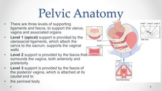
Uterovaginal prolapse is caused by failure of pelvic floor muscles and ligaments to support pelvic organs. Symptoms include a sensation of vaginal bulge or protrusion, as well as pain and discomfort relieved by lying down. Pelvic organs are supported by three levels of ligaments and fascia, with defects at different levels causing prolapse of different organs. Clinical assessment involves examination of the prolapse severity and other symptoms. Management includes pelvic floor exercises, pessaries, and surgery depending on the severity and patient preferences.
2. PROLAPSE
• Uterovaginal prolapse is caused by failure of the interaction between
the levator ani muscles and the ligaments and fascia that support
the pelvic organs.
• Pelvic organ prolapse can cause symptoms directly due to the
prolapsed organ or indirectly due to organ dysfunction secondary to
displacement from the anatomical position.
• Risk factors predisposing to prolapse are very much similar to those
predisposing to stress incontinence.
3. Symptoms from pelvic organ prolapse
• Proplase symptoms include
• A sensation of vaginal bulge,
• heaviness or a visible protrusion at or beyond the introitus.
• lower abdominal or back pain
• dragging discomfort relieved by lying or sitting.
• Indirect symptoms will depend on which other organs are involved in the
prolapse
• difficulty voiding or emptying the bowel (obstructive defaecation)
• sensations of incomplete emptying of bladder or rectum.
• Urinary or faecal incontinence
• bleeding from a prolapse that is external
5. Pelvic Anatomy
• There are three levels of supporting
ligaments and fascia, to support the uterus,
vagina and associated organs
• Level 1 (apical) support is provided by the
uterosacral ligaments, which attach the
cervix to the sacrum. supports the vaginal
walls
• Level 2 support is provided by the fascia that
surrounds the vagina, both anteriorly and
posteriorly.
• Level 3 support is provided by the fascia of
the posterior vagina, which is attached at its
caudal end to
• the perineal body
6. Pelvic Anatomy
• Defects in level 1 support can be seen on examination by the
descent of the uterus within the vagina. manifests as vaginal vault
prolapse
• Defects in level 2 support will lead to prolapse of the vaginal wall
into the vaginal lumen (causing anterior or posterior vaginal
prolapse). The bladder or rectum will prolapse behind the vaginal
wall. On examination, the affected vaginal wall will be seen bulging
into the vagina.
• Defects of the perineal body (level 3) usually cause the development
of lower posterior vaginal wall prolapse
7.
8. Clinical assessment of prolapse
• The history should elicit the presenting symptom(s) and severity,
and include questions to ascertain if the patient has any coexisting
urinary, faecal or sexual symptoms.
• Clinical examination should ideally be done in the lithotomy position
with a Sims speculum
9. Clinical Examination
• Prolapse is described in three stages of descent,
• note whether it occurs at patient straining or at rest and whether
traction has been applied
• Stage I where the prolapse does not reach the hymen.
• Stage II where the prolapse reaches the hymen.
• Stage III when the prolapse is mostly or wholly outside the hymen.
• When the uterus prolapses wholly outside this is termed procidentia.
13. Management
• Surgery for pelvic organ prolapse
• Offered if conservative treatments have failed or if the patient
chooses surgery from the outset.
• The procedure chosen depends on:
- which compartment is affected
- whether the woman wishes to retain her uterus
- whether the vaginal or abdominal route of surgery is chosen.
• Vaginal route is used for: anterior repair, posterior repair, and repair
of the perineal body.