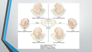
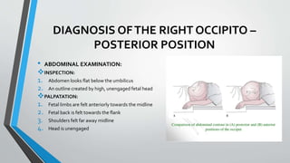
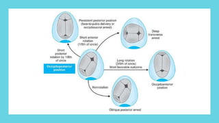
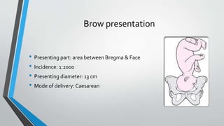
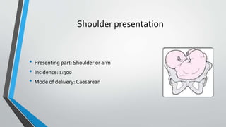
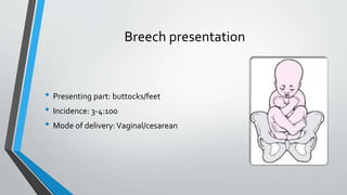
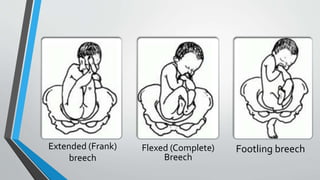
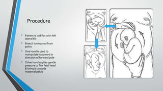
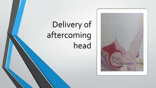
MALPRESENTATION &
MALPOSITIONING
AHMED ALI
AREESHA MOIZ ALVI
MALPOSITION
DEFINITIONS:
POSITION:
"Relationship between the presenting part of the fetus and the maternal pelvis.The
normal is the OCCIPITO ANTERIOR position."
MALPOSITION:
"It is the vertex position where is occiput is placed posteriorly over the sacroiliac joint
or directly over the sacrum, it is called the OCCIPITO POSTERIOR position."
TYPES OF OCCIPITO POSTERIOR POSITION
1. RIGHT OCCIPITO POSTERIOR (ROP)
2. LEFT OCCIPITO POSTERIOR (LOP)
3. DIRECT OCCIPITO POSTERIOR (OP)
CAUSES
• SHAPE OFTHE PELVIS:An anthropoid or android pelvic brim predisposes
to the OP position of the fetal head.
• ANTERIORLY PLACED PLACENTA
• ABNORMAL UTERINE CONTRACTIONS
• MULTIPLE PREGNANCIES
• FETAL BACK ONTHE RIGHT SIDE
DIAGNOSIS OFTHE RIGHT OCCIPITO –
POSTERIOR POSITION
• ABDOMINAL EXAMINATION:
INSPECTION:
1. Abdomen looks flat below the umbilicus
2. An outline created by high, unengaged fetal head
PALPATATION:
1. Fetal limbs are felt anteriorly towards the midline
2. Fetal back is felt towards the flank
3. Shoulders felt far away midline
4. Head is unengaged
…...
vAUSCULTATION:
1. Fetal heart sounds heard in the flank region
• VAGINAL EXAMINATION:
1. Elongated bag of membranes.
2. Sagittal suture in the right oblique diameter of the pelvis
3. Posterior fontanelle is felt in the sacroiliac joint.
4. Anterior fontanelle felt more easily
MECHANISM OF LABOUR
The mechanism of labor depends on whether the head is well flexed or
incompletely flexed.
The well flexed head:
• The occiput will be at lower level than the sinciput
• It will hit the pelvic floor first.
• Undergoing long anterior rotation through three-eighths of a circle to lie behind the
symphysis pubis.
• The rest of the mechanism is the same as the right occipitoanterior position.
When the head is incompletely flexed:
• If the head is incompletely flexed the occipitofrontal diameter which measure 11.5
cm has to pass through the pelvis instead of the sub occipitobregmatic diameter
which measure 9.5 cm.
• It is this that explains why some cases of occipitoposterior position have difficult
and prolonged labor.
• With incomplete flexion the sinciput will meet the pelvic floor first and rotate
anteriorly to lie behind the symphysis
• While the occiput rotate backward by one-eighth of the circle to lie in the hallow of
the sacrum
•The head may now be born with the face towards the
posterior surface of the symphysis pubis (face to pubis).
•The root of the nose is pressed against the bone.
•The vertex is born by flexion and followed by the occiput.
•Then the head extends, so the face and chin emerging
from under the pubic arch.
•The vulval orifice is stretched by the occipitofrontal
instead of the sub-occipitofrontal diameter with a
difference in size of 1.5 cm and a severe perineal tear may
result.
DeepTransverse Arrest
• • In some cases me head becomes arrested with its long axis in the transverse
diameter of the pelvis.
• •The degree of extension being such, that neither the occiput nor the forehead is
sufficiently in advance to influence rotationThis is called deep transverse arrest of
the head
• • It result from either:
1. Incomplete forward rotation of occipitoposterior position.
2. The majority are the result of failure of the head which enter the pelvis
within occipitotransverse position to rotate anteriorly
COURSE OF LABOUR IN OPP
• Prolongation of the 1' and 2 stages of labor is common.
• Ineffective uterine contraction is common because the poorty flexed head
fails to press down upon the cervix.
• In 70% of cases there will be spontaneous rotation of the occiput to the
anterior position.
• In about 10% there the occiput undergoes short back ward rotation and
delivered in direct occipto-posterior position Mace-to-pubes).
MANAGEMENT
Management of the first stage of labor:
• The 1st stage is managed as in a normal case.
• Nothing can be done to correct the Malposition or to influence the
rotation of the head at this stage.
• A partogram is done to monitor the .
1. Uterine contraction (frequency, duration and strength).
2. Fetal heart
3. Dilatation of the cervix
• If progressive cervical dilatation does not occur augmentation with
an oxytocin drip may be toed.
• If still no progress obtained in a few hours or in case of
cepahlopelvic dispropotion caesarian section (C/S) is performed.
• Also if there is fetal distress US is done.
Management
of the 2nd
stage of labor
• In most cases (70%) provided that the uterine
contractions are strong and the woman is able to make
good expulsive efforts the occiput rotates forward and
normal delivery takes place.
• In other cases (10%) the baby may be delievered face-
to-pubes without difficulty but there is a great risk of a
perineal tear.
• In about 20% of cases there is failure of the presenting
part to notate and descend and such cases delivered by
C/S or rotation can be enhanced by assistance via
manual rotations and forcep delivery, Kiellancrs fprceps
or a vaccumm extractor
MANAGEMENT OF DEEPTRANSVERSE
ARREST
CAESAREAN SECTION
1. The pelvis should be reassessed and if the pelvis is android or there is
evidence of disproportion CAESAREAN SECTION should be done.
2. Increasing use of caesarean for deep transverse arrest is to avoid the
intracranial haemorrhage due to traumatic vaginal delivery
VACCUM EXTRACTION
1. This is an alternative in the absence of cephalopelvic disproportion.
2. It promotes flexion thereby reducing the diameter presenting to the outlet
from occipitofrontal to smaller suboccipitobrgmatic.
3. It is less traumatic and does not need general anaesthesia.
4. The cup should be applied as near posterior fontanelle as possible as in
order to promote flexion and smooth descent .
MANUAL ROTATION
1. This procedure can be employed if the obstetrician is well versed in this
technique.
2. Under the general anaesthesia, the right hand grasps the sinciput
displacing it thereby increasing flexion.
3. The smaller bitemporal diameter allows more space for the thumb and
finger to have a firm grasp across the temple with middle finger on the
frontal suture. In LOP, the left hand is used.Then the sinciput is rotated
and forceps or vaccum is applied .
FORCEPS
ROTATION
1. In deep transverse arrest Keilland forceps is used.
2. It should be used only by the obstetrician who are
expert in its use.
3. Keilland forceps is applied under General
Anaesthesia in the anteroposterior direction and
rotation carried out.
Presentation
Definition
• Portion of the fetus overlying the pelvic inlet
• Cephalic presentation is the normal presentation
Risk
Factors
Maternal Facto
Uterine surgery
Fibroids
Uterine abnormalities, e.g. Bicornate uterus
Fetal
Multiple pregnancy
Abnormality e.g. hydrocephalus & anencephaly
Oligohydramnios
polyhydramnios
Malpresentations
Face presentation Brow presentation Shoulder presentation
Breech presentation Compound presentation
Face presentation
• Presenting part: area between chin & glabella
• Incidence: 1:500
• Presenting diameter: 9.5 cm
• Mode of delivery:Vaginal/Caesarean
Brow presentation
• Presenting part: area between Bregma & Face
• Incidence: 1:2000
• Presenting diameter: 13 cm
• Mode of delivery: Caesarean
Shoulder presentation
• Presenting part: Shoulder or arm
• Incidence: 1:300
• Mode of delivery: Caesarean
Breech presentation
• Presenting part: buttocks/feet
• Incidence: 3-4:100
• Mode of delivery:Vaginal/cesarean
Extended (Frank)
breech
Footling breech
Flexed (Complete)
Breech
Complications
• Premature rupture of membranes
• Premature labour
• Prolonged/obstructed labour
• Ruptured uterus
• Postpartum haemorrhage
• Fetal and maternal distress
Diagnosis
Abdominal Examination
Vaginal Examination during
labour
Ultrasound
Management
Caesarean
Vaginal
delivery
ECV
External CephalicVersion (ECV)
• Relatively safe technique
• Performed at 0r after 37 week
• Under ultrasound guidance
• Should last under 10 min
Procedure
• Patient is laid flat with left
lateral tilt
• Breech is elevated from
pelvis
• One hand is used to
manipulate it upward in
direction of forward pole
• Other hand applies gentle
pressure to flex fetal head
& bring it towards
maternal pelvis
Contraindications
• Fetal abnormalities
• Placenta previa
• Oligohydramnios or polyhydramnios
• Caesarean or myotomy scar on uterus
• Pre-eclampsia or hypertension
• Multiple gestation
Complications
• Placental abruptions
• Premature Rupture of membranes
• Cord accidents
• Fetal bradycardia
• Transplacental haemorrhage
Vaginal Breech
Delivery
Prerequisites
• Presentation should be extended or Flexed
• Estimated Fetal weight < 3500 g
• No evidence of fetal head extension
• No evidence of fetal abnormalities eg Hydrocephalus
• Should be performed by experienced obstetrician
Procedure
• Delivery of buttocks
• Delivery of legs and lower body (Pinard's Manoeuvre)
• Delivery of shoulders (Loveset's Manoeuvre)
• Delivery of the head (Mauriceau-SmellieVeit manoeuvres)
Delivery of buttocks
Delivery of shoulders (Loveset's Manoeuvre)
Delivery of the head (Mauriceau-
SmellieVeit manoeuvres)
Delivery of
aftercoming
head
Complication
Risks of obstructed Labour increases because of inappropriate use of oxytocic
agents or by breach extraction.
ThankYou

Malpresentation & Malpositioning

  • 1.
  • 2.
    MALPOSITION DEFINITIONS: POSITION: "Relationship between thepresenting part of the fetus and the maternal pelvis.The normal is the OCCIPITO ANTERIOR position." MALPOSITION: "It is the vertex position where is occiput is placed posteriorly over the sacroiliac joint or directly over the sacrum, it is called the OCCIPITO POSTERIOR position."
  • 3.
    TYPES OF OCCIPITOPOSTERIOR POSITION 1. RIGHT OCCIPITO POSTERIOR (ROP) 2. LEFT OCCIPITO POSTERIOR (LOP) 3. DIRECT OCCIPITO POSTERIOR (OP)
  • 6.
    CAUSES • SHAPE OFTHEPELVIS:An anthropoid or android pelvic brim predisposes to the OP position of the fetal head. • ANTERIORLY PLACED PLACENTA • ABNORMAL UTERINE CONTRACTIONS • MULTIPLE PREGNANCIES • FETAL BACK ONTHE RIGHT SIDE
  • 7.
    DIAGNOSIS OFTHE RIGHTOCCIPITO – POSTERIOR POSITION • ABDOMINAL EXAMINATION: INSPECTION: 1. Abdomen looks flat below the umbilicus 2. An outline created by high, unengaged fetal head PALPATATION: 1. Fetal limbs are felt anteriorly towards the midline 2. Fetal back is felt towards the flank 3. Shoulders felt far away midline 4. Head is unengaged
  • 8.
    …... vAUSCULTATION: 1. Fetal heartsounds heard in the flank region • VAGINAL EXAMINATION: 1. Elongated bag of membranes. 2. Sagittal suture in the right oblique diameter of the pelvis 3. Posterior fontanelle is felt in the sacroiliac joint. 4. Anterior fontanelle felt more easily
  • 9.
    MECHANISM OF LABOUR Themechanism of labor depends on whether the head is well flexed or incompletely flexed. The well flexed head: • The occiput will be at lower level than the sinciput • It will hit the pelvic floor first. • Undergoing long anterior rotation through three-eighths of a circle to lie behind the symphysis pubis. • The rest of the mechanism is the same as the right occipitoanterior position.
  • 10.
    When the headis incompletely flexed: • If the head is incompletely flexed the occipitofrontal diameter which measure 11.5 cm has to pass through the pelvis instead of the sub occipitobregmatic diameter which measure 9.5 cm. • It is this that explains why some cases of occipitoposterior position have difficult and prolonged labor. • With incomplete flexion the sinciput will meet the pelvic floor first and rotate anteriorly to lie behind the symphysis • While the occiput rotate backward by one-eighth of the circle to lie in the hallow of the sacrum
  • 11.
    •The head maynow be born with the face towards the posterior surface of the symphysis pubis (face to pubis). •The root of the nose is pressed against the bone. •The vertex is born by flexion and followed by the occiput. •Then the head extends, so the face and chin emerging from under the pubic arch. •The vulval orifice is stretched by the occipitofrontal instead of the sub-occipitofrontal diameter with a difference in size of 1.5 cm and a severe perineal tear may result.
  • 12.
    DeepTransverse Arrest • •In some cases me head becomes arrested with its long axis in the transverse diameter of the pelvis. • •The degree of extension being such, that neither the occiput nor the forehead is sufficiently in advance to influence rotationThis is called deep transverse arrest of the head • • It result from either: 1. Incomplete forward rotation of occipitoposterior position. 2. The majority are the result of failure of the head which enter the pelvis within occipitotransverse position to rotate anteriorly
  • 14.
    COURSE OF LABOURIN OPP • Prolongation of the 1' and 2 stages of labor is common. • Ineffective uterine contraction is common because the poorty flexed head fails to press down upon the cervix. • In 70% of cases there will be spontaneous rotation of the occiput to the anterior position. • In about 10% there the occiput undergoes short back ward rotation and delivered in direct occipto-posterior position Mace-to-pubes).
  • 15.
    MANAGEMENT Management of thefirst stage of labor: • The 1st stage is managed as in a normal case. • Nothing can be done to correct the Malposition or to influence the rotation of the head at this stage. • A partogram is done to monitor the . 1. Uterine contraction (frequency, duration and strength). 2. Fetal heart 3. Dilatation of the cervix • If progressive cervical dilatation does not occur augmentation with an oxytocin drip may be toed. • If still no progress obtained in a few hours or in case of cepahlopelvic dispropotion caesarian section (C/S) is performed. • Also if there is fetal distress US is done.
  • 16.
    Management of the 2nd stageof labor • In most cases (70%) provided that the uterine contractions are strong and the woman is able to make good expulsive efforts the occiput rotates forward and normal delivery takes place. • In other cases (10%) the baby may be delievered face- to-pubes without difficulty but there is a great risk of a perineal tear. • In about 20% of cases there is failure of the presenting part to notate and descend and such cases delivered by C/S or rotation can be enhanced by assistance via manual rotations and forcep delivery, Kiellancrs fprceps or a vaccumm extractor
  • 17.
    MANAGEMENT OF DEEPTRANSVERSE ARREST CAESAREANSECTION 1. The pelvis should be reassessed and if the pelvis is android or there is evidence of disproportion CAESAREAN SECTION should be done. 2. Increasing use of caesarean for deep transverse arrest is to avoid the intracranial haemorrhage due to traumatic vaginal delivery
  • 18.
    VACCUM EXTRACTION 1. Thisis an alternative in the absence of cephalopelvic disproportion. 2. It promotes flexion thereby reducing the diameter presenting to the outlet from occipitofrontal to smaller suboccipitobrgmatic. 3. It is less traumatic and does not need general anaesthesia. 4. The cup should be applied as near posterior fontanelle as possible as in order to promote flexion and smooth descent .
  • 19.
    MANUAL ROTATION 1. Thisprocedure can be employed if the obstetrician is well versed in this technique. 2. Under the general anaesthesia, the right hand grasps the sinciput displacing it thereby increasing flexion. 3. The smaller bitemporal diameter allows more space for the thumb and finger to have a firm grasp across the temple with middle finger on the frontal suture. In LOP, the left hand is used.Then the sinciput is rotated and forceps or vaccum is applied .
  • 20.
    FORCEPS ROTATION 1. In deeptransverse arrest Keilland forceps is used. 2. It should be used only by the obstetrician who are expert in its use. 3. Keilland forceps is applied under General Anaesthesia in the anteroposterior direction and rotation carried out.
  • 21.
    Presentation Definition • Portion ofthe fetus overlying the pelvic inlet • Cephalic presentation is the normal presentation
  • 22.
    Risk Factors Maternal Facto Uterine surgery Fibroids Uterineabnormalities, e.g. Bicornate uterus Fetal Multiple pregnancy Abnormality e.g. hydrocephalus & anencephaly Oligohydramnios polyhydramnios
  • 23.
    Malpresentations Face presentation Browpresentation Shoulder presentation Breech presentation Compound presentation
  • 24.
    Face presentation • Presentingpart: area between chin & glabella • Incidence: 1:500 • Presenting diameter: 9.5 cm • Mode of delivery:Vaginal/Caesarean
  • 25.
    Brow presentation • Presentingpart: area between Bregma & Face • Incidence: 1:2000 • Presenting diameter: 13 cm • Mode of delivery: Caesarean
  • 26.
    Shoulder presentation • Presentingpart: Shoulder or arm • Incidence: 1:300 • Mode of delivery: Caesarean
  • 27.
    Breech presentation • Presentingpart: buttocks/feet • Incidence: 3-4:100 • Mode of delivery:Vaginal/cesarean
  • 28.
  • 29.
    Complications • Premature ruptureof membranes • Premature labour • Prolonged/obstructed labour • Ruptured uterus • Postpartum haemorrhage • Fetal and maternal distress
  • 30.
  • 31.
  • 32.
    External CephalicVersion (ECV) •Relatively safe technique • Performed at 0r after 37 week • Under ultrasound guidance • Should last under 10 min
  • 33.
    Procedure • Patient islaid flat with left lateral tilt • Breech is elevated from pelvis • One hand is used to manipulate it upward in direction of forward pole • Other hand applies gentle pressure to flex fetal head & bring it towards maternal pelvis
  • 34.
    Contraindications • Fetal abnormalities •Placenta previa • Oligohydramnios or polyhydramnios • Caesarean or myotomy scar on uterus • Pre-eclampsia or hypertension • Multiple gestation
  • 35.
    Complications • Placental abruptions •Premature Rupture of membranes • Cord accidents • Fetal bradycardia • Transplacental haemorrhage
  • 36.
    Vaginal Breech Delivery Prerequisites • Presentationshould be extended or Flexed • Estimated Fetal weight < 3500 g • No evidence of fetal head extension • No evidence of fetal abnormalities eg Hydrocephalus • Should be performed by experienced obstetrician
  • 37.
    Procedure • Delivery ofbuttocks • Delivery of legs and lower body (Pinard's Manoeuvre) • Delivery of shoulders (Loveset's Manoeuvre) • Delivery of the head (Mauriceau-SmellieVeit manoeuvres)
  • 38.
  • 39.
    Delivery of shoulders(Loveset's Manoeuvre)
  • 40.
    Delivery of thehead (Mauriceau- SmellieVeit manoeuvres)
  • 41.
  • 42.
    Complication Risks of obstructedLabour increases because of inappropriate use of oxytocic agents or by breach extraction.
  • 43.

Editor's Notes

  • #25 Vaginal if chin is anterior. Nose, mouth & eyes can be palpated during vaginal exam during labour
  • #26 Ant. Frontanelle, supra orbital ridges and nose can be palpated