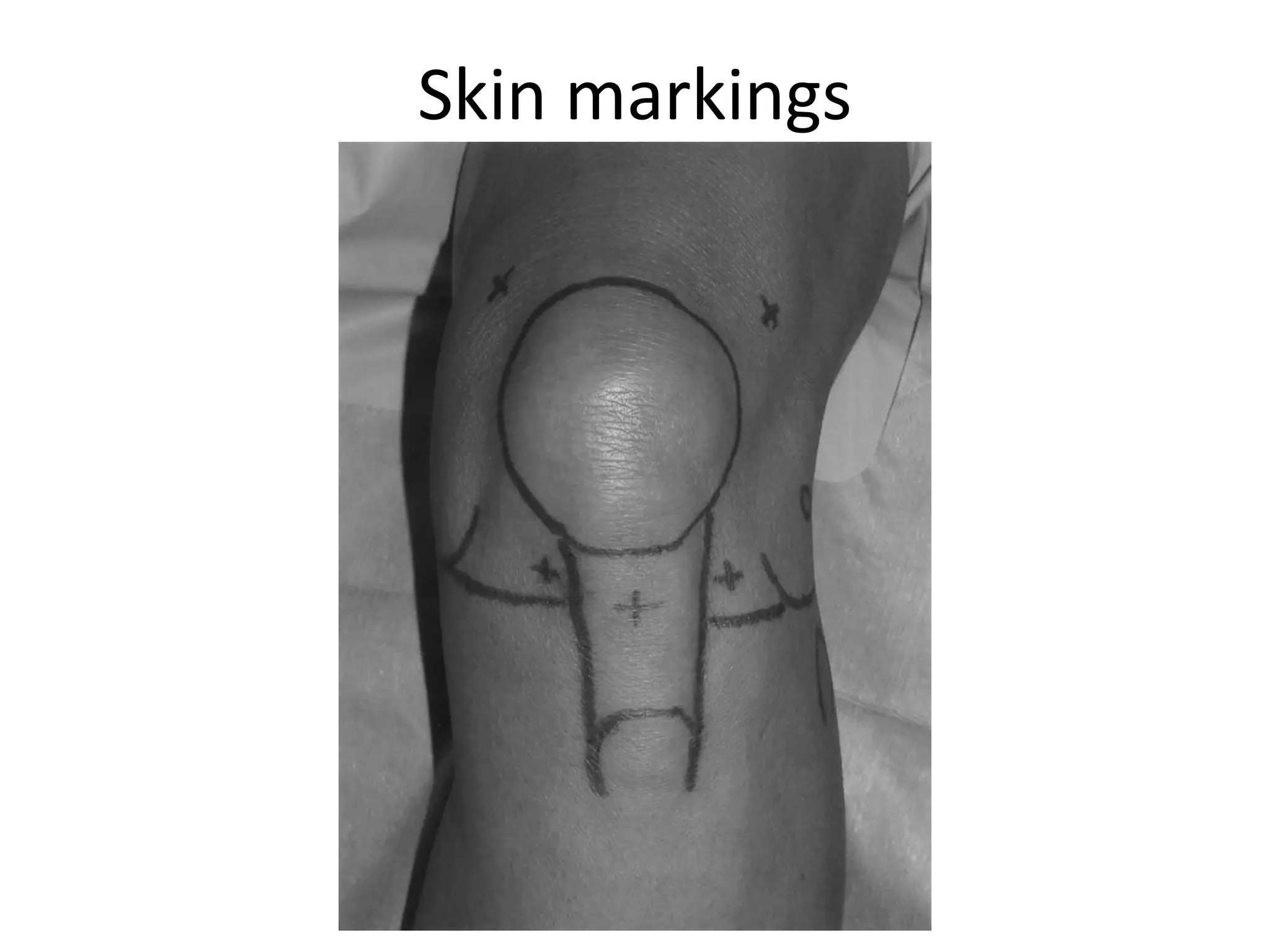
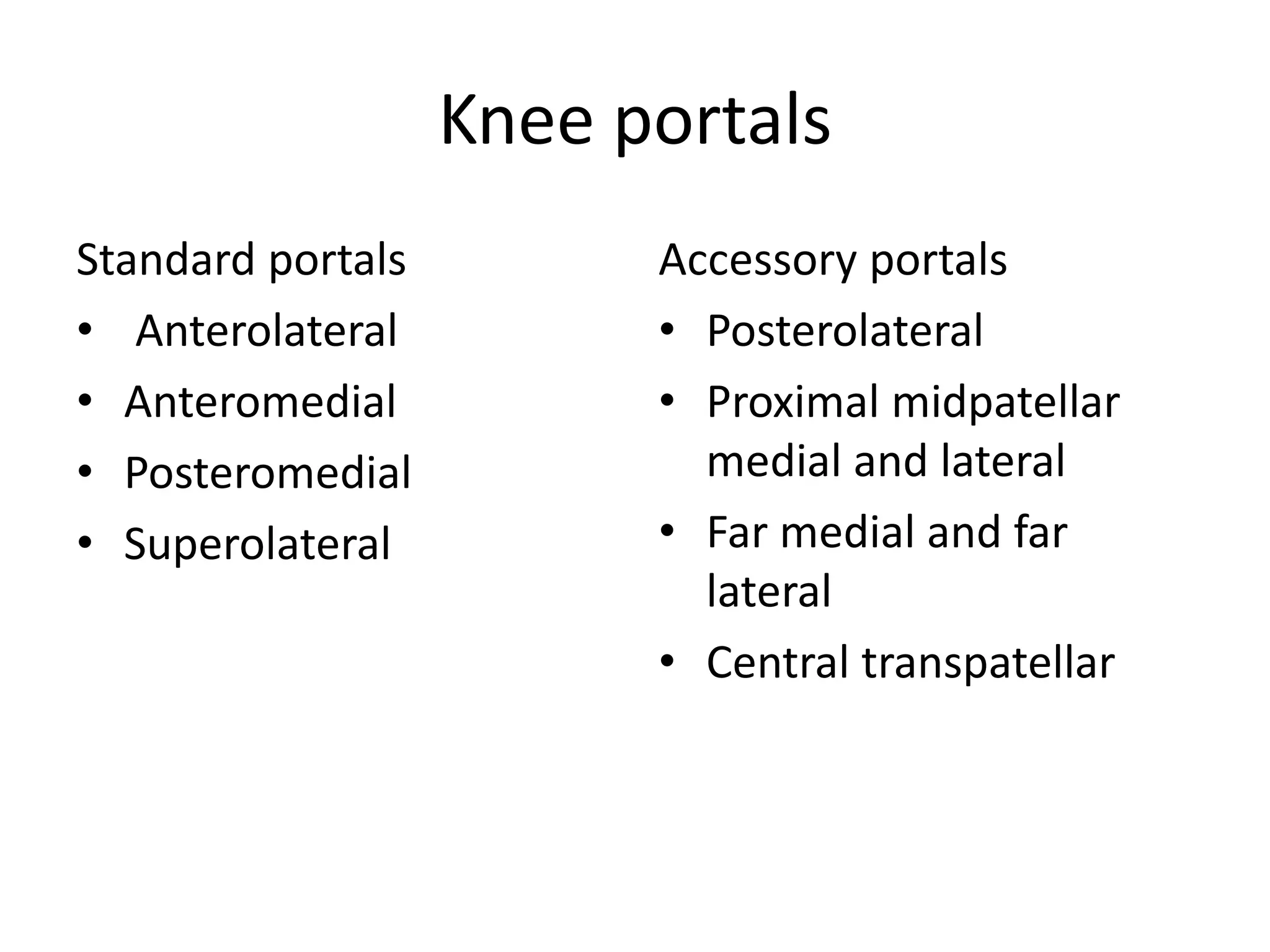
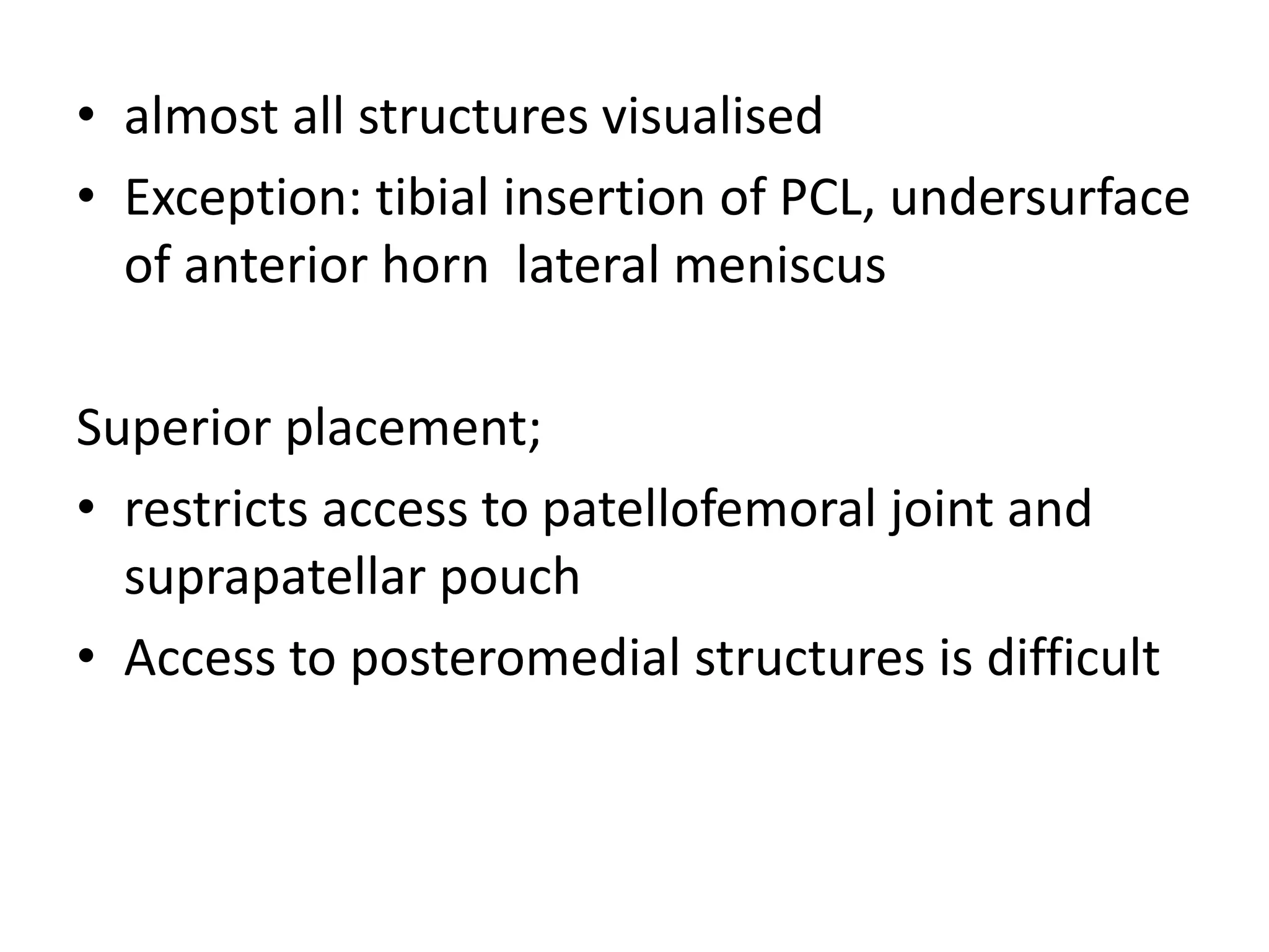
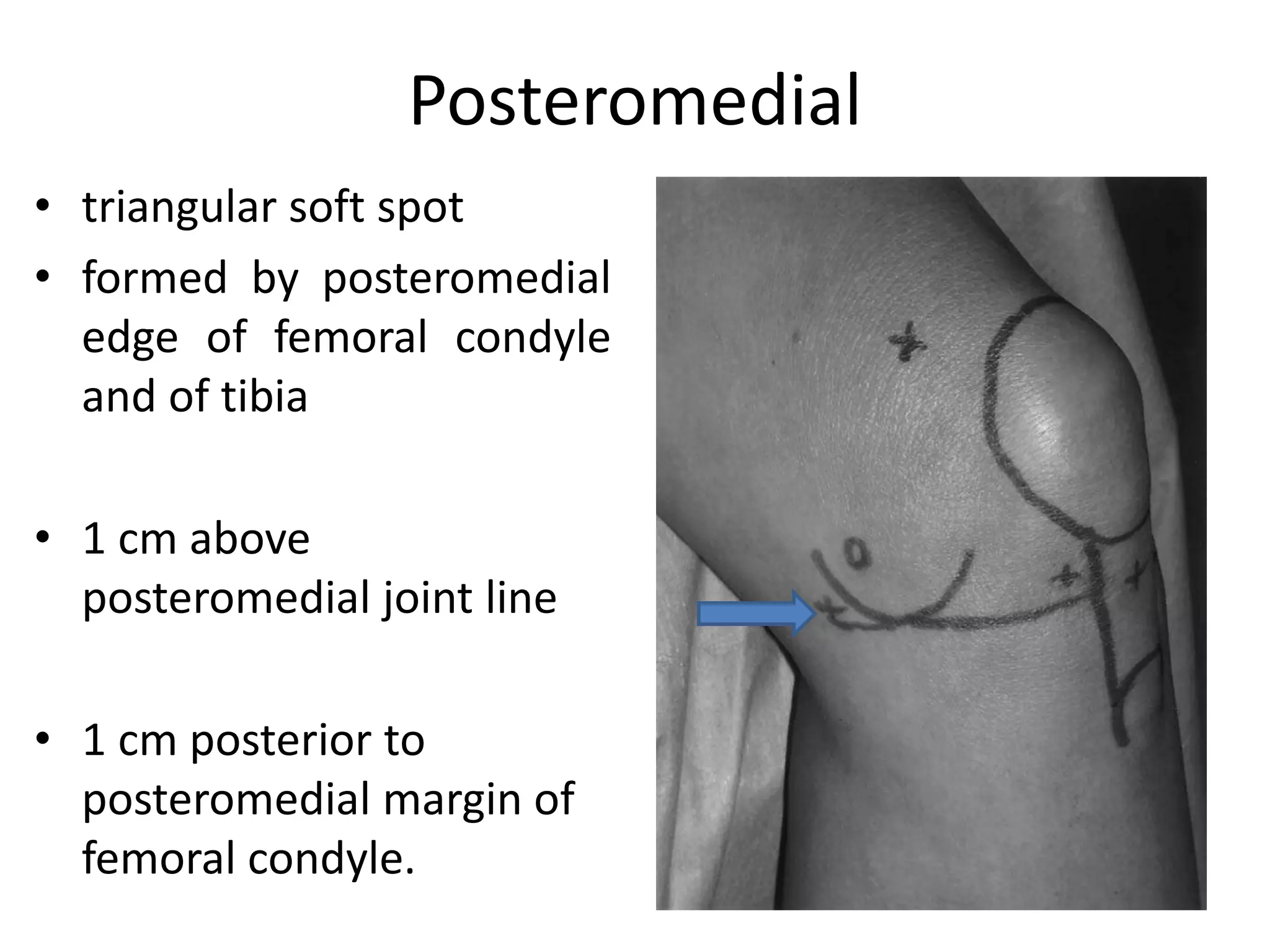
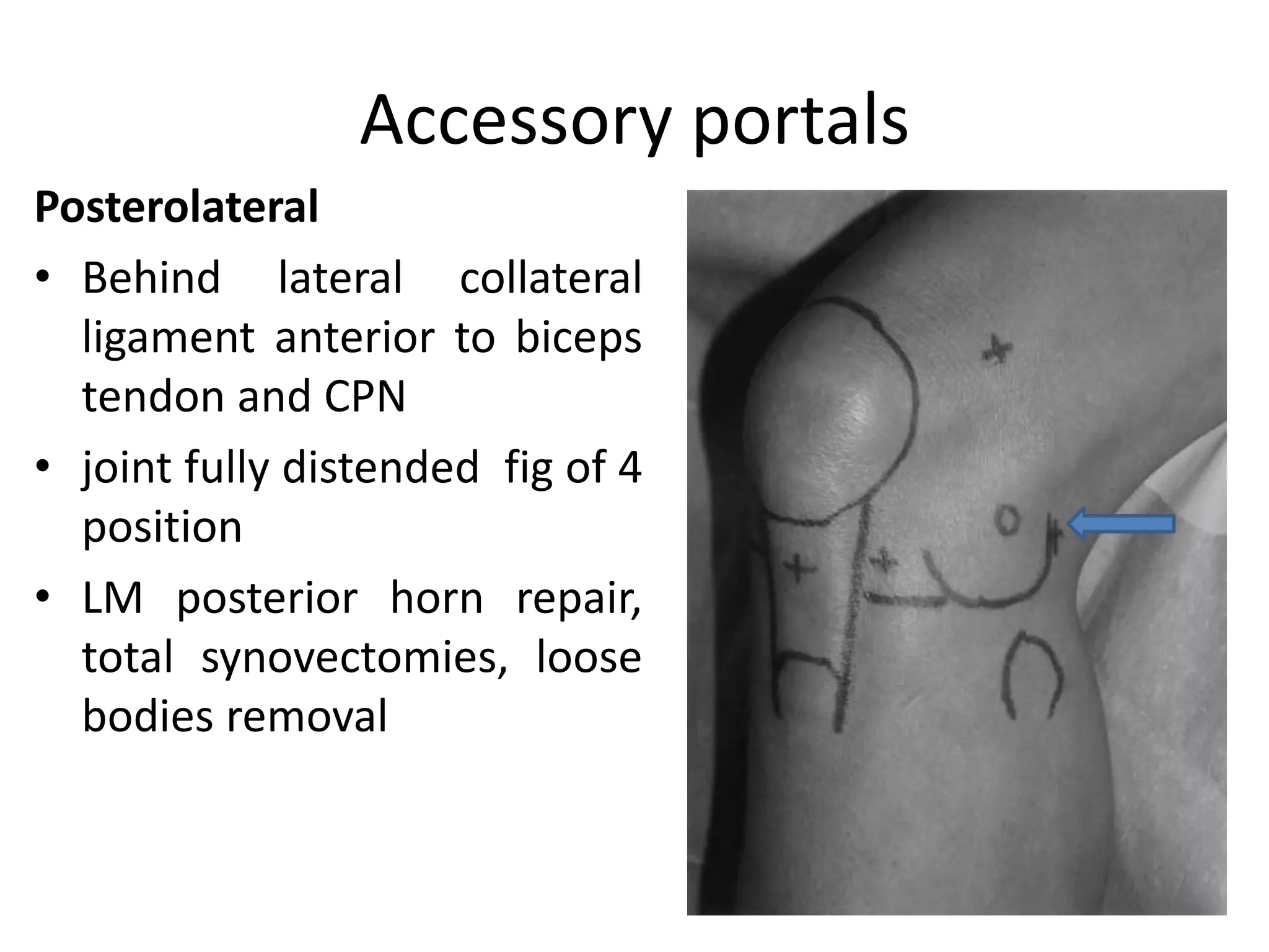
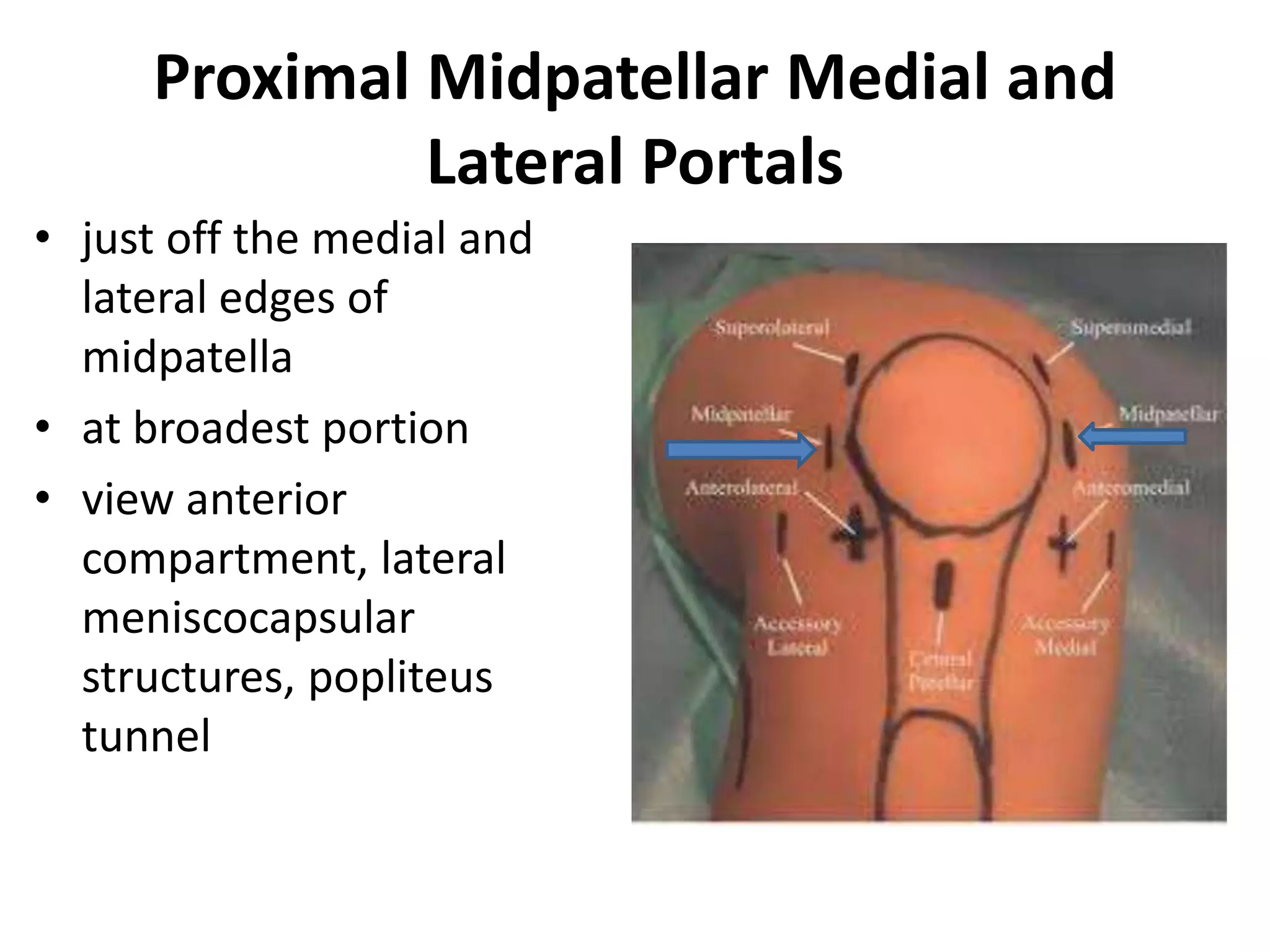
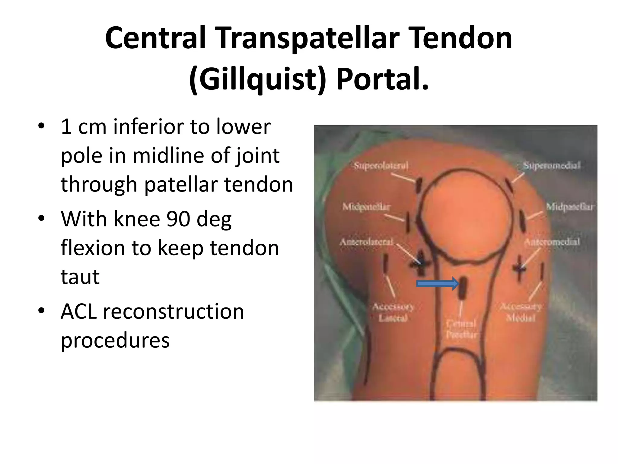
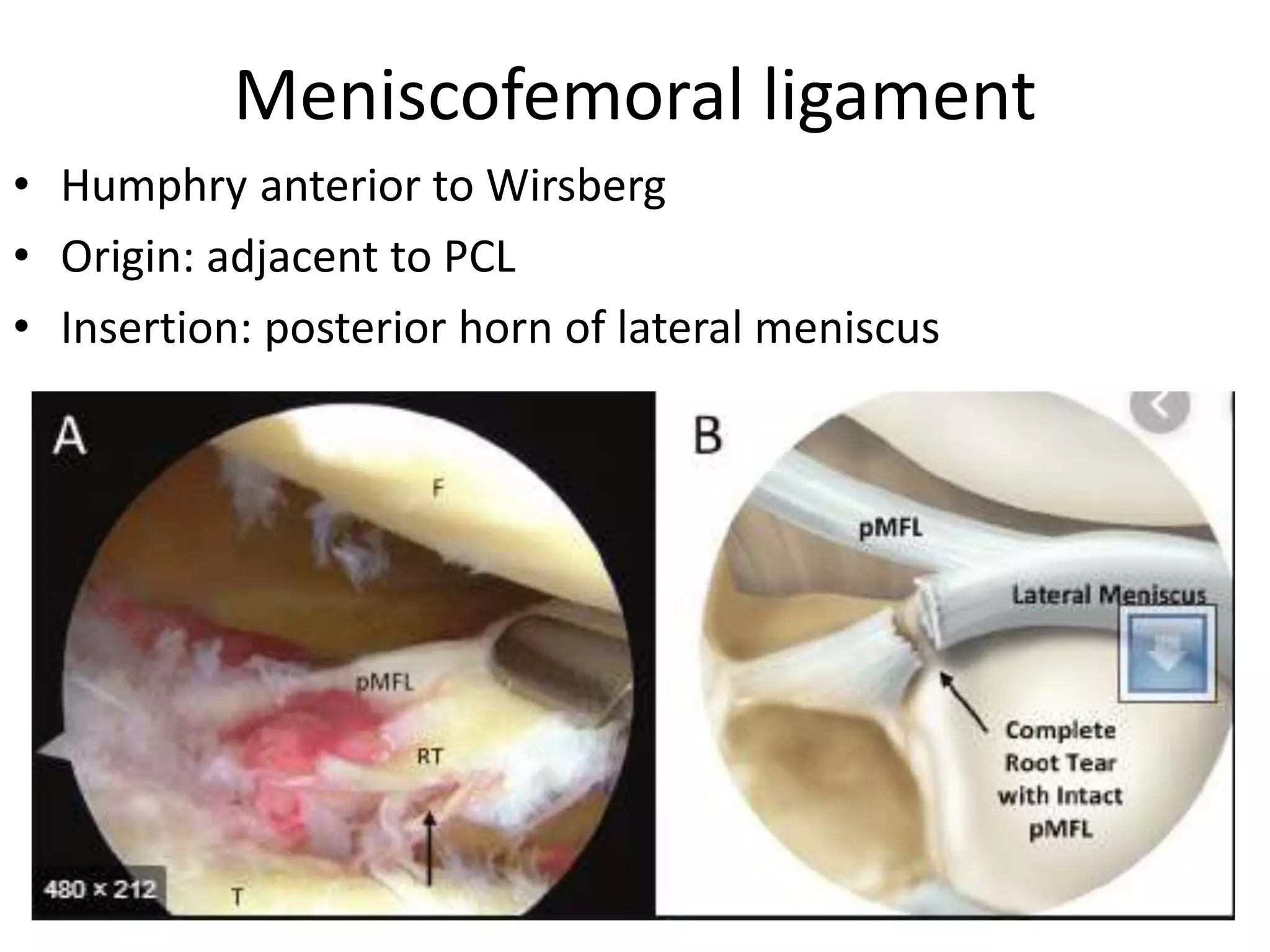
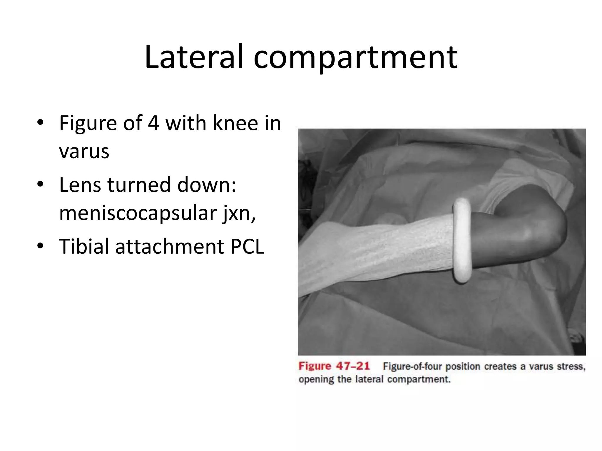
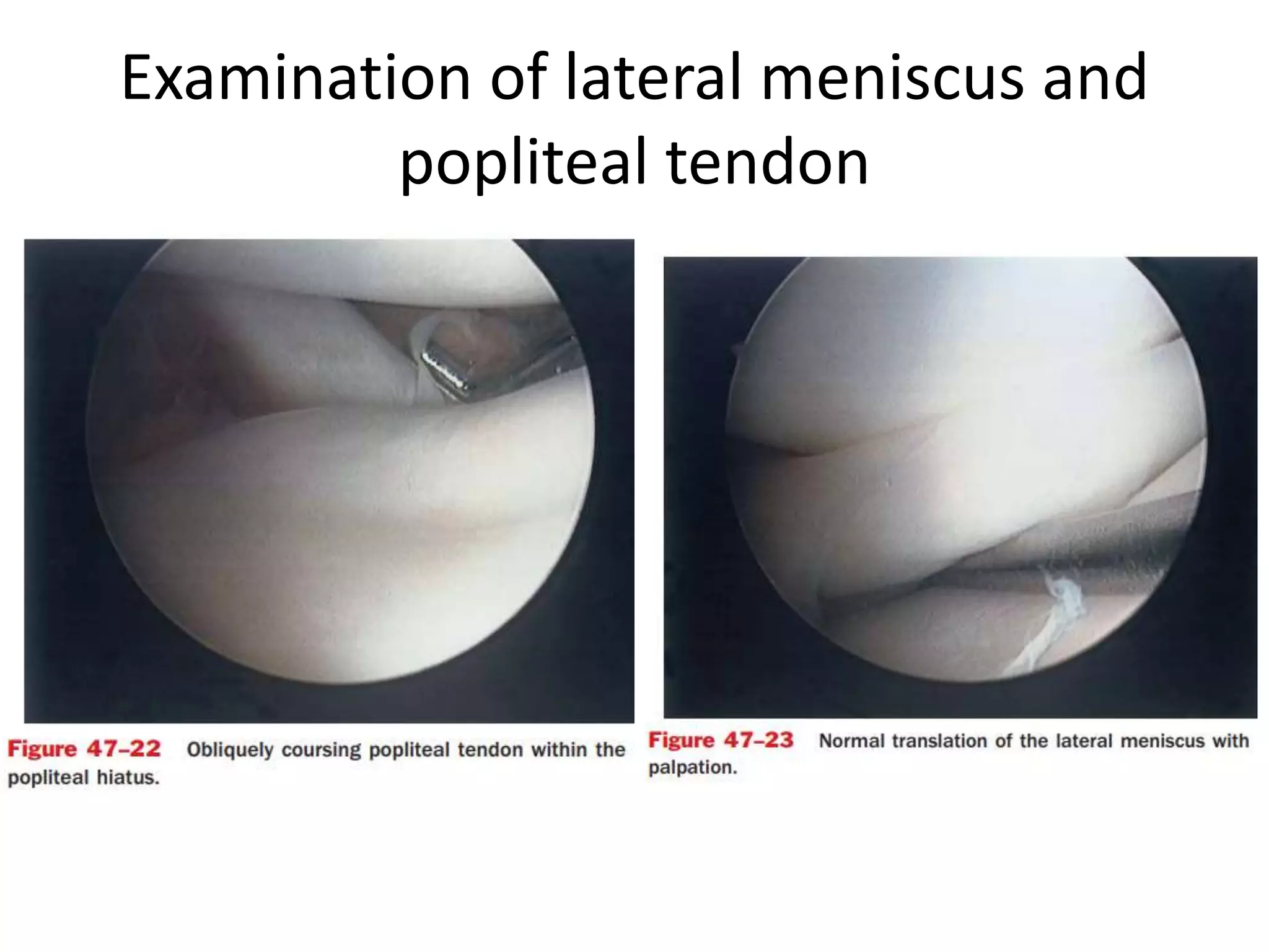
This document discusses knee arthroscopy portals and procedures. It outlines standard and accessory portals for the knee including anterolateral, anteromedial, posteromedial, and superolateral portals. It details patient positioning and skin markings. The document describes examining each compartment of the knee through the portals including the suprapatellar pouch, patellofemoral joint, medial and lateral gutters, medial and lateral menisci, intercondylar notch, ACL, PCL, and meniscofemoral ligaments. Procedures like diagnostic arthroscopy, creating portals, and visualizing structures are outlined.
6. Knee portals
Standard portals
• Anterolateral
• Anteromedial
• Posteromedial
• Superolateral
Accessory portals
• Posterolateral
• Proximal midpatellar
medial and lateral
• Far medial and far
lateral
• Central transpatellar
7. Standard portals
Antero lateral
• viewing portal:
arthroscope first inserted
• most versatile
• palpable lateral “soft spot”
• 1 cm above LJL adjacent to
lateral margin of PT
• 1 cm inferior to inferior
pole
8. • almost all structures visualised
• Exception: tibial insertion of PCL, undersurface
of anterior horn lateral meniscus
Superior placement;
• restricts access to patellofemoral joint and
suprapatellar pouch
• Access to posteromedial structures is difficult
9. • Inferior placement: anterior
horn of meniscus at risk of
laceration
• thickest infrapatellar fat pad
• medial placement:
penetration of patellar
tendon
10. Anteromedial portal
• 1 cm above medial joint
line,
• 1 cm inferior to tip of
patella
• adjacent to medial edge
of patellar tendon
• medial soft spot
11. • made at same time as anterolateral
• or under arthroscopic visualization after
localization with a spinal needle
• primary working portal.
• introduce instruments
• alternative viewing portal
13. • view dynamics of patellofemoral articulation.
• excision of medial plicae.
• evaluation of patellar tracking, congruity,
lateral overhang
• Viewed from extension to varying degrees of
flexion
14. Posteromedial
• triangular soft spot
• formed by posteromedial
edge of femoral condyle
and of tibia
• 1 cm above
posteromedial joint line
• 1 cm posterior to
posteromedial margin of
femoral condyle.
15. • Land marks drawn before distention
• Knee flexed to 90 degrees
• Make portal after distention
• repair or removal of displaced posterior horn
meniscal tears
• removal of posterior loose bodies
16. Accessory portals
Posterolateral
• Behind lateral collateral
ligament anterior to biceps
tendon and CPN
• joint fully distended fig of 4
position
• LM posterior horn repair,
total synovectomies, loose
bodies removal
17. Proximal Midpatellar Medial and
Lateral Portals
• just off the medial and
lateral edges of
midpatella
• at broadest portion
• view anterior
compartment, lateral
meniscocapsular
structures, popliteus
tunnel
18. Central Transpatellar Tendon
(Gillquist) Portal.
• 1 cm inferior to lower
pole in midline of joint
through patellar tendon
• With knee 90 deg
flexion to keep tendon
taut
• ACL reconstruction
procedures
19. Accessory Far Medial and Lateral
Portals
• 2.5 cm medial or lateral
to standard
anteromedial and
anterolateral portals
• insert spinal needle
under vision
• enter above superior
surface of meniscus
• accessory instruments
20. Diagnostic arthroscopy
Making of portal
• number 11 blade
• Aim: femoral notch
Vertical:
• Point blade superiorly, vertically: prevent
injury to anterior horn of lateral meniscus
21. Horizontal portal
• blade is oriented away from patellar tendon
• Avoid injury to the fibers
• penetrate capsule
• Introduce sheath with a blunt trocar
• twisting motion toward intercondylar notch
• knee flexed 60 to 90 degrees.
22.
23.
24. Suprapatellar pouch
• Examine from medial to
lateral and superior to
inferior
• Synovium: adhesions,
plicae, loose bodies,
crystals
27. Lateral gutter
• Entry: knee full extension
• relax the soft tissues on
lateral aspect
• posterior horn LM
• meniscosynovial capsular
reflection
• Popliteal tendon
• posterior limits of
popliteal hiatus
• posterior surface of
lateral femoral condyle
28. Medial gutter
• 40%: medial synovial
plica
• run medial and distal to
patella
• Origin: medial wall of
suprapatellar pouch and
inserting into the fat pad
• Ocassionally cause
patellofemoral symptoms
29. Medial compartment
To enter
• Leg holder: 30 deg flex,
valgus stress
• Lateral post: 90deg flex
over the side
To visualise
• Valgus stress, ER
30. Creating antero medial portal
• lens directed medially and anteriorly
• 18-gauge spinal needle inserted
31.
32. Medial meniscus examination
• Anterior, middle and
posterior
• Visual, probing
• wrinkling along the
length: peripheral
detachment
36. Intercondylar notch
• infrapatellar fat pad,
• ligamentum mucosum
• medial and lateral tibial
spines
• attachments of both
menisci,
• ACL, PCL
• Humphry and Wrisberg
(meniscofemoral
ligaments)
• intermeniscal ligament.
37. ACL
• knee flexed 60 to 90
degrees
• rotate lens until medial
aspect of lateral femoral
condyle is visualized
• 10 o’clock on right, 2
o’clock on left knees