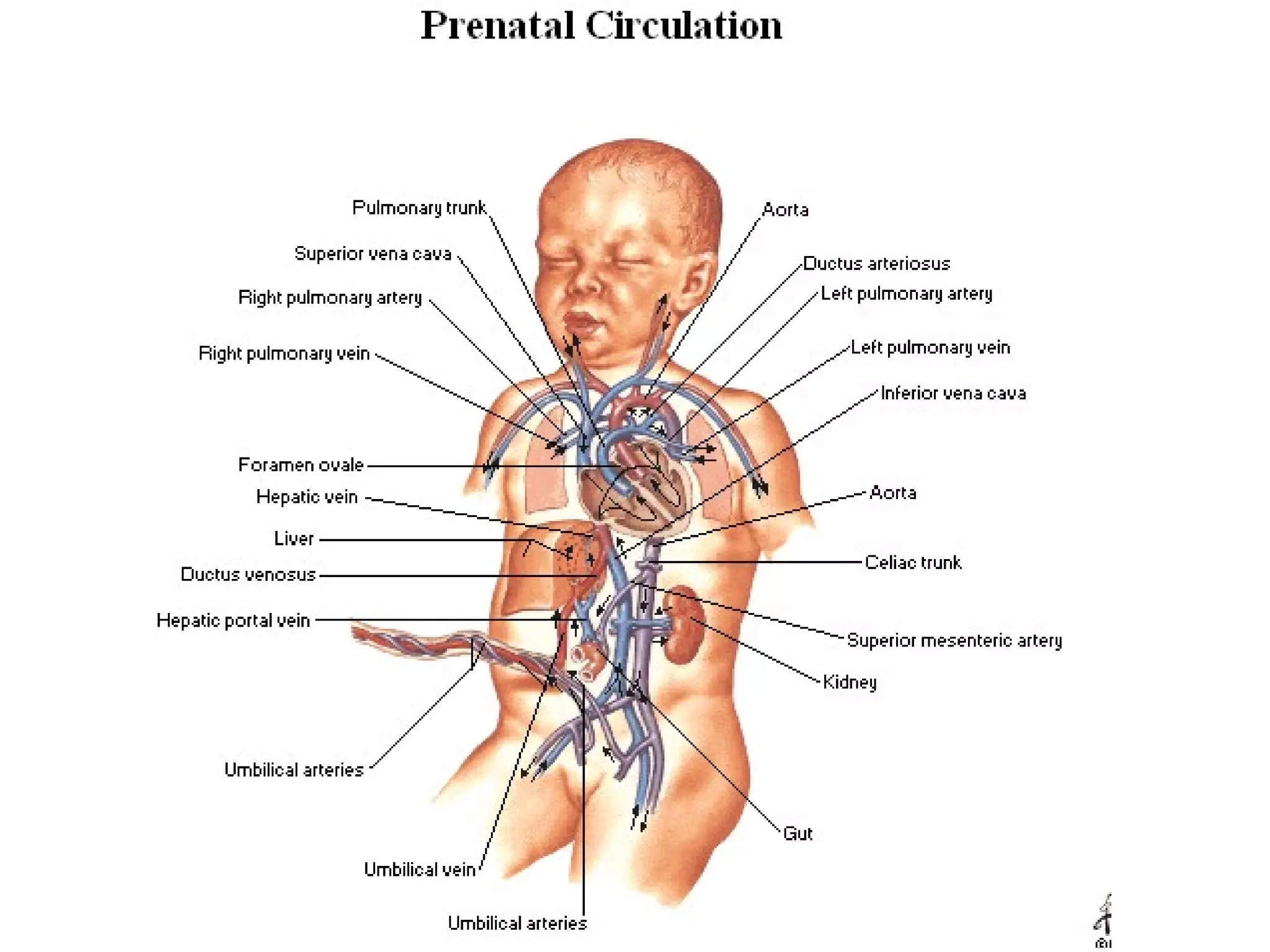
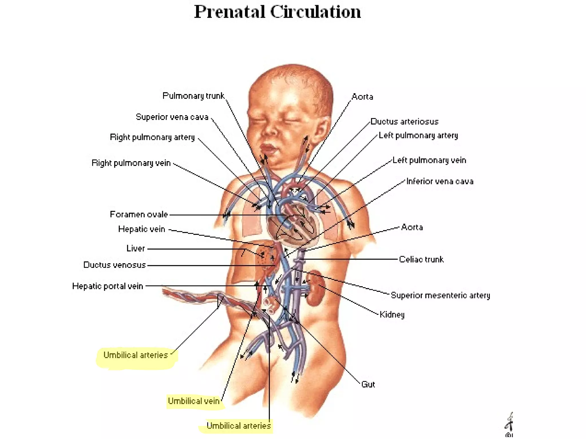
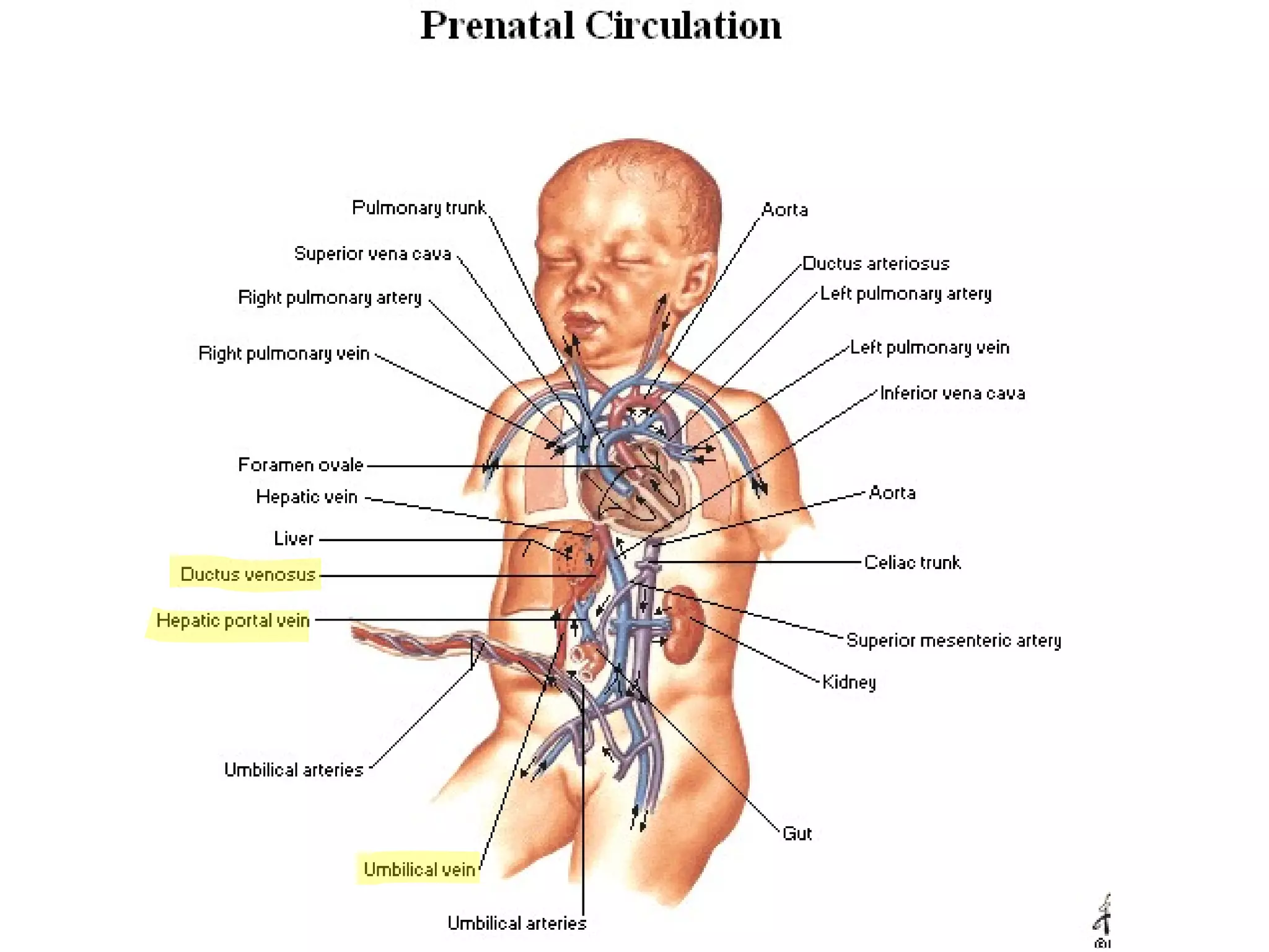
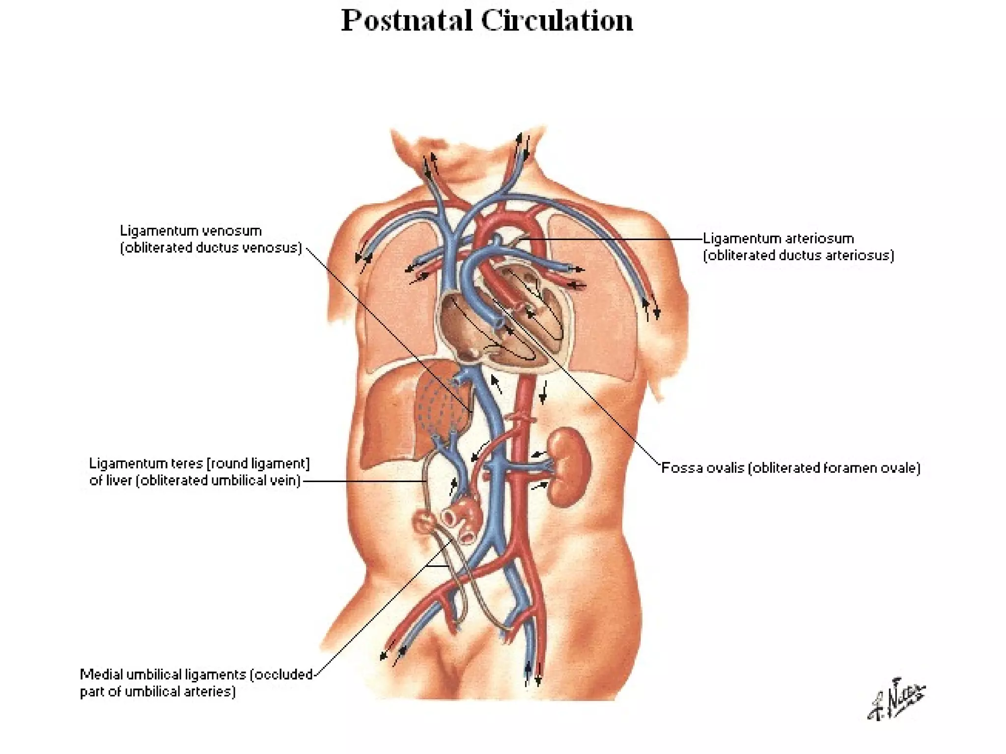
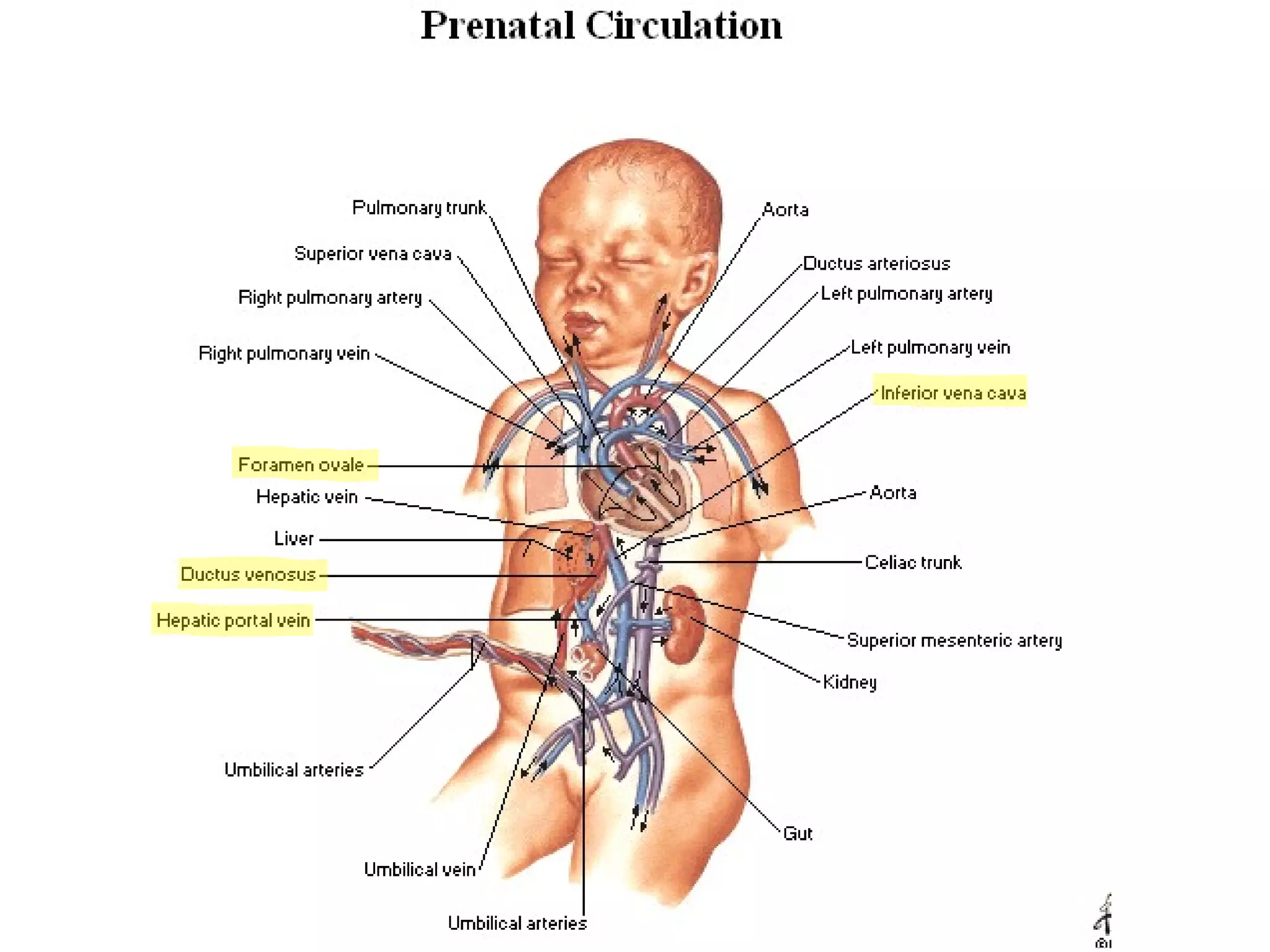
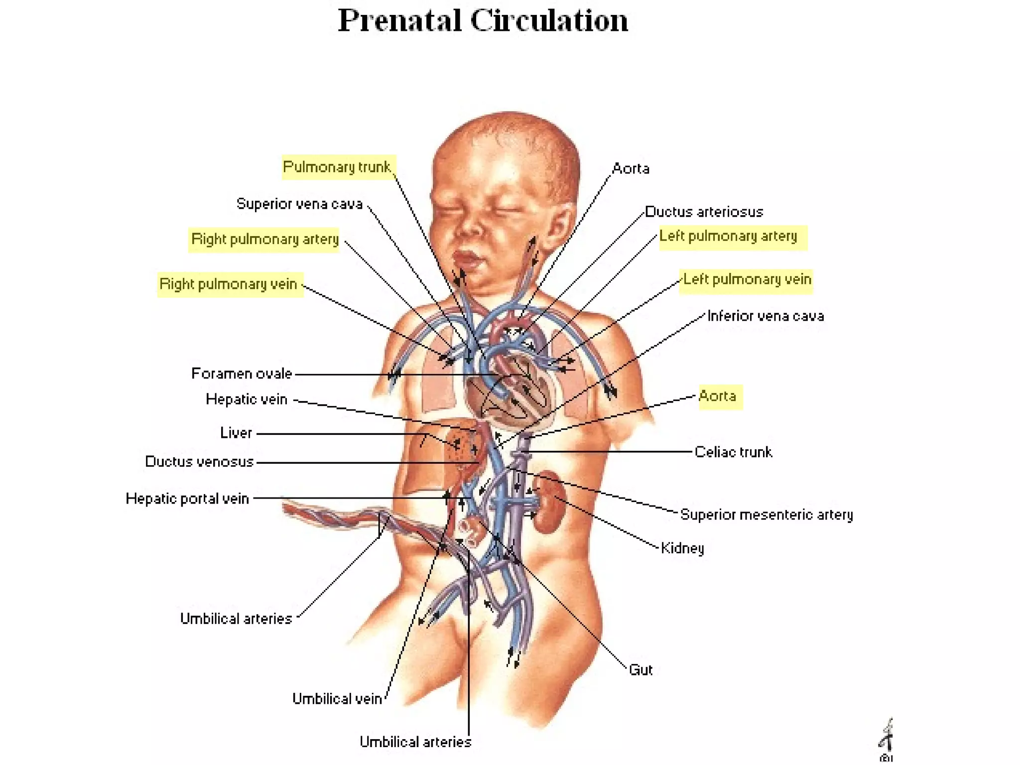
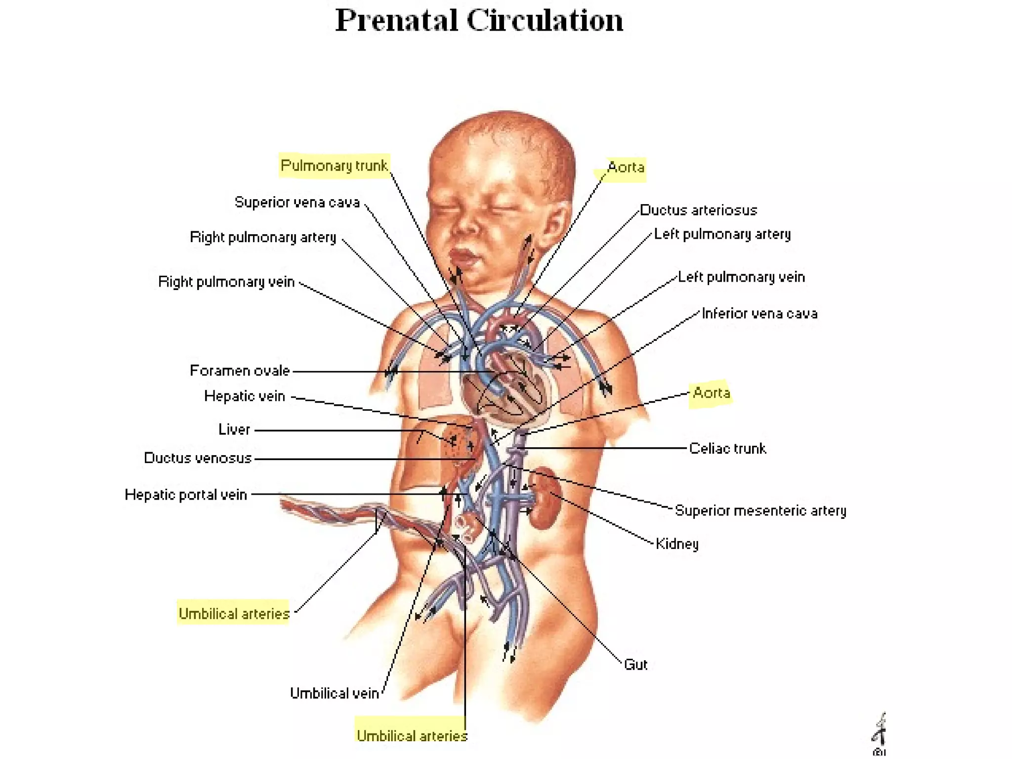
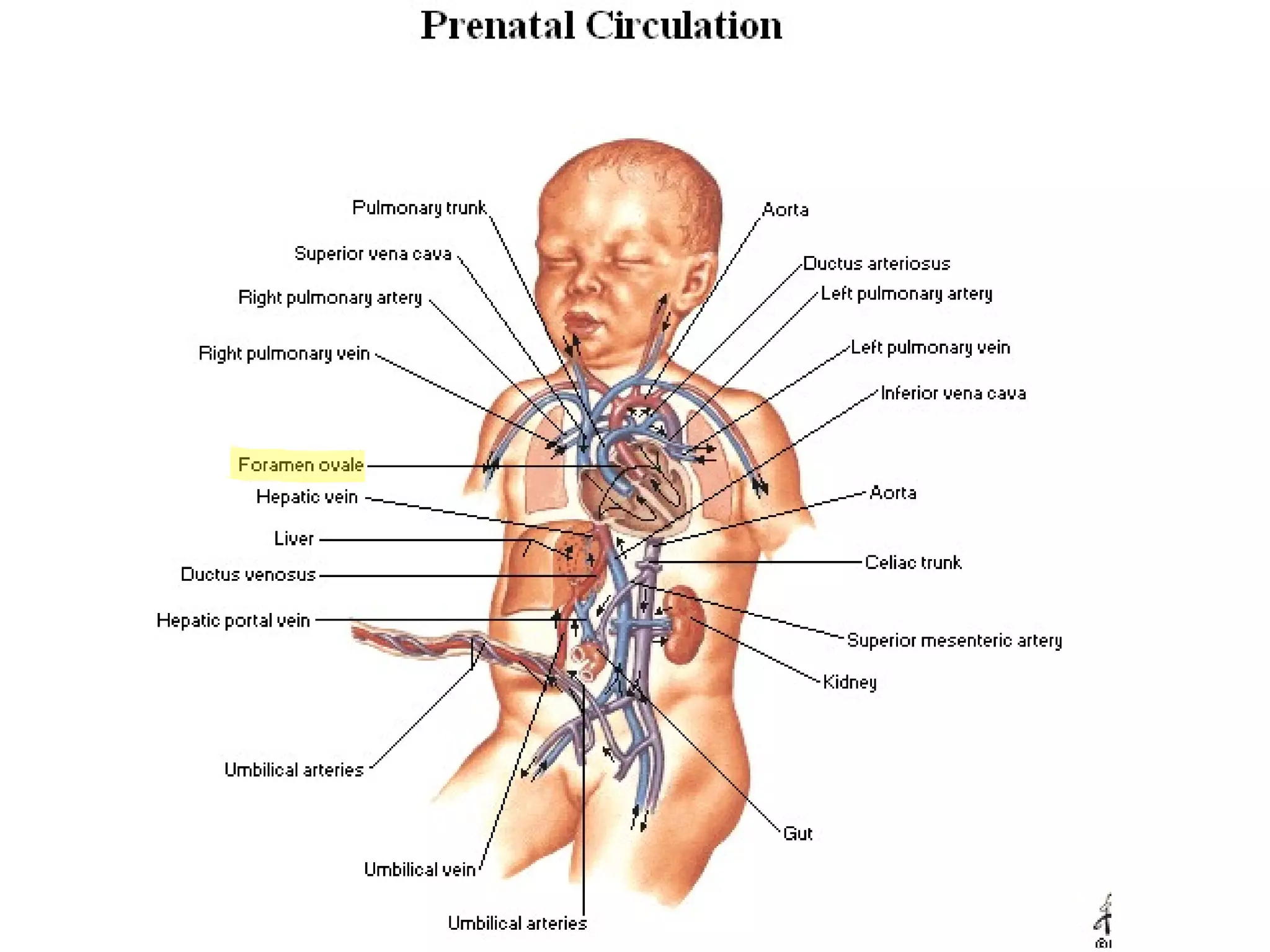
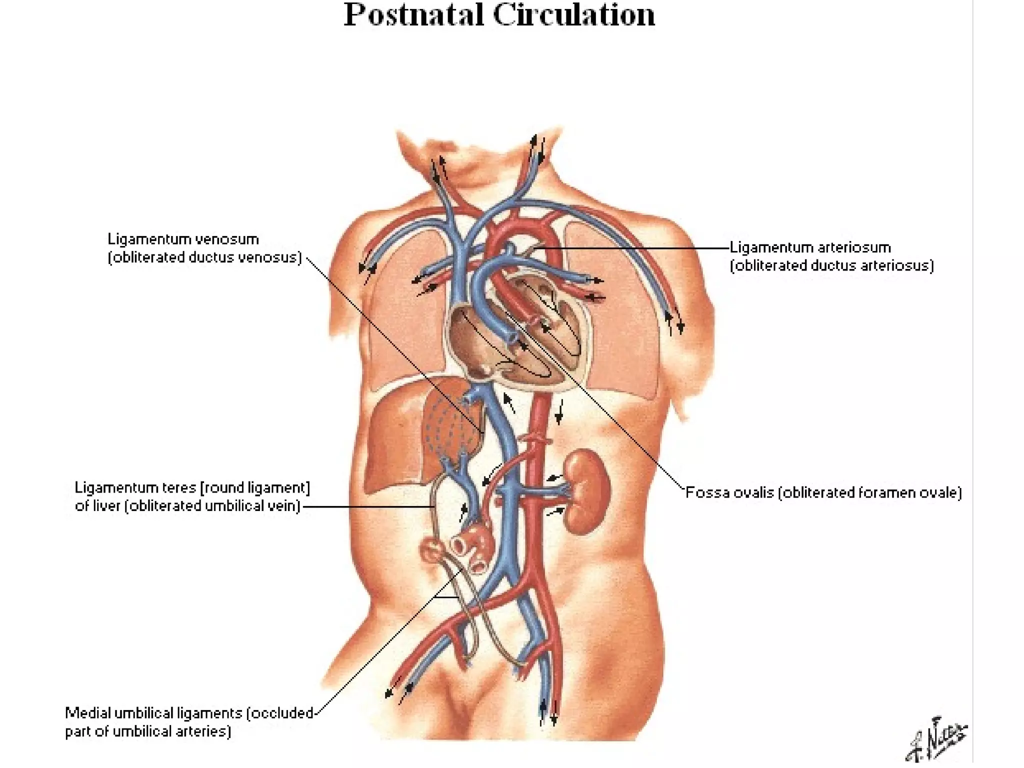
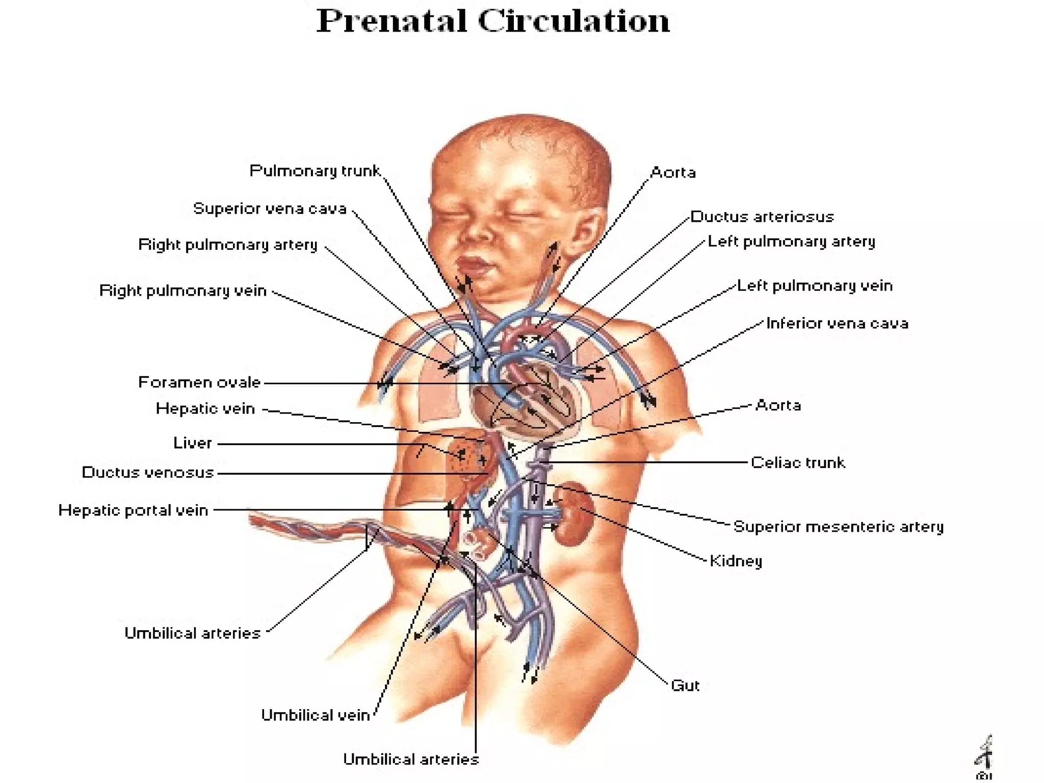
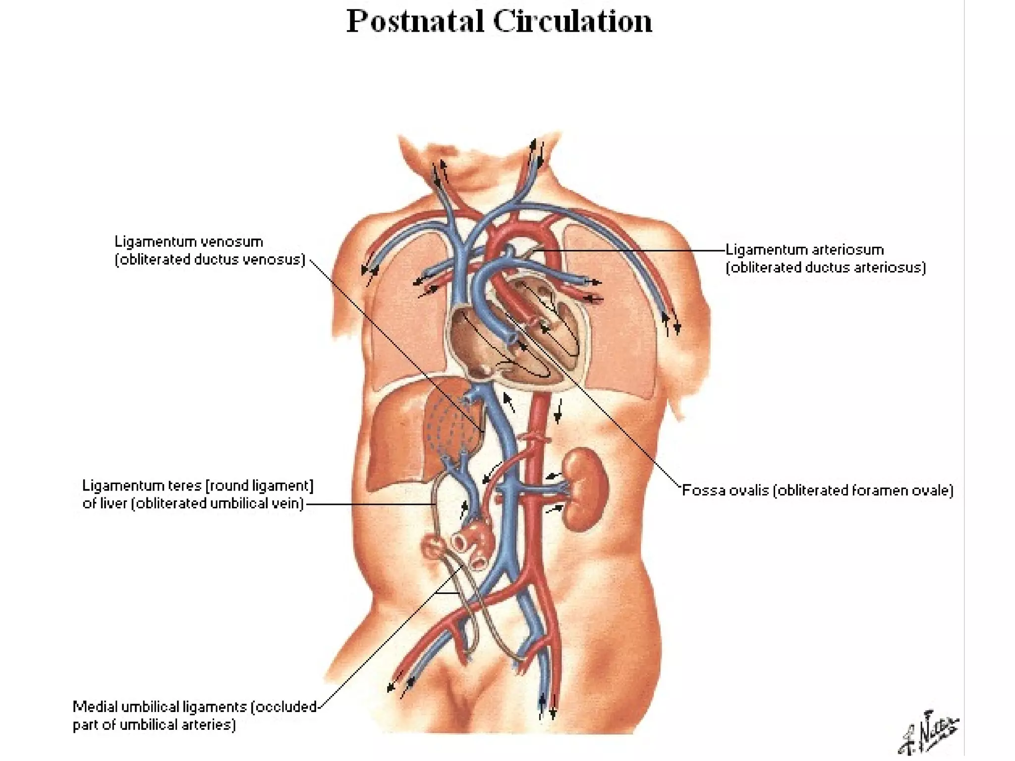
The document summarizes the fetal circulation system and how it differs from the newborn circulation. In the fetus, oxygen-rich blood from the placenta travels through the umbilical vein to the liver and mixes with venous blood before entering the heart. It then passes through the foramen ovale into the left side of the heart and mixes with pulmonary blood to be pumped throughout the body. After birth, the foramen ovale, ductus arteriosus, and umbilical vessels close as the lungs inflate and the placental circulation is lost. This allows the cardiovascular system to transition to adult circulation.
Blood from the placenta is carried to the fetus by the umbilical vein. In humans, less than a third of this enters the fetal ductus venosus and is carried to the inferior vena cava, while the rest enters the liver proper from the inferior border of the liver. The branch of the umbilical vein that supplies the right lobe of the liver first joins with the portal vein. The blood then moves to the right atrium of the heart. In the fetus, there is an opening between the right and left atrium (the foramen ovale), and most of the blood flows through this hole directly into the left atrium from the right atrium, thus bypassing pulmonary circulation. The continuation of this blood flow is into the left ventricle, and from there it is pumped through the aorta into the body. Some of the blood moves from the aorta through the internal iliac arteries to the umbilical arteries, and re-enters the placenta, where carbon dioxide and other waste products from the fetus are taken up and enter the maternal circulation.
Fetal Circulation by Barkha Devi,Lecturer,Sikkim Manipal College of NursingBarkha Devi
This PowerPoint will provide you a short a sweet lecture about fetal circulation. Please give me your feed back .
-Discuss anatomy and physiology of fetal circulation
-Compare and contrast fetal circulation to infant circulation
-Define specialized structures of fetal circulation
Describe the normal fetal circulation and mention the changes that occur in it is placental stage and after birth. Fetal circulation is composed of placenta, umbilical cord, heart and systemic blood vessels.
A major difference between the fetal circulation and postnatal circulation is that the lungs are not used during the fetal stage resulting in the presence of shunts to move oxygenated blood and nutrients from the placenta to the fetal tissue.
At birth, the start of breathing and the severance of the umbilical cord prompt various changes that quickly transform fetal circulation into postnatal circulation.
When the embryo develops into the fetus, it creates a functional cardiovascular system that cooperates with the mother's system.
During birth, there are functional physiological changes that transform the shared system into an individual one for the fetus.
In the fetus main filtration site for plasma nutrients and wastes in the placenta, which is outside of the body cavity.
In adults, the circulation occurs entirely inside the body.
The blood that flow to through the fetus is actually more complicated than after the baby is born (normal heart).
This is because the mother (the placenta) is doing the work that the baby's lungs will do after birth.
The placenta accepts the blood without oxygen from the fetus through blood vessels that leave the fetus through the umbilical cord (Umbilical arteries , there are two of them).
When blood goes through the placenta it pick up oxygwn.
The oxygen rich blood then returns to the fetus via the third vessels in the umbilical cord (Umbilical vein).
The oxygen rich blood that enters the fetus passes through the fetal liver and enters the right side of the heart.
The oxygen rich blood goes through one of the two extra connections in the fetal heart that will close after the baby is born.
The hole between the top two heart chmbers (right and left atrium) is called "Patent Foramen Ovale (PFO).
This hole allows the oxygen rich blood to go form the right atrium to left atrium and then to the left ventricle and out the aorta.
As a result the blood with the most oxygen gets to the brain.
Blood coming back from the fetus's body also enters the right atrium, but the fetus is able to send this oxygen poor blood from the right atrium to the right ventricle (the chamber that normally pumps blood to the lungs).
most of the blood that leaves the right ventricle in the fetus bypass the lungs through the second of the extra fetal connections known as the ductus arteriosus.
The ductus arteriosus sends the oxygen poor blood to the organs in the lower half of the fetal body. This also allows for the oxygen poor blood to leave the fetus through the umbilical arteries and get back to the placenta to pick up oxygen.
Since the patent foramen ovale and ductus arteriosus are normal findings in the fetus, it is impossible to predict whether or not these connections will close normally after birth in a normal fetal heart.
Embark on a captivating exploration of #FetalCirculation in this presentation. Delve into the intricacies of the developing cardiovascular system, understanding how the fetus receives oxygen and nutrients for optimal growth within the womb. Uncover the role of critical structures such as the ductus venosus and foramen ovale in facilitating unique circulatory patterns. Gain insights into the transition from fetal to neonatal circulation and its crucial significance for newborns. This presentation provides a comprehensive overview of the physiological marvel that sustains life before the first breath.
This is a presentation I had made for giving a seminar on Fetal Circulation in the first year of my MBBS course in Maharashtra.
Please share it with your juniors and colleagues.Thank You
Presentation By Tashif Jilani
These lecture slides, by Dr Sidra Arshad, offer a quick overview of physiological basis of a normal electrocardiogram.
Learning objectives:
1. Define an electrocardiogram (ECG) and electrocardiography
2. Describe how dipoles generated by the heart produce the waveforms of the ECG
3. Describe the components of a normal electrocardiogram of a typical bipolar leads (limb II)
4. Differentiate between intervals and segments
5. Enlist some common indications for obtaining an ECG
Study Resources:
1. Chapter 11, Guyton and Hall Textbook of Medical Physiology, 14th edition
2. Chapter 9, Human Physiology - From Cells to Systems, Lauralee Sherwood, 9th edition
3. Chapter 29, Ganong’s Review of Medical Physiology, 26th edition
4. Electrocardiogram, StatPearls - https://www.ncbi.nlm.nih.gov/books/NBK549803/
5. ECG in Medical Practice by ABM Abdullah, 4th edition
6. ECG Basics, http://www.nataliescasebook.com/tag/e-c-g-basics
Title: Sense of Smell
Presenter: Dr. Faiza, Assistant Professor of Physiology
Qualifications:
MBBS (Best Graduate, AIMC Lahore)
FCPS Physiology
ICMT, CHPE, DHPE (STMU)
MPH (GC University, Faisalabad)
MBA (Virtual University of Pakistan)
Learning Objectives:
Describe the primary categories of smells and the concept of odor blindness.
Explain the structure and location of the olfactory membrane and mucosa, including the types and roles of cells involved in olfaction.
Describe the pathway and mechanisms of olfactory signal transmission from the olfactory receptors to the brain.
Illustrate the biochemical cascade triggered by odorant binding to olfactory receptors, including the role of G-proteins and second messengers in generating an action potential.
Identify different types of olfactory disorders such as anosmia, hyposmia, hyperosmia, and dysosmia, including their potential causes.
Key Topics:
Olfactory Genes:
3% of the human genome accounts for olfactory genes.
400 genes for odorant receptors.
Olfactory Membrane:
Located in the superior part of the nasal cavity.
Medially: Folds downward along the superior septum.
Laterally: Folds over the superior turbinate and upper surface of the middle turbinate.
Total surface area: 5-10 square centimeters.
Olfactory Mucosa:
Olfactory Cells: Bipolar nerve cells derived from the CNS (100 million), with 4-25 olfactory cilia per cell.
Sustentacular Cells: Produce mucus and maintain ionic and molecular environment.
Basal Cells: Replace worn-out olfactory cells with an average lifespan of 1-2 months.
Bowman’s Gland: Secretes mucus.
Stimulation of Olfactory Cells:
Odorant dissolves in mucus and attaches to receptors on olfactory cilia.
Involves a cascade effect through G-proteins and second messengers, leading to depolarization and action potential generation in the olfactory nerve.
Quality of a Good Odorant:
Small (3-20 Carbon atoms), volatile, water-soluble, and lipid-soluble.
Facilitated by odorant-binding proteins in mucus.
Membrane Potential and Action Potential:
Resting membrane potential: -55mV.
Action potential frequency in the olfactory nerve increases with odorant strength.
Adaptation Towards the Sense of Smell:
Rapid adaptation within the first second, with further slow adaptation.
Psychological adaptation greater than receptor adaptation, involving feedback inhibition from the central nervous system.
Primary Sensations of Smell:
Camphoraceous, Musky, Floral, Pepperminty, Ethereal, Pungent, Putrid.
Odor Detection Threshold:
Examples: Hydrogen sulfide (0.0005 ppm), Methyl-mercaptan (0.002 ppm).
Some toxic substances are odorless at lethal concentrations.
Characteristics of Smell:
Odor blindness for single substances due to lack of appropriate receptor protein.
Behavioral and emotional influences of smell.
Transmission of Olfactory Signals:
From olfactory cells to glomeruli in the olfactory bulb, involving lateral inhibition.
Primitive, less old, and new olfactory systems with different path
Lung Cancer: Artificial Intelligence, Synergetics, Complex System Analysis, S...Oleg Kshivets
RESULTS: Overall life span (LS) was 2252.1±1742.5 days and cumulative 5-year survival (5YS) reached 73.2%, 10 years – 64.8%, 20 years – 42.5%. 513 LCP lived more than 5 years (LS=3124.6±1525.6 days), 148 LCP – more than 10 years (LS=5054.4±1504.1 days).199 LCP died because of LC (LS=562.7±374.5 days). 5YS of LCP after bi/lobectomies was significantly superior in comparison with LCP after pneumonectomies (78.1% vs.63.7%, P=0.00001 by log-rank test). AT significantly improved 5YS (66.3% vs. 34.8%) (P=0.00000 by log-rank test) only for LCP with N1-2. Cox modeling displayed that 5YS of LCP significantly depended on: phase transition (PT) early-invasive LC in terms of synergetics, PT N0—N12, cell ratio factors (ratio between cancer cells- CC and blood cells subpopulations), G1-3, histology, glucose, AT, blood cell circuit, prothrombin index, heparin tolerance, recalcification time (P=0.000-0.038). Neural networks, genetic algorithm selection and bootstrap simulation revealed relationships between 5YS and PT early-invasive LC (rank=1), PT N0—N12 (rank=2), thrombocytes/CC (3), erythrocytes/CC (4), eosinophils/CC (5), healthy cells/CC (6), lymphocytes/CC (7), segmented neutrophils/CC (8), stick neutrophils/CC (9), monocytes/CC (10); leucocytes/CC (11). Correct prediction of 5YS was 100% by neural networks computing (area under ROC curve=1.0; error=0.0).
CONCLUSIONS: 5YS of LCP after radical procedures significantly depended on: 1) PT early-invasive cancer; 2) PT N0--N12; 3) cell ratio factors; 4) blood cell circuit; 5) biochemical factors; 6) hemostasis system; 7) AT; 8) LC characteristics; 9) LC cell dynamics; 10) surgery type: lobectomy/pneumonectomy; 11) anthropometric data. Optimal diagnosis and treatment strategies for LC are: 1) screening and early detection of LC; 2) availability of experienced thoracic surgeons because of complexity of radical procedures; 3) aggressive en block surgery and adequate lymph node dissection for completeness; 4) precise prediction; 5) adjuvant chemoimmunoradiotherapy for LCP with unfavorable prognosis.
Pulmonary Thromboembolism - etilogy, types, medical- Surgical and nursing man...VarunMahajani
Disruption of blood supply to lung alveoli due to blockage of one or more pulmonary blood vessels is called as Pulmonary thromboembolism. In this presentation we will discuss its causes, types and its management in depth.
Ethanol (CH3CH2OH), or beverage alcohol, is a two-carbon alcohol
that is rapidly distributed in the body and brain. Ethanol alters many
neurochemical systems and has rewarding and addictive properties. It
is the oldest recreational drug and likely contributes to more morbidity,
mortality, and public health costs than all illicit drugs combined. The
5th edition of the Diagnostic and Statistical Manual of Mental Disorders
(DSM-5) integrates alcohol abuse and alcohol dependence into a single
disorder called alcohol use disorder (AUD), with mild, moderate,
and severe subclassifications (American Psychiatric Association, 2013).
In the DSM-5, all types of substance abuse and dependence have been
combined into a single substance use disorder (SUD) on a continuum
from mild to severe. A diagnosis of AUD requires that at least two of
the 11 DSM-5 behaviors be present within a 12-month period (mild
AUD: 2–3 criteria; moderate AUD: 4–5 criteria; severe AUD: 6–11 criteria).
The four main behavioral effects of AUD are impaired control over
drinking, negative social consequences, risky use, and altered physiological
effects (tolerance, withdrawal). This chapter presents an overview
of the prevalence and harmful consequences of AUD in the U.S.,
the systemic nature of the disease, neurocircuitry and stages of AUD,
comorbidities, fetal alcohol spectrum disorders, genetic risk factors, and
pharmacotherapies for AUD.
micro teaching on communication m.sc nursing.pdfAnurag Sharma
Microteaching is a unique model of practice teaching. It is a viable instrument for the. desired change in the teaching behavior or the behavior potential which, in specified types of real. classroom situations, tends to facilitate the achievement of specified types of objectives.
Report Back from SGO 2024: What’s the Latest in Cervical Cancer?bkling
Are you curious about what’s new in cervical cancer research or unsure what the findings mean? Join Dr. Emily Ko, a gynecologic oncologist at Penn Medicine, to learn about the latest updates from the Society of Gynecologic Oncology (SGO) 2024 Annual Meeting on Women’s Cancer. Dr. Ko will discuss what the research presented at the conference means for you and answer your questions about the new developments.
3. • All of the respiratory, excretory,
and nutritional needs of the fetus
are provided for by diffusion across
the placenta instead of by the fetal
lungs, kidneys, and
gastrointestinal tract.
• Fetal circulation is adaptive to these
conditions.
4. Objective :
• Describe the fetal circulation
to and from the placenta.
• Describe the structure and
function of the foramen
ovale, ductus venosus, and
ductus arteriosus.
5. • The circulation of blood through
a fetus is by necessity different
from blood circulation in a
newborn .
• Respiration, the procurement of
nutrients, and the elimination of
metabolic wastes occur through
the maternal blood instead of
through the organs of the fetus.
6. • The capillary exchange between
the maternal and fetal
circulation occurs within the
placenta .
• This remarkable structure, which
includes maternal and fetal
capillary beds, is discharged
following delivery as the
afterbirth.
7. • The umbilical cord is the
connection between the placenta
and the fetal umbilicus.
• It includes one umbilical vein and
two umbilical arteries,
surrounded by a gelatinous
substance.
8.
9. • Oxygenated and nutrient
rich blood flows through the
umbilical vein toward the
inferior surface of the liver.
• At this point, the umbilical
vein divides into two
branches.
10.
11.
12. • One branch merges with the
portal vein, while the other
branch, called the ductus
venosus , enters the inferior
vena cava.
13. • Thus, oxygenated blood is
mixed with venous blood
returning from the lower
extremities of the fetus before it
enters the heart.
• The umbilical vein is the only
vessel of the fetus that carries
fully oxygenated blood.
14. • The inferior vena cava
empties into the right atrium
of the fetal heart.
• Most of the blood passes from
the right atrium into the left
atrium through the foramen
ovale, an opening between
the two atria.
15.
16. • Here, it mixes with a small
quantity of blood returning
through the pulmonary
circulation.
• The blood then passes in to
the left ventricle, from which
it is pumped into the aorta and
through the body of the fetus.
17.
18. • Some blood entering the right
atrium passes into the right
ventricle and out of the heart via
the pulmonary trunk.
• Since the lungs of the fetus are
not functional, only a small portion
of blood continues through the
pulmonary circulation (the
resistance to blood flow is very
high in the collapsed fetal lungs).
19. • Most of the blood in the pulmonary
trunk passes through the ductus
arteriosus into the aortic arch,
where it mixes with blood coming from
the left ventricle.
• Blood is returned to the placenta by
the two umbilical arteries that arise
from the internal iliac arteries.
20.
21. • Notice that, in the fetus,
oxygen-rich blood is
transported by the inferior
vena cava to the heart, and
via the foramen ovale and
ductus arteriosus to the
systemic circulation.
22. • Important changes occur in
the cardiovascular system at
birth.
• The foramen ovale, ductus
arteriosus, ductus venosus,
and the umbilical vessels are
no longer necessary.
23.
24. • The foramen ovale abruptly
close with the first breath of
air because the reduced
pressure in the right side of
the heart causes a flap to
cover the opening.
25. • This reduction in pressure occurs
because the vascular resistance to
blood flow in the pulmonary
circulation falls far below that of the
systemic circulation when the lungs
fill with air.
• The pressure in the inferior vena
cava and right atrium falls as a result
of the loss of the placental
circulation.
26.
27. • The constriction of the
ductus arteriosus occurs
gradually over a period of
about 6 weeks after birth as
the vascular smooth muscle
fibers constrict in response to
the higher oxygen
concentration in the
postnatal blood.
28. • The remaining structure of the
ductus arteriosus gradually
atrophies and becomes
nonfunctional as a blood vessel.
• Transformation of the unique fetal
cardiovascular system is
summarized in table 16.6.