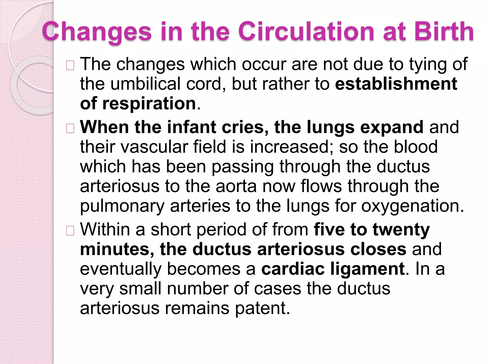
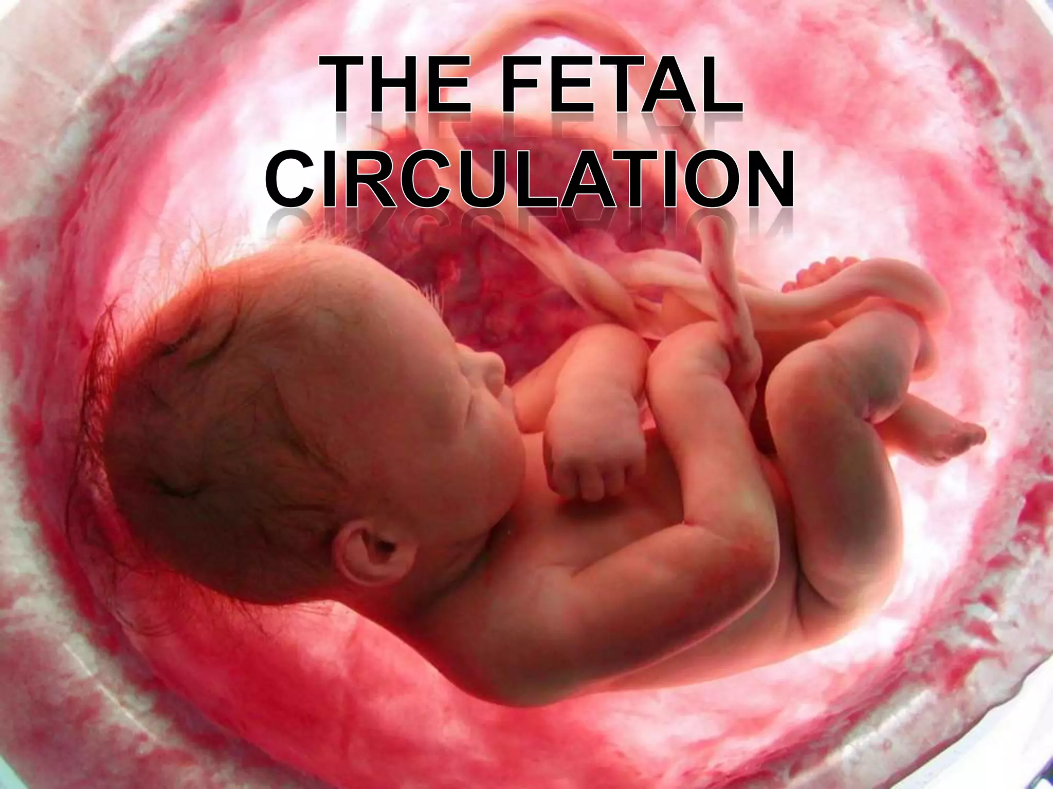
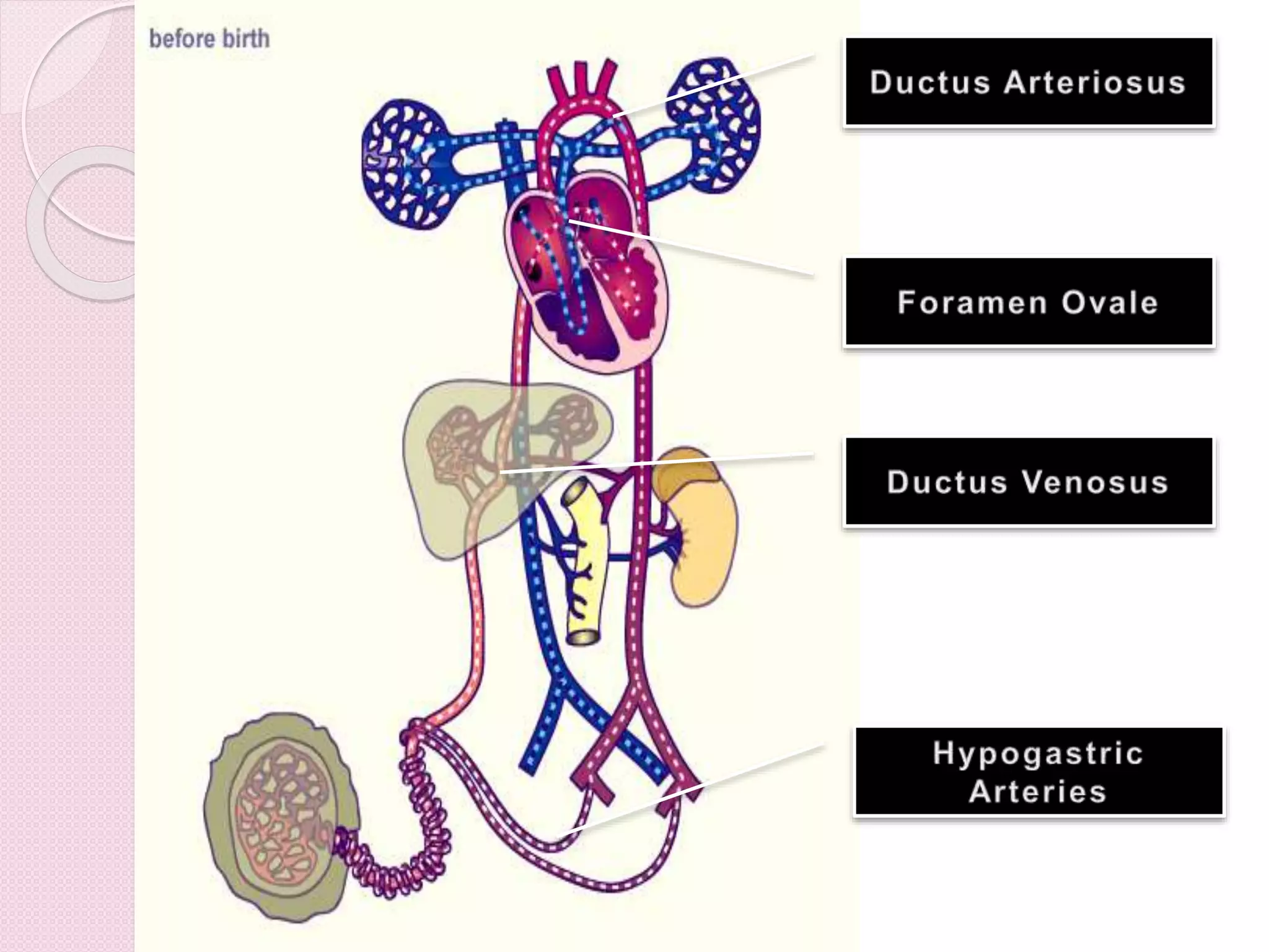
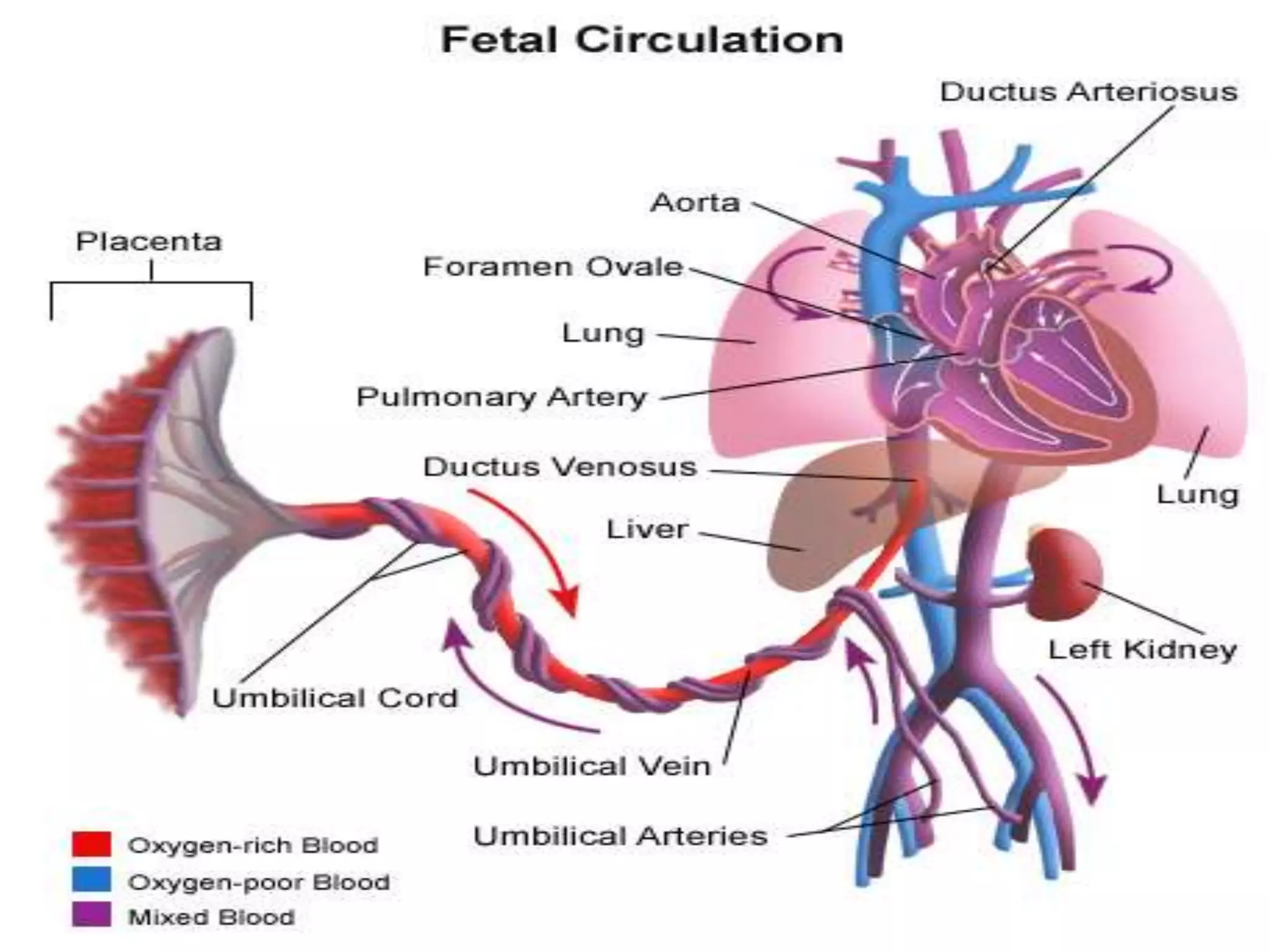
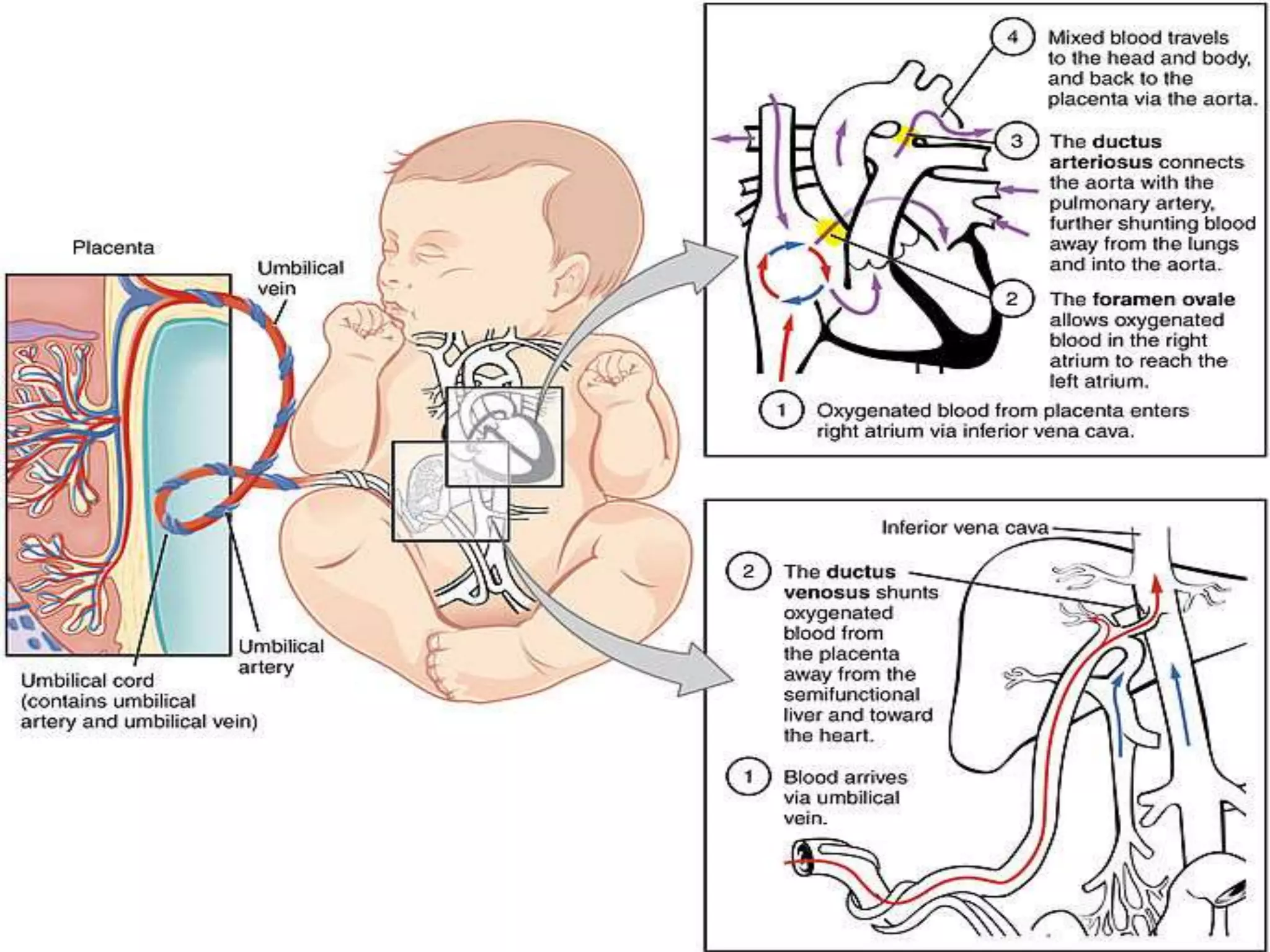
The fetal circulation differs from adult circulation in that the fetus receives oxygen and nutrients from the mother via the placenta and umbilical cord, as the lungs are not functional. Blood from the placenta enters the umbilical vein and most of it bypasses the liver through the ductus venosus to the inferior vena cava. It then flows through the foramen ovale into the left atrium and ventricle before being pumped through the aorta to supply the fetus. At birth, crying causes lung expansion which redirects blood flow through the pulmonary arteries, closing the ductus arteriosus and foramen ovale to establish postnatal circulation.
Fetal Circulation by Barkha Devi,Lecturer,Sikkim Manipal College of NursingBarkha Devi
This PowerPoint will provide you a short a sweet lecture about fetal circulation. Please give me your feed back .
-Discuss anatomy and physiology of fetal circulation
-Compare and contrast fetal circulation to infant circulation
-Define specialized structures of fetal circulation
DEVELOPMENT OF PLACENTA,PLACENTA AT TERM , DECIDUA,PLACENTAL MEMBRANE , PLACENTAL CICULATION,PLACENTAL ENDOCRINE SYNTHESIS,ABNORMAL PLACENTA,FUNCTIONS.
Describe the flow of oxygenated blood from the placenta to the fe.pdffasttrackscardecors
Describe the flow of oxygenated blood from the placenta to the fetus\' systemic circulation and
deoxygenated blood from the fetus to the placenta. Be sure to include all five structures that
allow for fetal circulation to take place effectively.
Solution
Placenta receives blood from two systems: a uteroplacental system that delivers maternal blood
to the placenta; and the fetoplacental system that receives from the fetus.
The umbilical vein carries blood from the placenta to the fetus. A majority of it enters liver and
1/3rd is diverted to ductus venosus and carried to inferior vena cava. In liver proper, the blood
enters from its inferior border. A branch of the umbilical vein joins the portal vein. The blood is
then supplied to the right atrium of the heart. The heart has an opening connecting left and right
atrium called foramen ovale. Due to this opening, the blood bypasses the pulmonary circulation
and is directly circulated to the body of the fetus, via the aorta. Some of the blood moves to the
placenta through iliac and then umbilical arteries into the mother\'s blood circulation for the
exchange of gasses and removal of waste products.
A part of the blood that enters the atrium of the fetus enters left ventricle and is pumped to the
pulmonary artery The pulmonary artery of the fetus is connected to the aorta via ductus atriosus
and thus the blood enters aorta and escapes from entering the lungs. The blood in aorta takes the
same route as described earlier..
Describe the normal fetal circulation and mention the changes that occur in it is placental stage and after birth. Fetal circulation is composed of placenta, umbilical cord, heart and systemic blood vessels.
A major difference between the fetal circulation and postnatal circulation is that the lungs are not used during the fetal stage resulting in the presence of shunts to move oxygenated blood and nutrients from the placenta to the fetal tissue.
At birth, the start of breathing and the severance of the umbilical cord prompt various changes that quickly transform fetal circulation into postnatal circulation.
When the embryo develops into the fetus, it creates a functional cardiovascular system that cooperates with the mother's system.
During birth, there are functional physiological changes that transform the shared system into an individual one for the fetus.
In the fetus main filtration site for plasma nutrients and wastes in the placenta, which is outside of the body cavity.
In adults, the circulation occurs entirely inside the body.
The blood that flow to through the fetus is actually more complicated than after the baby is born (normal heart).
This is because the mother (the placenta) is doing the work that the baby's lungs will do after birth.
The placenta accepts the blood without oxygen from the fetus through blood vessels that leave the fetus through the umbilical cord (Umbilical arteries , there are two of them).
When blood goes through the placenta it pick up oxygwn.
The oxygen rich blood then returns to the fetus via the third vessels in the umbilical cord (Umbilical vein).
The oxygen rich blood that enters the fetus passes through the fetal liver and enters the right side of the heart.
The oxygen rich blood goes through one of the two extra connections in the fetal heart that will close after the baby is born.
The hole between the top two heart chmbers (right and left atrium) is called "Patent Foramen Ovale (PFO).
This hole allows the oxygen rich blood to go form the right atrium to left atrium and then to the left ventricle and out the aorta.
As a result the blood with the most oxygen gets to the brain.
Blood coming back from the fetus's body also enters the right atrium, but the fetus is able to send this oxygen poor blood from the right atrium to the right ventricle (the chamber that normally pumps blood to the lungs).
most of the blood that leaves the right ventricle in the fetus bypass the lungs through the second of the extra fetal connections known as the ductus arteriosus.
The ductus arteriosus sends the oxygen poor blood to the organs in the lower half of the fetal body. This also allows for the oxygen poor blood to leave the fetus through the umbilical arteries and get back to the placenta to pick up oxygen.
Since the patent foramen ovale and ductus arteriosus are normal findings in the fetus, it is impossible to predict whether or not these connections will close normally after birth in a normal fetal heart.
2. Fetal Circulation
The fetal circulation is the circulatory
system of a human fetus, often
encompassing the entire fetoplacental
circulation which includes the umbilical
cord and the blood vessels within
the placenta that carry fetal blood.
The foetal circulation works differently
from that of born humans, mainly because
the lungs are not in use:
the foetus obtains oxygen and nutrients
from the mother through the placenta and
the umbilical cord.
3. Placental Role
The core concept behind foetal circulation is that
foetal hemoglobin has a higher affinity for oxygen
than does adult hemoglobin, which allows a diffusion
of oxygen from the mother's circulatory system to the
foetus.
The circulatory system of the mother is not directly
connected to that of the fetus, so the placenta
functions as the respiratory center for the fetus as
well as a site of filtration for plasma nutrients and
wastes.
Water, glucose, amino acids, vitamins, and inorganic
salts freely diffuse across the placenta along with
oxygen.
The umbilical arteries carry blood to the
placenta, and the blood permeates the sponge-like
material there. Oxygen then diffuses from the
placenta to the chorionic villus, an alveolus-like
structure, where it is then carried to the umbilical
4. Connection between the right and left
atria via the foramen ovale
Connection between the truncus
pulmonalis and the aorta via the ductus
arteriosus
The ductus venosus shunts most of the
left umbilical vein blood flow directly to the
inferior vena cava. Thus, it allows
oxygenated blood from the placenta to
bypass the liver.
The hypogastric arteries enter the
umbilical cord and then known as the
5.
6. Circuit
Blood from the placenta as carried to the foetus by
the umbilical vein. Less than a third of this enters
the foetal ductus venosus and is carried to
the inferior venacava, while the rest enters
the liver proper from the inferior border of the liver.
The branch of the umbilical vein that supplies the
right lobe of the liver first joins with the portal vein.
The blood then moves to the right atrium of
the heart. In the foetus, there is an opening
between the right and left atrium (the foramen
ovale), and most of the blood flows through this
hole directly into the left atrium from the right
atrium, thus bypassing pulmonary circulation.
The continuation of this blood flow is into the left
ventricle, and from there it is pumped through
the aorta into the body.
7. Some of the blood moves from the aorta
through the internal iliac arteries to the ,
umbilical arteries and re-enters the
placenta, where carbon dioxide and other
waste products from the fetus are taken up
and enter the maternal circulation.
Some of the blood entering the right atrium
does not pass directly to the left
atrium through the foramen ovale, but enters
the right ventricle and is pumped into
the pulmonary artery. In the foetus, there is a
special connection between the pulmonary
artery and the aorta, called the ductus
arteriosus, which directs most of this blood
away from the lungs (which aren't being used
for respiration at this point as the fetus is
suspended in amniotic fluid).
8.
9.
10. Blood Pressure
It is the fetal heart and not the mother's heart that
builds up the fetal blood pressure to drive its blood
through the fetal circulation.
Intracardiac pressure remains identical between the
right and left ventricles of the human fetus.
The blood pressure in the fetal aorta is
approximately 30 mmHg at 20 weeks of gestation,
and increases to ca 45 mmHg at 40 weeks of
gestation.The fetal pulse pressure is ca 20 mmHg
at 20 weeks of gestation, increasing to ca 30 mmHg
at 40 weeks of gestation.
The blood pressure decreases when passing through
the placenta. In the arteria umbilicalis, it is ca 50
mmHg. It falls to 30 mmHg in the capillaries in the
villi. Subsequently, the pressure is 20 mm Hg in the
umbilical vein, returning to the heart
11. Flow
The blood flow through the umbilical cord is
approximately 35 mL/min at 20 weeks, and
240 mL/min at 40 weeks of gestation.
Adapted to the weight of the fetus, this
corresponds to 115 mL/min/kg at 20 weeks
and 64 mL/min/kg at 40 weeks. It corresponds
to 17% of the combined cardiac output of the
fetus at 10 weeks, and 33% at 20 weeks of
gestation.[6]
Endothelin and prostanoids
cause vasoconstriction in placental arteries,
while nitric oxide causes vasodilation. On the
other hand, there is no neural vascular
regulation, and catecholamines have only
little effect.
12. Changes in the Circulation at Birth
The changes which occur are not due to tying of
the umbilical cord, but rather to establishment
of respiration.
When the infant cries, the lungs expand and
their vascular field is increased; so the blood
which has been passing through the ductus
arteriosus to the aorta now flows through the
pulmonary arteries to the lungs for oxygenation.
Within a short period of from five to twenty
minutes, the ductus arteriosus closes and
eventually becomes a cardiac ligament. In a
very small number of cases the ductus
arteriosus remains patent.
13. The increased flow of blood to the lungs
reduces the pressure in the right side of the
heart and increases the tension in the left side,
causing the valve-like foramen ovale to
close. If this does not occur, the venous blood
in the right atrium will mix with arterial blood in
the left atrium of the heart.
The part of the umbilical vein lying under the
abdominal wall becomes thrombosed and
occluded soon after the cord is tied and forms
a fibrous cord, the ligamentumteres of the liver.
The ductus venosus becomes the
ligamentum venosum which supports the
attachment of the portal vein to the inferior
vena cava. The hypogastric arteries atrophy
and form a ligament between the bladder and
umbilicus.