Downloaded 693 times


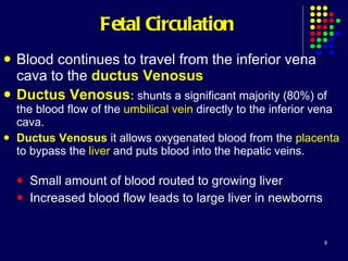


The document summarizes fetal circulation and how it differs from circulation after birth. In the fetus, oxygenated blood flows from the placenta to the umbilical vein and into the liver and heart. Most blood shunts past the lungs through openings like the foramen ovale and ductus arteriosus. After birth, clamping the umbilical cord and lung inflation cause structures like the ductus venosus to close as the infant transition to getting oxygen from the lungs.