Downloaded 824 times
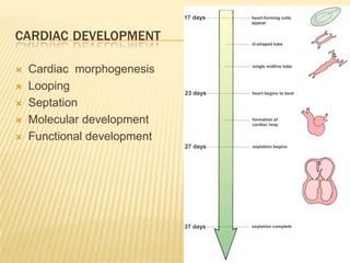
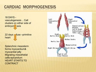
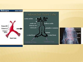
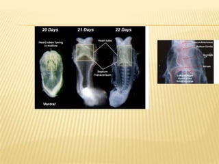
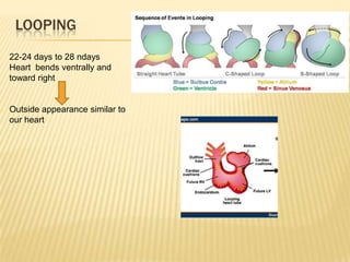
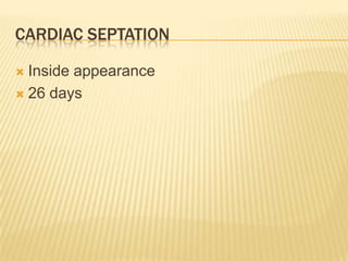
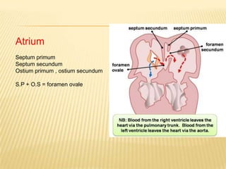
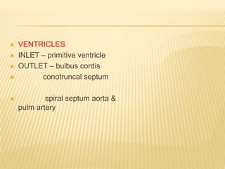
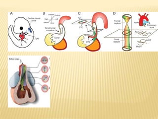
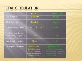
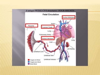
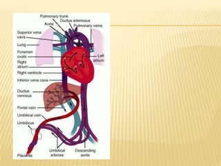
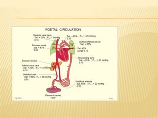
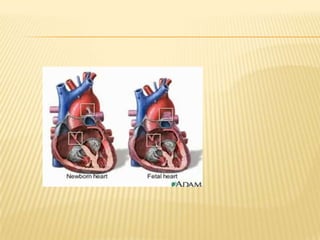
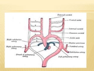
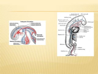
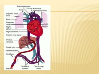
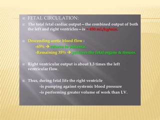


The document summarizes cardiac development and fetal circulation. During cardiac morphogenesis, the heart forms from clusters of cells on either side of the embryo that fuse to form the primitive heart by day 22. The heart then undergoes looping and septation to form the four chambers. In fetal circulation, blood bypasses the lungs via the ductus arteriosus and bypasses the liver via the ductus venosus to reach the placenta for gas and nutrient exchange. Blood then returns to the heart via the umbilical vein and inferior vena cava before mixing in the atria via the foramen ovale.


