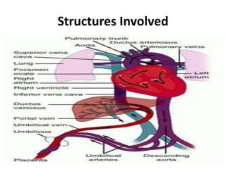
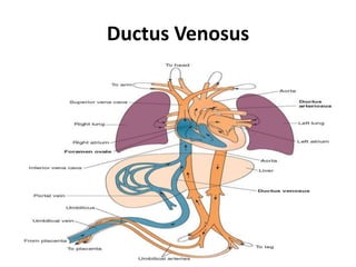
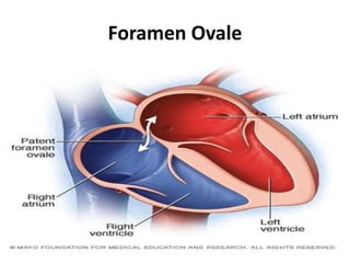
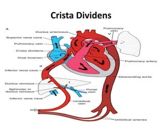
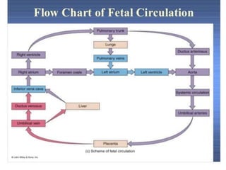
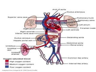
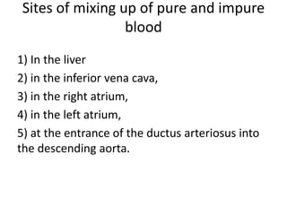
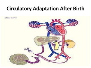
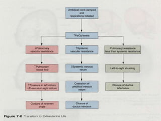
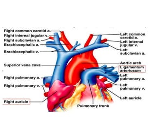
Unit 2.3
Fetal Circulation
PREPARED BY:
Sapana Dahal
MNC,IOM,TU
General Objective
At the end of the session,learners will be able to
explain about Fetal Circulation.
Specific Objectives
At the end of this session, all participants will be
able to:
introduce fetal circulation.
 state structures involved.
explain process of fetal circulation.
state sites of blood mixing.
explain circulatory changes after birth
Introduction
The circulation of blood within the cardiovascular
system of the fetus is called fetal circulation.
The oxygenated blood from maternal circulation is
passed through placenta to fetus via umbilical vein and
deoxygenated blood is returned to maternal circulation
through umbilical arteries.
Structures Involved
1. Single Umbilical Vein
2. Ductus Venosus
Ductus Venosus
3. Inferior Venacava
4. Fetal heart
5. Superior Venacava
6.Foramen Ovale
Foramen Ovale
7. Crista Dividens
Crista Dividens
8.Ascending aorta, coronary and carotid artery and
its branches
9. Pulmonary trunk or arteries
10. Ductus arteriosus
11.Descending aorta
12.2 umbilical arteries
Physiology of Fetal Circulation
Physiology of fetal circulation
Sites of mixing up of pure and impure
blood
1) In the liver
2) in the inferior vena cava,
3) in the right atrium,
4) in the left atrium,
5) at the entrance of the ductus arteriosus into
the descending aorta.
Circulatory Adaptation After Birth
1. Closure of umbilical arteries :
• Distal part forms medial umbilical ligaments and proximal
portion remains open as superior vesicle arteries.
• functionally closes within a few minutes after birth.
• Actual obliteration may take after 2-3 months.
• Caused by the thermal and mechanical stimuli and change
in the oxygen tension
2. Closure of umbilical veins and ductus venosus:
• Closes after the umbilical arteries closure.
• Blood from placenta may enter newborn for some time
after birth.
• After obliteration, umbilical vein forms the ligamentum
teres hepatis in the lower margin of the falciform
ligament.
Ligamentum teres Ligamentum venosum
• The ductus venosus forms ligamentum
venosum.
3. Closure of Ductus Arteriosus:
• It is mediated by the release of bradykinin during initial inflation.
• Functionally it closes by the 4th day after birth but complete obliteration
takes place around 1-3 months.
• The obliterated ductus arteriosus forms ligamentum arteriosum.
4. Closure of Foramen Ovale:
• It is caused by increased pressure in the left
atrium.
• It functionally closes at or soon after birth but
anatomically occurs in about 1 year.
• In 20% of the individuals, however, perfect
anatomically closure may never be obtained.
Summary:
References:
1. T.W.saddler ,langman’s medical embryology , 12th edition,Lippincott Williams and wilkins, south asian
edition published by wolters kluwer.
2.adhikari tulsi , essentials of pediatric nursing, 1st edition,vidhyarthi pustak bhandar
3. Subedi durga , Gautam saraswoti, midwifery nuring part-1, 2nd edition, medhabi publication.
Jacob, textbook of midwifery nd gynaecology nursing,
4. D.C. Dutta, textbook of obstetrics, 5th edition, new central book agency Pvt ltd, India.
5. Subedi maya devi, manual of midwifery A, Makalu publication house, Kathmandu.l
6. http:/www.embryology.med.unsw.edu.aw
7.www.embryology.ch/.../etape
. Tuitui roshani, manual of midwifery A, vidyarthi pustak bhandar, Kathmandu.
8.http://www.med.umich.edu/lrc/coursepages/m1/embryology/embryo/05embryonicperiod
HOME ASSIGNMENT
NEXT CLASS
POLYHYDRAMINOUS
Fetal circulation
Fetal circulation

Fetal circulation

  • 1.
    Unit 2.3 Fetal Circulation PREPAREDBY: Sapana Dahal MNC,IOM,TU
  • 2.
    General Objective At theend of the session,learners will be able to explain about Fetal Circulation.
  • 3.
    Specific Objectives At theend of this session, all participants will be able to: introduce fetal circulation.  state structures involved. explain process of fetal circulation. state sites of blood mixing. explain circulatory changes after birth
  • 4.
    Introduction The circulation ofblood within the cardiovascular system of the fetus is called fetal circulation. The oxygenated blood from maternal circulation is passed through placenta to fetus via umbilical vein and deoxygenated blood is returned to maternal circulation through umbilical arteries.
  • 5.
  • 6.
    1. Single UmbilicalVein 2. Ductus Venosus
  • 7.
  • 8.
    3. Inferior Venacava 4.Fetal heart 5. Superior Venacava
  • 9.
  • 10.
  • 11.
  • 12.
  • 13.
    8.Ascending aorta, coronaryand carotid artery and its branches 9. Pulmonary trunk or arteries 10. Ductus arteriosus 11.Descending aorta 12.2 umbilical arteries
  • 14.
  • 15.
  • 17.
    Sites of mixingup of pure and impure blood 1) In the liver 2) in the inferior vena cava, 3) in the right atrium, 4) in the left atrium, 5) at the entrance of the ductus arteriosus into the descending aorta.
  • 19.
  • 21.
    1. Closure ofumbilical arteries : • Distal part forms medial umbilical ligaments and proximal portion remains open as superior vesicle arteries. • functionally closes within a few minutes after birth. • Actual obliteration may take after 2-3 months. • Caused by the thermal and mechanical stimuli and change in the oxygen tension
  • 23.
    2. Closure ofumbilical veins and ductus venosus: • Closes after the umbilical arteries closure. • Blood from placenta may enter newborn for some time after birth. • After obliteration, umbilical vein forms the ligamentum teres hepatis in the lower margin of the falciform ligament.
  • 24.
  • 25.
    • The ductusvenosus forms ligamentum venosum.
  • 26.
    3. Closure ofDuctus Arteriosus: • It is mediated by the release of bradykinin during initial inflation. • Functionally it closes by the 4th day after birth but complete obliteration takes place around 1-3 months. • The obliterated ductus arteriosus forms ligamentum arteriosum.
  • 28.
    4. Closure ofForamen Ovale: • It is caused by increased pressure in the left atrium. • It functionally closes at or soon after birth but anatomically occurs in about 1 year. • In 20% of the individuals, however, perfect anatomically closure may never be obtained.
  • 29.
  • 30.
    References: 1. T.W.saddler ,langman’smedical embryology , 12th edition,Lippincott Williams and wilkins, south asian edition published by wolters kluwer. 2.adhikari tulsi , essentials of pediatric nursing, 1st edition,vidhyarthi pustak bhandar 3. Subedi durga , Gautam saraswoti, midwifery nuring part-1, 2nd edition, medhabi publication. Jacob, textbook of midwifery nd gynaecology nursing, 4. D.C. Dutta, textbook of obstetrics, 5th edition, new central book agency Pvt ltd, India. 5. Subedi maya devi, manual of midwifery A, Makalu publication house, Kathmandu.l 6. http:/www.embryology.med.unsw.edu.aw 7.www.embryology.ch/.../etape . Tuitui roshani, manual of midwifery A, vidyarthi pustak bhandar, Kathmandu. 8.http://www.med.umich.edu/lrc/coursepages/m1/embryology/embryo/05embryonicperiod
  • 31.
  • 32.