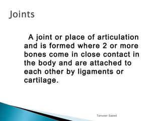
A joint orplace of articulation
and is formed where 2 or more
bones come in close contact in
the body and are attached to
each other by ligaments or
cartilage.
Tanveer Saeed
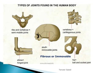
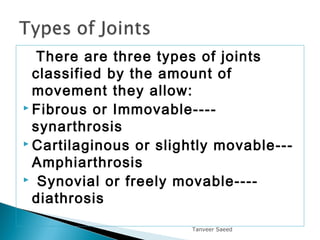
There are threetypes of joints
classified by the amount of
movement they allow:
Fibrous or Immovable----
synarthrosis
Cartilaginous or slightly movable---
Amphiarthrosis
Synovial or freely movable----
diathrosis
Tanveer Saeed
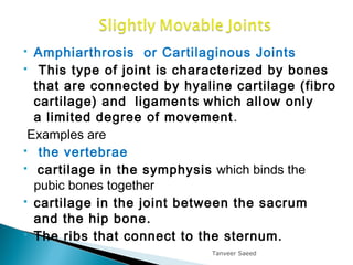
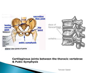
Amphiarthrosis orCartilaginous Joints
This type of joint is characterized by bones
that are connected by hyaline cartilage (fibro
cartilage) and ligaments which allow only
a limited degree of movement.
Examples are
the vertebrae
cartilage in the symphysis which binds the
pubic bones together
cartilage in the joint between the sacrum
and the hip bone.
The ribs that connect to the sternum.
Tanveer Saeed
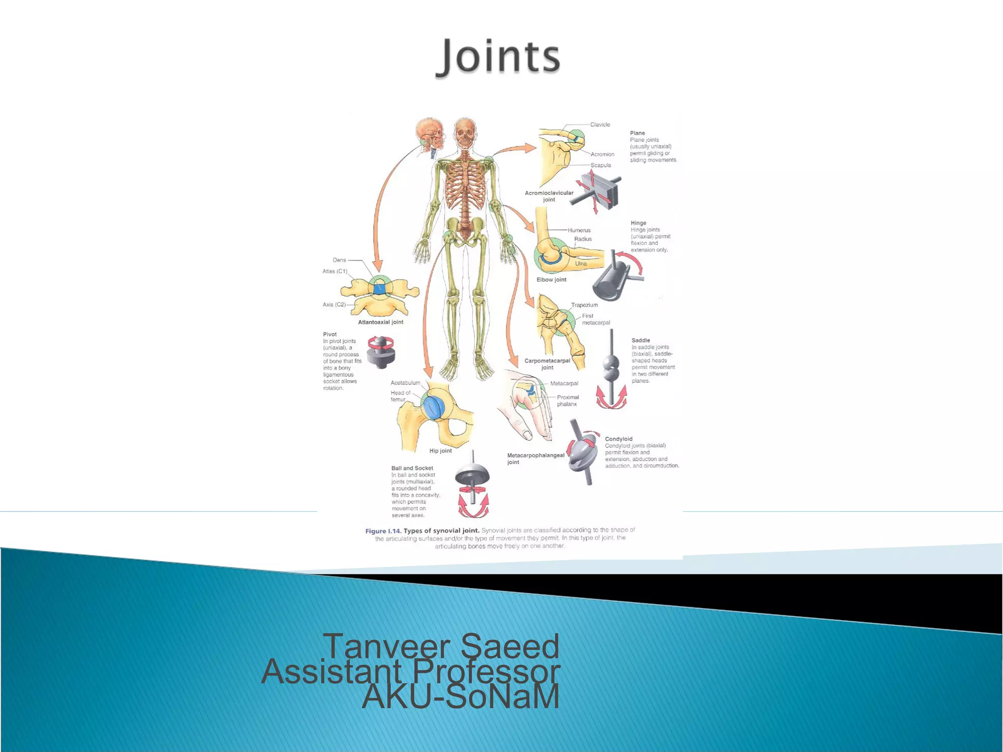
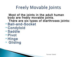
Most of thejoints in the adult human
body are freely movable joints.
There are six types of diarthroses joints:
Ball-and-Socket
Condyloid
Saddle
Pivot
Hinge
Gliding
Tanveer Saeed
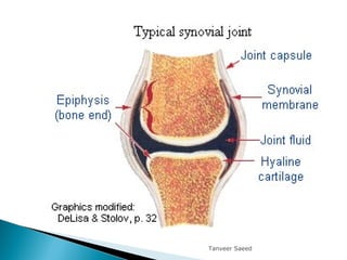
The capsular ligament islined with
a synovial membrane.
This membrane secretes synovial fluid into
the synovial cavity
acts as a seal, waterproofing the
joint,lubricates the joint.
In addition to the capsule, the bones are
also attached and held together by strong,
tough ligaments made of dense connective
tissue. These ligaments prevent
dislocation during normal movement.
Tanveer Saeed
12.
Synovial jointscan be subdivided into the
following groups according to the type of
movement they carry out.
All combinations of movements, including
circumduction and rotationcan be
performed.
Tanveer Saeed
13.
Tanveer Saeed+
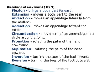
Directions ofmovement ( ROM)
Flexion - brings a body part forward.
Extension - moves a body part to the rear.
Abduction - moves an appendage laterally from
the midline.
Adduction - moves an appendage toward the
midline.
Circumduction - movement of an appendage in a
circle around a joint.
Pronation - rotating the palm of the hand
downward.
Supination - rotating the palm of the hand
upward.
Inversion - turning the toes of the foot inward.
Eversion - turning the toes of the foot outward.
14.
Tanveer Saeed
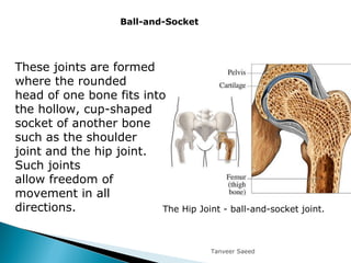
These jointsare formed
where the rounded
head of one bone fits into
the hollow, cup-shaped
socket of another bone
such as the shoulder
joint and the hip joint.
Such joints
allow freedom of
movement in all
directions.
Ball-and-Socket
The Hip Joint - ball-and-socket joint.
15.
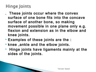
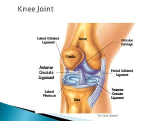
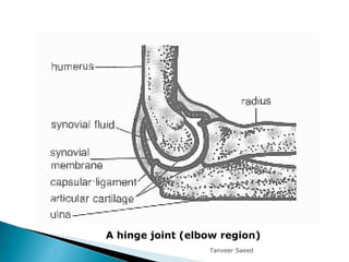
. These jointsoccur where the convex
surface of one bone fits into the concave
surface of another bone, so making
movement possible in one plane only e.g.
flexion and extension as in the elbow and
knee joints.
Examples of these joints are the :
knee ,ankle and the elbow joints.
Hinge joints have ligaments mainly at the
sides of the joints.
Tanveer Saeed
Oval shapedcondyle fits into oval cup
shaped end of another bone allowing
angular motion but not rotation.
Flexion,extension,adduction,abduction
and circumduction but no axial rotation.
Examples include:
wrist joint
Metacarpophalangeal joints(Knuckles)
Meta tarsophalangeal joints (toes)
Tanveer Saeed
19.
This typeof joint occurs when the
touching surfaces of two bones have
both concave and convex regions
allowing rotation in two directions.
The only saddle joint in the body is in
the thumb.
Tanveer Saeed
20.
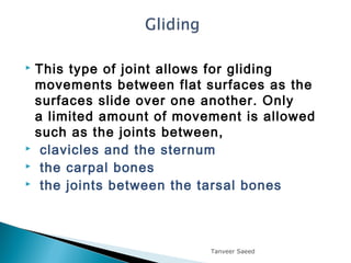
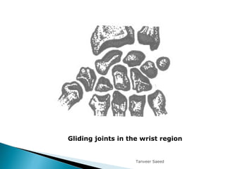
This typeof joint allows for gliding
movements between flat surfaces as the
surfaces slide over one another. Only
a limited amount of movement is allowed
such as the joints between,
clavicles and the sternum
the carpal bones
the joints between the tarsal bones
Tanveer Saeed
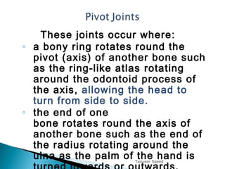
These joints occurwhere:
◦ a bony ring rotates round the
pivot (axis) of another bone such
as the ring-like atlas rotating
around the odontoid process of
the axis, allowing the head to
turn from side to side.
◦ the end of one
bone rotates round the axis of
another bone such as the end of
the radius rotating around the
ulna as the palm of the hand isTanveer Saeed
23.
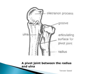
A pivot jointbetween the radius
and ulna
Tanveer Saeed
24.
Tanveer Saeed
Self quizto check what you have learned.
1.A point where one or two bones meet is__________.
2. Ball and socket, hinge, gliding and pivot joints are
example of _______.
3. Elbows, knees and fingers use what type of
joints________.
4. What type of joint can be found between
vertebrae________.
5. What type of movement is possible at Joint between
atlas and axis___________.
6. What type of joints are present in below mentioned
diagram___________.