Download to read offline
![International Research Journal of Engineering and Technology (IRJET) e-ISSN: 2395-0056
Volume: 06 Issue: 07 | July 2019 www.irjet.net p-ISSN: 2395-0072
© 2019, IRJET | Impact Factor value: 7.211 | ISO 9001:2008 Certified Journal | Page 1074
R Peak Detection with Diagnosis of Arrhythmia using Adaptive Filter
and Hilbert Transform
Saudagar1, Bhawna Jindal2
1Student, M.Tech, Department of ECE, UIET, Kurukshetra University, Kurukshetra, Haryana, India
2Student, M.Tech, Department of ECE, UIET, Kurukshetra University, Kurukshetra, Haryana, India
---------------------------------------------------------------------***---------------------------------------------------------------------
Abstract - R peak of electrocardiogram (ECG) signal
detected with good accuracy by combining adaptive filterand
Hilbert transform. In proposed algorithm adaptive filter
reduces the mean square error to an extent and estimates the
fundamental signal even in nonappearance of prior data
about statistical properties of the signal and noise, and gives
improved detection outcomes. AllpasscharacteristicofHilbert
transform eliminatesunnecessarysignal distortionandexhibit
the property of time dependency. Arrhythmiahappeneddueto
irregular heart rhythm which diagnosed by extracting
numerous ECG features such as RR interval, Heart rate, QRS
width, PR interval. A graphical user interface (GUI) also
developed which is a convenient way to represent output
waveform, features and type of abnormalities in a single
window screen with more simplicity, consistency and
familiarity. The signal has been taken from MIT-BIH
arrhythmia database. Sensitivity of 99.22% and positive
predictivity of 99.34% achieved by proposed methodology.
Key Words: Electrocardiogram, adaptive filtering, Hilbert
transform, features, GUI, arrhythmia.
1. INTRODUCTION
Electrocardiogram (ECG) also abbreviatedasEKGsignal.For
diagnosis electrocardiogram remainsthereferencestandard
in spite of the development of many other diagnostic
procedures. EKG signal comprises six wave i.e. P wave, QRS
complex (Q wave, S wave and peak R wave), T wave and U
wave. P wave generated due to depolarization of atria, QRS
complex indicates the depolarization of left and right
ventricles, R wave has peak amplitudeinQRS complex which
is a point of interest in detection process and contains most
of the important clinical information regarding heart [1], PR
interval estimated from beginning of P-wave to the starting
of QRS complex, RR interval is the difference between two
successive R peaks, also named as Inter-beat- interval (IBI).
Electrocardiographyisanimportanttool usedfordiagnosing
heart condition. Deviation from normal sinus rhythm
(normal heart cycle) are the symptoms of arrhythmia that
denotes some form of cardiovascular disease (CVD) [2].
Cardiac arrhythmia displays a situation of abnormal
electrical movement in the heart which is a danger to
humans [3]. The heart produces miniature electrical
impulses which spread over the heart muscle to make the
heart contract. These impulses can be identified by the ECG
machine. The electrocardiogram signal and heart rate
reveals the cardiac health of human heart. Any disorder in
heart rate or rhythm or variation in the morphological
pattern of this signal is a sign of cardiac arrhythmia. It is
detected and diagnosed by scrutiny of the recorded
waveform. The amplitude and duration of the P-QRS-T-U
wave holds beneficial information about the nature of
disease associated to heart. An electrocardiogram (EKG) is
completed to determine the heart electrical activity,tocheck
exactly how rapid your heart is beating and to find the
reason of symptoms of heart diseaselikeshortnessofbreath,
dizziness, unbalances heartbeat [1]. Various noise or
artifacts such as power line interference, baseline
wandering, motion artifacts, and electrode contact noise
distort the signal quality, frequency resolution, which
generates signals with large amplitudethatcanbeanalogous
to PQRST waveforms and masks miniature features that are
essential for clinical diagnosis and monitoring [4].
Elimination of these artifacts is a significant task for better
diagnosis. For that purpose wavelet denoising used in
proposed work. R-Peak is the most noticeable part during
signal examination which relates to the contraction of the
ventricles for the duration of a heartbeat. QRS complex
detected more accurately with presented algorithm that
escalates detection sensitivity by processing the RR interval
as compared to some previous methods such as wavelet
Transform method in which accuracythrowsdownto77.3%
for large muscle artifact signals [5], Template matching
technique [6], Wavelet Transform with histogram, moving
average and two thresholds technique [7], Wavelets and
artificial neural networks based method [8]. So the main
objective of this study to detect R peak with good accuracy
and to extract electrocardiogram features along with
arrhythmia diagnosis by using an algorithm formed with
combination of adaptive filter and Hilbert transform.
2. MATERIAL AND METHODS
The processing of ECG signal involve of de-noising, baseline
correction, filtering, thresholding, feature extraction and
arrhythmia detection. Flow chart of proposed algorithm
represented inFig.1.Thewholeexplanationof preprocessing
steps and evaluation of performance parameters discussed
here.](https://image.slidesharecdn.com/irjet-v6i7117-191104035614/75/IRJET-R-Peak-Detection-with-Diagnosis-of-Arrhythmia-using-Adaptive-Filter-and-Hilbert-Transform-1-2048.jpg)
![International Research Journal of Engineering and Technology (IRJET) e-ISSN: 2395-0056
Volume: 06 Issue: 07 | July 2019 www.irjet.net p-ISSN: 2395-0072
© 2019, IRJET | Impact Factor value: 7.211 | ISO 9001:2008 Certified Journal | Page 1075
Fig -1: Flowchart of proposed algorithm
2.1 Original ECG signal
An offline data used for processing electrocardiogram
signals that can be taken from MIT-BIHarrhythmia database
available on Physionet ATM bank [9]. The raw data basically
contaminated with numerous noise artifacts which needsto
be eliminate to enhance detection performance. These
artifacts added to the signal while recording due to
movement in muscles, electrode etc.
2.2 Wavelet denoising and subtraction
Most of the signal corrupted with the baseline wandering
noise which shift the signal upward or downward and affect
the pattern of QRS recognition. Elimination of baseline drift
is hence mandatory in the analysis of the ECG signal to
decrease the fluctuations in beat morphology with no
physiological equivalent [10]. Electrode impedance and
respiration alters because of perspiration which is the
leading sources of baseline wander in utmost types of ECG
recordings.Thisbaselinewanderingcanbe removedwithout
troubling the ECG waveform features.Tokeep baselineatthe
correct position firstly signal denoise using waveletfunction
wden(db6) and then subtracted from the original signal.
2.3 Adaptive filtering
An adaptive filter uses iterative computations to decrease
the error “in forming the relationship among two signals in
real time”. Basic diagram of an adaptivefilterisshownbelow
in Fig.2. Here, the input denotes the ECG which is
observed with the additive noise n. The reference signal c is
either a pure noise generator or a signal associated to n.
Since the n and are uncorrelated, then
(1)
Fig -2: A general diagram of an adaptive filter
Let the N coefficients of the filter at the iteration be
represented as = . For an
input vector = ,
the output will be given in equation as:
d(k)= (2)
The working of filter is to adjust its weights W iteratively to
decrease the mean square error (MSE) within the primary
and the reference inputs [11]. Usually, LMS and RLS
algorithm has been used to bring up to date the coefficients
of predictor. In present work, RLS (Recursive least square)
have been used for the said purpose. Upon convergence, a
predictor is able to predict the gradually varying
components (low frequency content) accurately i.e.
producing a reduced amount of error in comparison to fast
moving components (high frequency content).
2.3.1 RLS Algorithm
Recursive least square(RLS)algorithmrecursivelydiscovers
the filter coefficients with an objective function of lessening
the cost function of summation of weighted linear least
squares linking to the input vector unlike LMS (least mean
square) and other analogous variants that aim to decrease
the mean square error. The RLS algorithm shows very fast
convergence at the price of biggercomputational complexity
[2]. Eigen value spread problem also eliminated by this
algorithm. Equation representing the relations for updating
weights is as follows:
(3)
(4)](https://image.slidesharecdn.com/irjet-v6i7117-191104035614/75/IRJET-R-Peak-Detection-with-Diagnosis-of-Arrhythmia-using-Adaptive-Filter-and-Hilbert-Transform-2-2048.jpg)
![International Research Journal of Engineering and Technology (IRJET) e-ISSN: 2395-0056
Volume: 06 Issue: 07 | July 2019 www.irjet.net p-ISSN: 2395-0072
© 2019, IRJET | Impact Factor value: 7.211 | ISO 9001:2008 Certified Journal | Page 1076
(5)
(6)
where c(n) is desired signal value of nth sample, λ is
exponential forgetting factor, P(n) is a k*k matrix, is a
1*k vector, is transposed input vector, ,
represent weight vectors at nth and (n-1)th
sample having dimension of 1*K , ande(n)arethestepsize
parameter and the instantaneous error respectively having
dimension of 1*1, is a 1*K input signal vector
containing signal values from c(n) to c(n-k).
2.4 Savitzky-Golay Filtering
A technique of data smoothing centered on local least
squares polynomial approximation was given by Savitzky
and Golay in 1964. They revealed that fitting a polynomial to
a set of input samples and then estimating the resulting
polynomial at a distinct point within the approximation
interval is corresponding to discrete convolution with a
static impulse response. The low pass filtersachievedbythis
process are broadly known as Savitzky Golay filters. Noisy
data gained from chemical spectrum analyzers smoothens
and least squares smoothing decreases noise while
preserving the shape and heightofwaveformpeaks[12]. For
N data points (N is odd, N=2M+1) a data vector X having M
points on either side of :
(7)
Then by a polynomial of degree d, N data samples fitted in X:
(8)
In this case , i=0,1,…,d, is defined to have components
which is a d+1 polynomial basis vectors:
(9)
The corresponding matrix S (N*(d+1)) is well-defined to
have as columns:
(10)
Then the design stages for the SG filters can be explained as
follows:
(11)
(12)
(13)
The steady state equation of SG filter for smoothing a noisy
sequence x(n) with length N, order d as:
= (14)
For proposed work the SG filter has order of 3 and window
length of 15.
2.5 Squaring and differentiation
The strength of the signal decreased after passing through
number of preprocessing steps. Hence it is required to
enhance the strength of signal so that squaring performed
[13]. This raises the amplitude of signal and all the
information available in the positive half which added
easiness in detection of correct peaks. Then the signal
differentiated to acquire the slope information of QRS
complex.
2.6 Hilbert transform
To detect R peaks proficiently the proposed technique is
described in a very suitable manner. A real valued time
function x(t) i.e. differentiated ECG, and the Hilbert
transform of the that signal is given by equation:
(15)
In accordance with transformationtheindependentvariable
do not altered so that Hilbert transform shows the property
of time dependency [14]. And this relation comes from the
convolution calculation:
(16)
(17)
where sgnf= (18)
The i/o correlation of Hilbert transform is the conversion of
linear function, therefore it act like a linear filter where all
the amplitudes of spectral components are lasting identical
where the phases altered with ±90°. The imaginary part of
the analytic signal is Hilbert transformanditsreal partactas
its input. Output waveform demonstrates the Hilbert
transform of a differentiated EKG segment. The R-peaks
happened due to the zero-crossings of the differentiated
EKG, because the Hilbert transform is an odd filterandin the
output of the transform represented as peaks. On the
differentiated electrocardiogram the effects of the Hilbert
transform described in terms of its signal envelope and odd
symmetry property. All pass characteristic of the Hilbert
transform avoids needless signal distortion, in comparison
to the additional derivative technique which tends to
diminish the signal at the lower frequencies. Therefore, the](https://image.slidesharecdn.com/irjet-v6i7117-191104035614/75/IRJET-R-Peak-Detection-with-Diagnosis-of-Arrhythmia-using-Adaptive-Filter-and-Hilbert-Transform-3-2048.jpg)
![International Research Journal of Engineering and Technology (IRJET) e-ISSN: 2395-0056
Volume: 06 Issue: 07 | July 2019 www.irjet.net p-ISSN: 2395-0072
© 2019, IRJET | Impact Factor value: 7.211 | ISO 9001:2008 Certified Journal | Page 1077
essential rectification of the differentiated EKG signal
delivered by the odd phase element of the filter in order to
recognize the QRS peaks while the uniform magnitudeofthe
filter confirms that essential information of the QRS
complexes is preserved[15]. TheHilberttransformedoutput
signal is stated as;
(19)
(20)
(21)
Where V(t) is the envelop of x(t) and is instantaneous
phase angle.
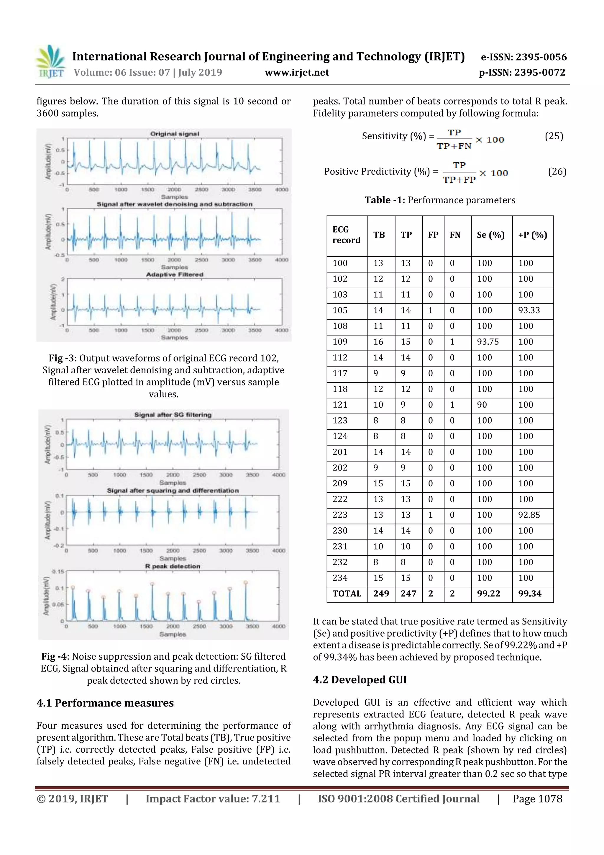
2.7 R peak detection
There is need of extraction of suitable metrics from the
signal. Before the extraction of these metrics from the ECG
signal, the Q, R, S peaks with their position in each beat were
identified. This is achieved with an algorithmic script with
the following approach: The first aimisthedetectionoftheR
Peak because one time the R-Peak is detected; it can be used
to notice the Q and S points effortlessly. Because of the
idiosyncratic nature of the QRS complex and the typical
characteristics of the R peak, this is willingly identified even
in the utmost distorted ECG readings. Thus it is castoff asthe
basis for ECG feature computations [16]. By choosing a
window of particular length, the peak with maximum value
detected. A threshold also applied for better detection,
distinguishing correctly detected and falselydetectedpeaks.
Then R peaks detected on the envelope of Hilbert
transformed signal represented by the small red circles as
shown in Fig.4.
2.8 Feature extraction
For the resolution of diagnosis after R peak detection, there
is need to extract several features from the preprocessed
ECG data, comprising QRS width, RR interval, PR intervals.
The deflection positional information provides the feature
such as RR interval which used in medical as a pointer of
Ventricular Heart Rate. Two R peaks in successive beats is
calculated and their difference is figuredfordetermining the
RR interval.Electrocardiogramfeatureextractedonthebasis
of given equations:
(22)
(23)
(24)
Where stand for sampling frequency specify
current R peak locations and indicates current P peak
locations. Here variable x shows instant 5 ms, added to
and are subtracted from .S and Q peak sitesrepresented
by and respectively [17]. Heart rate computed in
beats per minutes (bpm) and for a normal person in resting
state varies from 60 to 100 bpm.
2.9 Arrhythmia
Arrhythmia happened due to irregular heart rhythm and
various types of arrhythmias diagnosed in this paper. With
extractedfeaturesnumerouscardiovasculararrhythmiasare
noticed as Right bundle branch block. Within the right
bundle branch it act as a delay or block of conduction. An
interruption of conduction reveals as incomplete right
bundle branch block.Rightbundlebranchblock occurswhen
QRS duration > 0.14 sec. Sinus Bradycardia occurs when
heart rate is less than 60 beats per minute in resting state,
though it is rarely suggestive until the rate falls below 50
beat/min. Sinus Tachycardia states to quick beating of the
heart as a heart rate more than 100 beats per minute in
adult. Diseases were predicted from extracted features as
according to medical science condition for Tachycardia
fulfilled if heart rate > 100 bpm (beats per minute) [18].
Normal range of heart rhythm is 60 to 100 bpm. When QRS
width > 0.12 sec and heart rate varies from 101 to 250 bpm
then Ventricular tachycardia happened. Normal range QRS
width lies between 0.08 to 0.10 sec. Incomplete Bundle
Branch Block occurred when QRS width between 0.10sec to
0.12 sec and QRS width > 0.12 sec for Bundle Branch Block
[16]. First degree heart block take place when PR interval >
0.2 sec.
3. ECG DATABASE
MIT-BIH arrhythmia database used in this work which
available on Physionet ATM bank. This offline data firstly
collected in a directory which further used for
preprocessing. It consistsoftotal 48subjectssignal including
males and females. There are 25 men of age group 32-89
years and 23 women of age group 23-89 years. For
estimation of arrhythmia detection this database was firstly
usually accessible from the set of standard test material and
has been used for that purpose as well as for elementary
investigation into cardiac dynamics at larger than 500 sites
worldwide [9]. These signals have voltage of 10mV,
resolution of 10 bit and 360 Hz sampling frequency.
4. RESULTS
Output waveforms obtained from proposed methodology as
shown in Fig.3 and Fig.4. These waveforms produced in
MATLAB tool. An ECG record 102 has been taken from MIT-
BIH arrhythmia database and processed as illustrated in](https://image.slidesharecdn.com/irjet-v6i7117-191104035614/75/IRJET-R-Peak-Detection-with-Diagnosis-of-Arrhythmia-using-Adaptive-Filter-and-Hilbert-Transform-4-2048.jpg)
![International Research Journal of Engineering and Technology (IRJET) e-ISSN: 2395-0056
Volume: 06 Issue: 07 | July 2019 www.irjet.net p-ISSN: 2395-0072
© 2019, IRJET | Impact Factor value: 7.211 | ISO 9001:2008 Certified Journal | Page 1079
of arrhythmia diagnosed is first degree heartblock asshown
in Fig.5.
Fig -5: Developed GUI
4.3 Comparison with previous state of art methods
A quick survey of some previous studies has been provided
to compare the best performance observed in the present
study. Se and +P are listed in Table with methods, novelty of
particular study. In this paper a newalgorithmbycombining
adaptive filter and Hilbert transform for QRS complex
detection is proposed. This method achieves good accuracy
in term of sensitivity and positive predictivityincomparison
with other competing methods, offering an advantage of
minimized mean square error and low computational
complexity. A graphical user interface also developed which
provide unity, clarity, directness, alignment, integrity,
comfort and charm to the user. The user interface is a single
object with a certain personality to help a particular user
perform a particular task.
Table -2: Comparison with previous state of art methods
Methods Novelty Se
(%)
+P
(%)
Wavelet Transform
method [5]
Detect arrhythmia types
by relating the extracted
ECG features.
94.12 88.90
Template matching
algorithm [6]
Template waveform
produced using a short-
term autocorrelation
95.80 98.30
process for QRS
detection.
Wavelet Transform
with histogram,
moving average
and two thresholds
technique [7]
Two wavelet function
(haar &db10) with dual
thresholding for QRS
detection
98.68 97.24
Second derivative
method [15]
Bandpass filtering,
squaring and variable
thresholds comparison
for QRS detection
98.08 99.18
Wavelets and
artificial neural
networks method
[8]
Four wavelet basis
functions those are
appropriate in detection
of QRS complexes inside
dissimilar QRS
morphologies in the
signal.
97.20 98.52
Geometrical
matching algorithm
[19]
Use decision function in
a confined moving-
window process for QRS
detection.
97.94 99.13
Presented
methodology
Combination of
adaptive filter and
Hilbert transform for
R peak detection with
arrhythmia diagnosis.
99.22 99.34
5. CONCLUSION
This paper presents an improved R peak detection system
that work on adaptive filtering and Hilbert transform. Type
of arrhythmia also diagnosed on the basis of extracted ECG
features. By adopting a wavelet function for denoising and
Savitzky Golay filter for filtering occurrence of false positive
and false negative beats reduced. During recording extra
ventricular waveform formed due to which artifacts
generated that are also removed with adaptive filtering.
Presented technique achieves better-quality detection of
delayed potentials. So sensitivity of 99.22% and positive
predictivity of 99.34% has been attained by proposed
methodology.
REFERENCES
[1] Joshi AK, Tomar A, Tomar M. A Review Paper on
Analysis of Electrocardiograph (ECG) Signal for the
Detection of Arrhythmia Abnormalities.IntJAdvRes
Electr2014;3(10):12466–75.
http://dx.doi.org/10.15662/ijareeie.2014.0310028.
[2] Jain S, Ahirwal MK, Kumar A, Bajaj V, Singh GK. QRS
detection usingadaptivefilters:Acomparativestudy.
ISA Trans 2017;66:362–75.
http://dx.doi.org/10.1016/j.isatra.2016.09.023.
[3] Khadirnaikar S, Aparna P. A feasible QRS detection
algorithm for arrhythmia diagnosis. 2016 Int. Conf.](https://image.slidesharecdn.com/irjet-v6i7117-191104035614/75/IRJET-R-Peak-Detection-with-Diagnosis-of-Arrhythmia-using-Adaptive-Filter-and-Hilbert-Transform-6-2048.jpg)
![International Research Journal of Engineering and Technology (IRJET) e-ISSN: 2395-0056
Volume: 06 Issue: 07 | July 2019 www.irjet.net p-ISSN: 2395-0072
© 2019, IRJET | Impact Factor value: 7.211 | ISO 9001:2008 Certified Journal | Page 1080
Adv. Electr. Electron. Syst. Eng. ICAEES 2016;32–7.
http://dx.doi.org/10.1109/ICAEES.2016.7888004.
[4] Mr. HrishikeshLimayeMVVD.ECGNoiseSourcesand
Various Noise Removal Techniques: A Survey. Int J
Appl or Innov Eng Manag 2016;5(2):2319–4847.
[5] Priya PK, Reddy GU. MATLAB Based GUI for
Arrhythmia Detection Using Wavelet Transform.
Ijareeie 2015;4(2):807–16.
http://dx.doi.org/10.15662/ijareeie.2015.0402055.
[6] Nakai Y, Izumi S, Nakano M, Yamashita K, Fujii T,
Kawaguchi H, et al. Noise tolerant QRS detection
using template matching with short-term
autocorrelation. 2014 36th Annu Int Conf IEEE Eng
Med Biol Soc EMBC 2014;34–7.
http://dx.doi.org/10.1109/EMBC.2014.6943522.
[7] Chouakri SA, Bereksi-Reguig F, Taleb-Ahmed A. QRS
complex detection based on multi wavelet packet
decomposition. Appl Math Comput
2011;217(23):9508–25.
http://dx.doi.org/10.1016/j.amc.2011.03.001.
[8] Abibullaev B, Seo HD. A new QRS detection method
using wavelets and artificial neural networks. J Med
Syst 2011;35(4):683–91.
http://dx.doi.org/10.1007/s10916-009-9405-3.
[9] Moody RGM and GB. The MIT-BIH Arrhythmia
Database on CD-ROM and software for use with it.
Proc. Int. Conf. Comput. Cardiol., 1990, p. 185–8.
[10] Pokharkar S, Kulkarni A. ECG Real Time Feature
Extraction Using MATLAB. Int J Technol Sci
2015;V(1):2350–1111.
[11] AlMahamdy M, Riley HB. Performance study of
different denoising methods for ECG signals.
Procedia Comput Sci 2014;37:325–32.
http://dx.doi.org/10.1016/j.procs.2014.08.048.
[12] Nahiyan KMT, Amin A. Removal of ECG Baseline
Wander using Savitzky-Golay Filter Based Method.
Bangladesh J Med Phys 2015;8:32–45.
[13] Pan J, Tompkins WJ. A Real-Time QRS Detection
Algorithm. IEEE Trans Biomed Eng 1985;BME-
32(3):230–6.
http://dx.doi.org/10.1109/TBME.1985.325532.
[14] Sahoo S, Biswal P, Das T, Sabut S. De-noising of ECG
Signal and QRS Detection Using Hilbert Transform
and Adaptive Thresholding. Procedia Technol
2016;25:68–75.
http://dx.doi.org/10.1016/j.protcy.2016.08.082.
[15] Arzeno NM, Deng Z De, Poon CS. Analysis of first-
derivative based QRS detection algorithms. IEEE
Trans Biomed Eng 2008;55(2):478–84.
http://dx.doi.org/10.1109/TBME.2007.912658.
[16] Gothwal H, KedawatS,KumarR.Cardiacarrhythmias
detection in an ECG beat signal using fast fourier
transform and artificial neural network.JBiomedSci
Eng 2011;04:289–96.
http://dx.doi.org/10.4236/jbise.2011.44039.
[17] Umer M, Bhatti BA, Tariq MH, Zia-Ul-HassanM,Khan
MY, Zaidi T. Electrocardiogram Feature Extraction
and Pattern Recognition Using a Novel Windowing
Algorithm. Adv Biosci Biotechnol 2014;5:886–94.
http://dx.doi.org/10.4236/abb.2014.511103.
[18] Miller JM, Das MK, Yadav A V, Bhakta D, Nair G,
Alberte C. Value of the 12-Lead ECG in Wide QRS
Tachycardia. Cardiol Clin 2006;24:439–51.
http://dx.doi.org/10.1016/j.ccl.2006.03.003.
[19] Suárez K V., Silva JC, Berthoumieu Y, Gomis P, Najim
M. ECG beat detection using a geometrical matching
approach. IEEE Trans Biomed Eng 2007;54(4):641–
50. http://dx.doi.org/10.1109/TBME.2006.889944.
[20] Mhetre MR, Vaishampayan A, Raskar M. ECG
Processing & Arrhythmia Detection : An Attempt. Int
J Eng Innov Technol 2013;2(10):272–6.
[21] AL-Ziarjawey H. Heart Rate Monitoring and PQRST
Detection Based on Graphical User Interface with
Matlab. Int J Inf Electron Eng 2015;5(4):311–6.
http://dx.doi.org/10.7763/IJIEE.2015.V5.550.](https://image.slidesharecdn.com/irjet-v6i7117-191104035614/75/IRJET-R-Peak-Detection-with-Diagnosis-of-Arrhythmia-using-Adaptive-Filter-and-Hilbert-Transform-7-2048.jpg)

The document presents a method for detecting R peaks in electrocardiogram (ECG) signals with high accuracy by combining adaptive filtering and Hilbert transform. Adaptive filtering reduces noise and estimates the fundamental signal, while Hilbert transform eliminates signal distortion and shows time dependency. Features are then extracted from the ECG, including RR interval, heart rate, QRS width, and PR interval. These features can be used to diagnose arrhythmias based on irregular heart rhythms. A graphical user interface was also developed to conveniently display the output waveform, features, and type of arrhythmia diagnosis. When tested on data from the MIT-BIH arrhythmia database, the proposed method achieved a sensitivity of 99.22% and positive predict