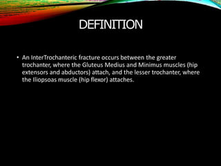
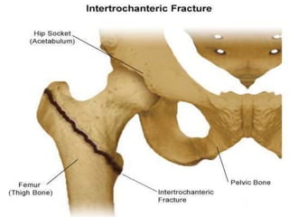
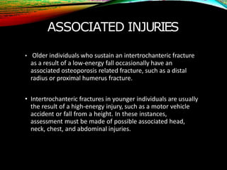
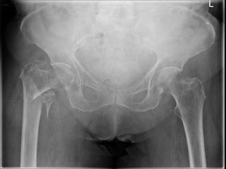
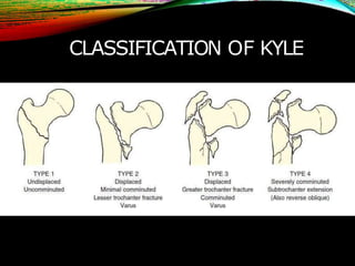
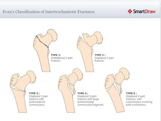
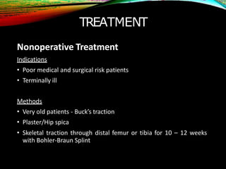
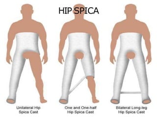
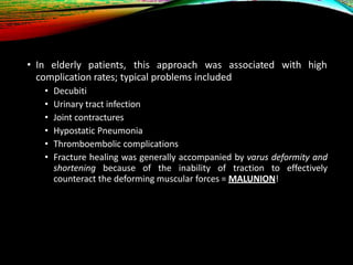
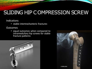
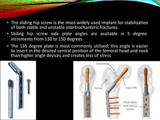
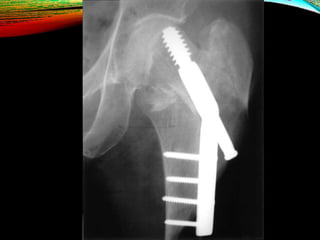
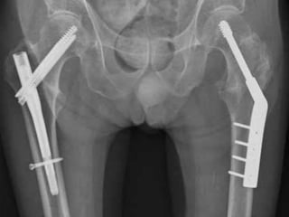
INTERTROCHANTERIC
FRACTURE
Dr
. Kevin Joseph Ambadan
DEFINITION
• An InterTrochanteric fracture occurs between the greater
trochanter, where the Gluteus Medius and Minimus muscles (hip
extensors and abductors) attach, and the lesser trochanter, where
the Iliopsoas muscle (hip flexor) attaches.
GENERAL FEATURES
• Completely ExtraCapsular fracture with variable comminution
• Common in elderly osteoporotic patient, usually women in their
80’s
• More common than IntraCapsular # - Neck of Femur
• Unites easily
• Rarely causes avascular necrosis
MECHANISM OF INJURY
• Intertrochanteric fractures in younger individuals are usually the
result of a high-energy injury, such as a motor vehicle accident
(MVA) or fall from a height
• In the elderly, it results from a simple fall (trivial trauma).
SIGNS AND SYMPTOMS
• Pain
• Marked shortening of lower limb
• Patient cannot lift their leg
• Complete External Rotation Deformity
• Swelling, ecchymosis and tenderness over the Greater Trochanter
• Displaced fractures are clearly symptomatic, such patients usually
cannot stand, much less ambulate
• Non-displaced fractures may be ambulatory and experience minimal
pain, and there are yet others who complain of thigh or groin pain
but have no history of antecedent trauma
• The amount of clinical deformity in patients with an intertrochanteric
fracture reflects the degree of fracture displacement
ASSOCIATED INJURIES
• Older individuals who sustain an intertrochanteric fracture
as a result of a low-energy fall occasionally have an
associated osteoporosis related fracture, such as a distal
radius or proximal humerus fracture.
• Intertrochanteric fractures in younger individuals are usually
the result of a high-energy injury, such as a motor vehicle
accident or fall from a height. In these instances,
assessment must be made of possible associated head,
neck, chest, and abdominal injuries.
DIAGNOSTIC IMAGING
• X-ray is the standard diagnostic tool.
• When a hip fracture is suspected but not apparent on standard x-
rays, a technetium bone scan or a MRI scan should be obtained.
• MRI has been shown to be at least as accurate as bone scanning in
identification of occult fractures of the hip, and it will reveal a
fracture within 24 hours of injury.
BOYD & GRIFFIN’S CLASSIFICATION
1. Linear IT line #
2. Linear IT line # with comminution
3. Subtrochanteric #
4. Inter-/Subtrochanteric # with extension into proximal
femoral shaft
CLASSIFICATION OF KYLE
EVAN’S CLASSIFICATION
TREATMENT
Nonoperative Treatment
Indications
• Poor medical and surgical risk patients
• Terminally ill
Methods
• Very old patients - Buck’s traction
• Plaster/Hip spica
• Skeletal traction through distal femur or tibia for 10 – 12 weeks
with Bohler-Braun Splint
BUCKS TRACTION
HIP SPICA
• In elderly patients, this approach was associated with high
complication rates; typical problems included
• Decubiti
• Urinary tract infection
• Joint contractures
• Hypostatic Pneumonia
• Thromboembolic complications
• Fracture healing was generally accompanied by varus deformity and
shortening because of the inability of traction to effectively
counteract the deforming muscular forces = MALUNION!
OPERATIVE TREATMENT
Intertrochanteric fractures are almost always treated
by early internal fixation – not because they fail to
unite with conservative treatment (they unite quite
readily), but
• Obtain the best possible position
• Early ambulation to reduce the complications associated with
prolonged recumbency.
SLIDING HIP COMPRESSION SCREW
Indications
• stable intertrochanteric fractures
Outcomes
• equal outcomes when compared to
intramedullary hip screws for stable
fracture patterns
• The sliding hip screw is the most widely used implant for stabilization
of both stable and unstable intertrochanteric fractures.
• Sliding hip screw side plate angles are available in 5 degree
increments from 130 to 150 degrees.
• The 135 degree plate is most commonly utilized; this angle is easier
to insert in the desired central position of the femoral head and neck
than higher angle devices and creates less of stress
TROCHANTERIC STABILIZING PLATES
• The trochanteric stabilizing plate and the
lateral buttress plate are modular
components that reinforces the greater
trochanter
• These plates are placed over a four-hole
sideplate and are used to prevent
excessive slide (and resulting deformity)
in unstable fracture patterns
• These devices prevent telescoping of the
lag screw within the plate barrel when
the proximal head and neck fragment
abuts the lateral buttress plate
INTRAMEDULLARY HIP SCREW
Also known as the Proximal Femoral Nail (PFN).
Indications
• stable fracture patterns
• unstable fracture patterns
• reverse obliquity fractures (56% failure when treated with sliding
hip screw)
• subtrochanteric extension
• lack of integrity of femoral wall
Outcome
• equivalent to sliding hip screw for stable fracture patterns
• use has significantly increased in last decade
COMPLICATIONS
EARLY
• The same as with femoral neck fractures, reflecting the fact
that most of these patients are in poor health.
LATE
• Failed fixation Screws may cut out of the osteoporotic bone if
reduction is poor or if the fixation device is incorrectly
positioned. If union is delayed, the implant itself may break. In
either event, reduction and fixation may have to be re-done.
• Malunion
• Coxa Vara and external rotation deformities are common
• Non-union (uncommon, unlike # NoF)
• Traumatic Osteoarthritis
• Avascular Necrosis (rare)
PATHOLOGICAL FRACTURE
• Due to metastatic disease or myeloma.
• Unless patients are terminally ill, fracture fixation is essential in
order to ensure an acceptable quality of life for their remaining
years.
• In addition to internal fixation, methylmethacrylate cement may
be packed in the defect to improve stability.
THANK YOU

intertrochantericfractures

  • 1.
  • 2.
    DEFINITION • An InterTrochantericfracture occurs between the greater trochanter, where the Gluteus Medius and Minimus muscles (hip extensors and abductors) attach, and the lesser trochanter, where the Iliopsoas muscle (hip flexor) attaches.
  • 4.
    GENERAL FEATURES • CompletelyExtraCapsular fracture with variable comminution • Common in elderly osteoporotic patient, usually women in their 80’s • More common than IntraCapsular # - Neck of Femur • Unites easily • Rarely causes avascular necrosis
  • 5.
    MECHANISM OF INJURY •Intertrochanteric fractures in younger individuals are usually the result of a high-energy injury, such as a motor vehicle accident (MVA) or fall from a height • In the elderly, it results from a simple fall (trivial trauma).
  • 6.
    SIGNS AND SYMPTOMS •Pain • Marked shortening of lower limb • Patient cannot lift their leg • Complete External Rotation Deformity • Swelling, ecchymosis and tenderness over the Greater Trochanter • Displaced fractures are clearly symptomatic, such patients usually cannot stand, much less ambulate • Non-displaced fractures may be ambulatory and experience minimal pain, and there are yet others who complain of thigh or groin pain but have no history of antecedent trauma • The amount of clinical deformity in patients with an intertrochanteric fracture reflects the degree of fracture displacement
  • 7.
    ASSOCIATED INJURIES • Olderindividuals who sustain an intertrochanteric fracture as a result of a low-energy fall occasionally have an associated osteoporosis related fracture, such as a distal radius or proximal humerus fracture. • Intertrochanteric fractures in younger individuals are usually the result of a high-energy injury, such as a motor vehicle accident or fall from a height. In these instances, assessment must be made of possible associated head, neck, chest, and abdominal injuries.
  • 8.
    DIAGNOSTIC IMAGING • X-rayis the standard diagnostic tool. • When a hip fracture is suspected but not apparent on standard x- rays, a technetium bone scan or a MRI scan should be obtained. • MRI has been shown to be at least as accurate as bone scanning in identification of occult fractures of the hip, and it will reveal a fracture within 24 hours of injury.
  • 10.
    BOYD & GRIFFIN’SCLASSIFICATION 1. Linear IT line # 2. Linear IT line # with comminution 3. Subtrochanteric # 4. Inter-/Subtrochanteric # with extension into proximal femoral shaft
  • 11.
  • 12.
  • 13.
    TREATMENT Nonoperative Treatment Indications • Poormedical and surgical risk patients • Terminally ill Methods • Very old patients - Buck’s traction • Plaster/Hip spica • Skeletal traction through distal femur or tibia for 10 – 12 weeks with Bohler-Braun Splint
  • 14.
  • 15.
  • 16.
    • In elderlypatients, this approach was associated with high complication rates; typical problems included • Decubiti • Urinary tract infection • Joint contractures • Hypostatic Pneumonia • Thromboembolic complications • Fracture healing was generally accompanied by varus deformity and shortening because of the inability of traction to effectively counteract the deforming muscular forces = MALUNION!
  • 17.
    OPERATIVE TREATMENT Intertrochanteric fracturesare almost always treated by early internal fixation – not because they fail to unite with conservative treatment (they unite quite readily), but • Obtain the best possible position • Early ambulation to reduce the complications associated with prolonged recumbency.
  • 18.
    SLIDING HIP COMPRESSIONSCREW Indications • stable intertrochanteric fractures Outcomes • equal outcomes when compared to intramedullary hip screws for stable fracture patterns
  • 19.
    • The slidinghip screw is the most widely used implant for stabilization of both stable and unstable intertrochanteric fractures. • Sliding hip screw side plate angles are available in 5 degree increments from 130 to 150 degrees. • The 135 degree plate is most commonly utilized; this angle is easier to insert in the desired central position of the femoral head and neck than higher angle devices and creates less of stress
  • 21.
    TROCHANTERIC STABILIZING PLATES •The trochanteric stabilizing plate and the lateral buttress plate are modular components that reinforces the greater trochanter • These plates are placed over a four-hole sideplate and are used to prevent excessive slide (and resulting deformity) in unstable fracture patterns • These devices prevent telescoping of the lag screw within the plate barrel when the proximal head and neck fragment abuts the lateral buttress plate
  • 23.
    INTRAMEDULLARY HIP SCREW Alsoknown as the Proximal Femoral Nail (PFN). Indications • stable fracture patterns • unstable fracture patterns • reverse obliquity fractures (56% failure when treated with sliding hip screw) • subtrochanteric extension • lack of integrity of femoral wall Outcome • equivalent to sliding hip screw for stable fracture patterns • use has significantly increased in last decade
  • 25.
    COMPLICATIONS EARLY • The sameas with femoral neck fractures, reflecting the fact that most of these patients are in poor health. LATE • Failed fixation Screws may cut out of the osteoporotic bone if reduction is poor or if the fixation device is incorrectly positioned. If union is delayed, the implant itself may break. In either event, reduction and fixation may have to be re-done. • Malunion • Coxa Vara and external rotation deformities are common • Non-union (uncommon, unlike # NoF) • Traumatic Osteoarthritis • Avascular Necrosis (rare)
  • 26.
    PATHOLOGICAL FRACTURE • Dueto metastatic disease or myeloma. • Unless patients are terminally ill, fracture fixation is essential in order to ensure an acceptable quality of life for their remaining years. • In addition to internal fixation, methylmethacrylate cement may be packed in the defect to improve stability.
  • 27.