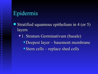
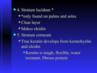
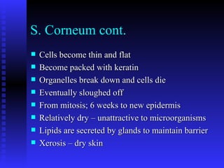
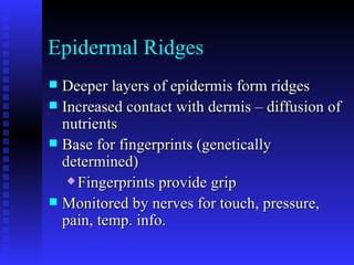
Integumentary System Tissues in combination
Functions of Integument Protection of underlying tissues Excretion of salts, water, organic waste Maintenance of normal body temp Synthesis of vitamin D Storage of nutrients Sensitivity to touch, pressure, pain, temp
Skin is the largest organ of your body – 16% by weight 2 Functional components Cutaneous membrane – layers of tissues Accessory structures – hair, nails, glands Cutaneous membrane has 3 regions Epidermis – epithelial layer Dermis – CT layers Hypodermis – fat layer (deep)
Epidermis Stratified squamous epithelium in 4 (or 5) layers 1. Stratum Germinativum (basale) Deepest layer – basement membrane Stem cells – replace shed cells
2. Stratum spinosum Bound together by desmosomes Several cells thick Spiky looking under microscope 3. Stratum granulosum Grainy layer Make keratohyalin
4. Stratum lucidum * *only found on palms and soles Clear layer Makes eleidin 5. Stratum corneum True keratin develops from keratohyalin and eleidin Keratin is tough, flexible, water resistant, fibrous protein
S. Corneum cont. Cells become thin and flat Become packed with keratin Organelles break down and cells die Eventually sloughed off From mitosis; 6 weeks to new epidermis Relatively dry – unattractive to microorganisms Lipids are secreted by glands to maintain barrier Xerosis – dry skin
Cornification Accumulation of keratin – keratinized  Occurs everywhere except the surface of the eyes Thin skin – 1.5 to 4mm; epidermis is .08mm, about 20 cell layers thick Palms and soles have 30+ layers
Epidermal Ridges Deeper layers of epidermis form ridges Increased contact with dermis – diffusion of nutrients Base for fingerprints (genetically determined) Fingerprints provide grip Monitored by nerves for touch, pressure, pain, temp. info.
UV Radiation Vitamin D synthesis – sterol altered by UV Stresses skin Breaks down underlying CT Wrinkles Skin cancer Destroys folate – folate helps protect against spina bifida Tanning beds are as risky as the sun
Color of epidermis 1. Underlying blood vessels – reddish In dermis 2. Carotene – orange yellow pigment In epithelia 3. Melanin – yellow brown to black In between stem cells
Melanin Secreted by melanocytes, passed to adjacent cells Everyone has approximately the same number of melanocytes; but activity, and color and size of granules of melanin differs Absorbs UV radiation, protects deeper layers Activity increases with exposure to UV Accelerates slowly – maxes about 10 days after  initial exposure
Dermis 1. Papillary (superficial) layer 2. Reticular (deep) layer Both contain fibers, blood vessels, lymphatic vessels, fat cells, muscle cells, nerve endings, and accessory structures
Papillary layer Loose CT Capillaries and nerves that supply epidermis Named for  dermal papillae  (bumps) that project between epidermal ridges
Reticular layer Dense irregular CT Collagen fibers extend into papillary layer Elastin fibers
Lines of Cleavage Bundles of collagen tend to run parallel Different areas of body are oriented differently Surgeons use these lines to minimize scarring, heal faster Stretch marks – bundles that have been pulled apart by rapid weight gain or pregnancy (perpendicular to lines of cleavage)
Hypodermis Subcutaneous layer Adipose tissue Insulates against heat loss Energy reserve Cushioning for kidneys and babies Collagen fibers extend into here from dermis Hypodermic needle introduces drugs into fat layer, slowly enters circulation
Skin cancers Basal cell carcinoma – S. germinativum cells are affected, most common type Squamous cell carcinoma – also epithelial, more deadly than basal cell Melanoma – cancer of melanocytes, most deadly form, more likely from severe burns Size, shape, color Moles – benign tumors of melanocytes
Accessory Structures Hair Found everywhere except: sides and soles of feet, palms of hands, sides of fingers and toes, lips, portions of external genitalia Color – variations in melanin Gray – pigment lessens White – air bubble in shaft
Hair Follicle Tube hair occupies  Deep into dermis  Structure Papilla – bump containing nerves and blood vessels Matrix – epithelium at base of follicle that forms hair, basal cells divide, cornification of outer layers
Hair Structure Medulla – soft core closest to center of papilla; soft keratin Cortex – farther out – hard keratin, stiffness Cuticle – superficial – hard keratin coats hair Root – enclosed by matrix, bulb of follicle Shaft – the hair itself Straight hair – round in cross section Curly hair – flat or oval in cross section
Hair types 1. Vellus hair – peach fuzz 2. Terminal hair – heavy, dark, hair on head, eyebrows, eyelashes, etc. 3. Intermediate hair – hair on arms and legs Hormones change hair from one form to another  Puberty Male pattern baldness
Hair loss may also be caused by  Drugs (chemo) Dietary factors Radiation High fever Stress Pregnancy hormones Hair grows 1/3mm per day Stays in scalp for 2 – 5 years 50 hairs lost per day is normal
Hair Functions Control heat loss Arrector pili – small smooth muscle that pulls on follicle to make hair stand up Increases trapped air insulation Causes goosebumps in humans Reduces friction
Nails Made of keratin at the nail root (not visible) Lunula – pale crescent at base of nail Blood vessels are obscured Protection of finger and toe tips (claws in some animals)
Glands  Sebaceous glands Holocrine glands found along hair follicles Secrete  sebum Inhibits bacterial growth Lubricates and protects hair shaft and skin  Contraction of arrector pili helps squeeze sebum onto skin surface Sebaceous follicles discharge directly onto skin
Sweat glands 1. Apocrine sweat glands – stinky sweat (bacteria that eat this sweat contribute to the smell) Found in armpits, around nipples, groin Associated with hair follicles Send social chemical messages in mammals about sex
2. Merocrine sweat glands – watery sweat (99% water, some salt, waste) Cools the body  “sensible perspiration” Discharge onto skin surface Palms and soles – 3000 per sq. in. Flushes out microorganisms Smaller than apocrine glands
Control of sweat glands Sebaceous and apocrine are hormonally controlled; all on or all off Merocrine are nerve controlled, can be regional  Sweaty palms
Ceruminous glands  Modified sweat glands in ear canal Secretions mix with sebum to make  cerumen  – ear wax Protection Bug repellent
Nerve endings Pain – free nerve endings in epidermis Touch Merkel’s discs – in epidermis Meissner’s corpuscles – in papillae Root hair plexus – around hair at base of follicle Pressure – Pacinian corpuscle – in hypodermis Cold – Krause corpuscle – in dermis, round Heat – Ruffini corpuscle – in dermis, flattened
 

Integumentary system

  • 1.
  • 2.
    Functions of IntegumentProtection of underlying tissues Excretion of salts, water, organic waste Maintenance of normal body temp Synthesis of vitamin D Storage of nutrients Sensitivity to touch, pressure, pain, temp
  • 3.
    Skin is thelargest organ of your body – 16% by weight 2 Functional components Cutaneous membrane – layers of tissues Accessory structures – hair, nails, glands Cutaneous membrane has 3 regions Epidermis – epithelial layer Dermis – CT layers Hypodermis – fat layer (deep)
  • 4.
    Epidermis Stratified squamousepithelium in 4 (or 5) layers 1. Stratum Germinativum (basale) Deepest layer – basement membrane Stem cells – replace shed cells
  • 5.
    2. Stratum spinosumBound together by desmosomes Several cells thick Spiky looking under microscope 3. Stratum granulosum Grainy layer Make keratohyalin
  • 6.
    4. Stratum lucidum* *only found on palms and soles Clear layer Makes eleidin 5. Stratum corneum True keratin develops from keratohyalin and eleidin Keratin is tough, flexible, water resistant, fibrous protein
  • 7.
    S. Corneum cont.Cells become thin and flat Become packed with keratin Organelles break down and cells die Eventually sloughed off From mitosis; 6 weeks to new epidermis Relatively dry – unattractive to microorganisms Lipids are secreted by glands to maintain barrier Xerosis – dry skin
  • 8.
    Cornification Accumulation ofkeratin – keratinized Occurs everywhere except the surface of the eyes Thin skin – 1.5 to 4mm; epidermis is .08mm, about 20 cell layers thick Palms and soles have 30+ layers
  • 9.
    Epidermal Ridges Deeperlayers of epidermis form ridges Increased contact with dermis – diffusion of nutrients Base for fingerprints (genetically determined) Fingerprints provide grip Monitored by nerves for touch, pressure, pain, temp. info.
  • 10.
    UV Radiation VitaminD synthesis – sterol altered by UV Stresses skin Breaks down underlying CT Wrinkles Skin cancer Destroys folate – folate helps protect against spina bifida Tanning beds are as risky as the sun
  • 11.
    Color of epidermis1. Underlying blood vessels – reddish In dermis 2. Carotene – orange yellow pigment In epithelia 3. Melanin – yellow brown to black In between stem cells
  • 12.
    Melanin Secreted bymelanocytes, passed to adjacent cells Everyone has approximately the same number of melanocytes; but activity, and color and size of granules of melanin differs Absorbs UV radiation, protects deeper layers Activity increases with exposure to UV Accelerates slowly – maxes about 10 days after initial exposure
  • 13.
    Dermis 1. Papillary(superficial) layer 2. Reticular (deep) layer Both contain fibers, blood vessels, lymphatic vessels, fat cells, muscle cells, nerve endings, and accessory structures
  • 14.
    Papillary layer LooseCT Capillaries and nerves that supply epidermis Named for dermal papillae (bumps) that project between epidermal ridges
  • 15.
    Reticular layer Denseirregular CT Collagen fibers extend into papillary layer Elastin fibers
  • 16.
    Lines of CleavageBundles of collagen tend to run parallel Different areas of body are oriented differently Surgeons use these lines to minimize scarring, heal faster Stretch marks – bundles that have been pulled apart by rapid weight gain or pregnancy (perpendicular to lines of cleavage)
  • 17.
    Hypodermis Subcutaneous layerAdipose tissue Insulates against heat loss Energy reserve Cushioning for kidneys and babies Collagen fibers extend into here from dermis Hypodermic needle introduces drugs into fat layer, slowly enters circulation
  • 18.
    Skin cancers Basalcell carcinoma – S. germinativum cells are affected, most common type Squamous cell carcinoma – also epithelial, more deadly than basal cell Melanoma – cancer of melanocytes, most deadly form, more likely from severe burns Size, shape, color Moles – benign tumors of melanocytes
  • 19.
    Accessory Structures HairFound everywhere except: sides and soles of feet, palms of hands, sides of fingers and toes, lips, portions of external genitalia Color – variations in melanin Gray – pigment lessens White – air bubble in shaft
  • 20.
    Hair Follicle Tubehair occupies Deep into dermis Structure Papilla – bump containing nerves and blood vessels Matrix – epithelium at base of follicle that forms hair, basal cells divide, cornification of outer layers
  • 21.
    Hair Structure Medulla– soft core closest to center of papilla; soft keratin Cortex – farther out – hard keratin, stiffness Cuticle – superficial – hard keratin coats hair Root – enclosed by matrix, bulb of follicle Shaft – the hair itself Straight hair – round in cross section Curly hair – flat or oval in cross section
  • 22.
    Hair types 1.Vellus hair – peach fuzz 2. Terminal hair – heavy, dark, hair on head, eyebrows, eyelashes, etc. 3. Intermediate hair – hair on arms and legs Hormones change hair from one form to another Puberty Male pattern baldness
  • 23.
    Hair loss mayalso be caused by Drugs (chemo) Dietary factors Radiation High fever Stress Pregnancy hormones Hair grows 1/3mm per day Stays in scalp for 2 – 5 years 50 hairs lost per day is normal
  • 24.
    Hair Functions Controlheat loss Arrector pili – small smooth muscle that pulls on follicle to make hair stand up Increases trapped air insulation Causes goosebumps in humans Reduces friction
  • 25.
    Nails Made ofkeratin at the nail root (not visible) Lunula – pale crescent at base of nail Blood vessels are obscured Protection of finger and toe tips (claws in some animals)
  • 26.
    Glands Sebaceousglands Holocrine glands found along hair follicles Secrete sebum Inhibits bacterial growth Lubricates and protects hair shaft and skin Contraction of arrector pili helps squeeze sebum onto skin surface Sebaceous follicles discharge directly onto skin
  • 27.
    Sweat glands 1.Apocrine sweat glands – stinky sweat (bacteria that eat this sweat contribute to the smell) Found in armpits, around nipples, groin Associated with hair follicles Send social chemical messages in mammals about sex
  • 28.
    2. Merocrine sweatglands – watery sweat (99% water, some salt, waste) Cools the body “sensible perspiration” Discharge onto skin surface Palms and soles – 3000 per sq. in. Flushes out microorganisms Smaller than apocrine glands
  • 29.
    Control of sweatglands Sebaceous and apocrine are hormonally controlled; all on or all off Merocrine are nerve controlled, can be regional Sweaty palms
  • 30.
    Ceruminous glands Modified sweat glands in ear canal Secretions mix with sebum to make cerumen – ear wax Protection Bug repellent
  • 31.
    Nerve endings Pain– free nerve endings in epidermis Touch Merkel’s discs – in epidermis Meissner’s corpuscles – in papillae Root hair plexus – around hair at base of follicle Pressure – Pacinian corpuscle – in hypodermis Cold – Krause corpuscle – in dermis, round Heat – Ruffini corpuscle – in dermis, flattened
  • 32.