Downloaded 449 times
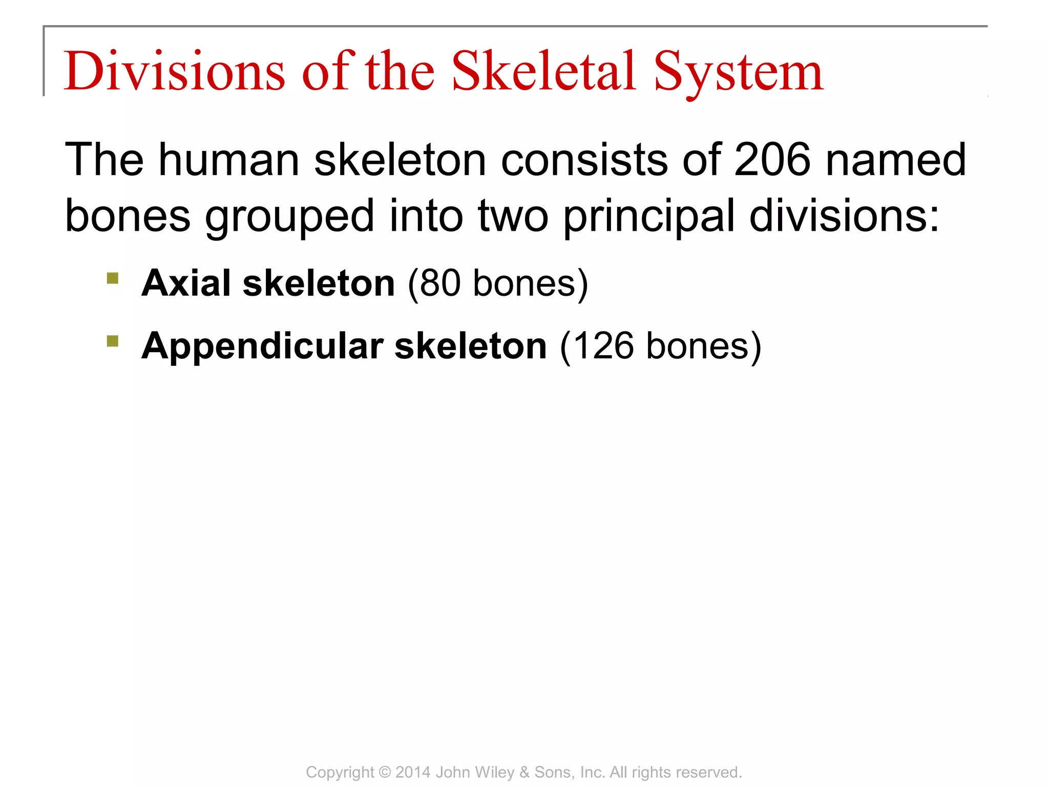
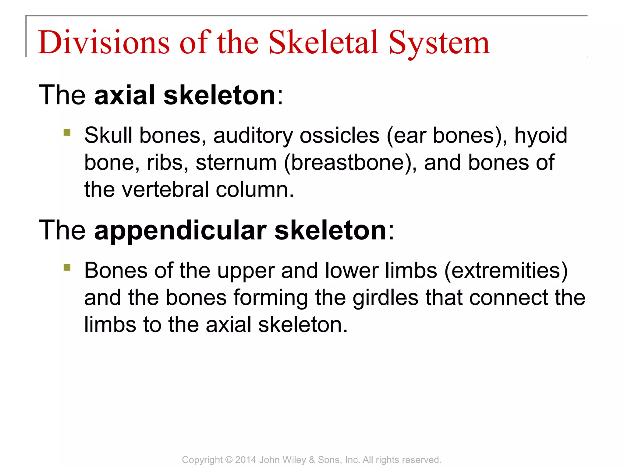

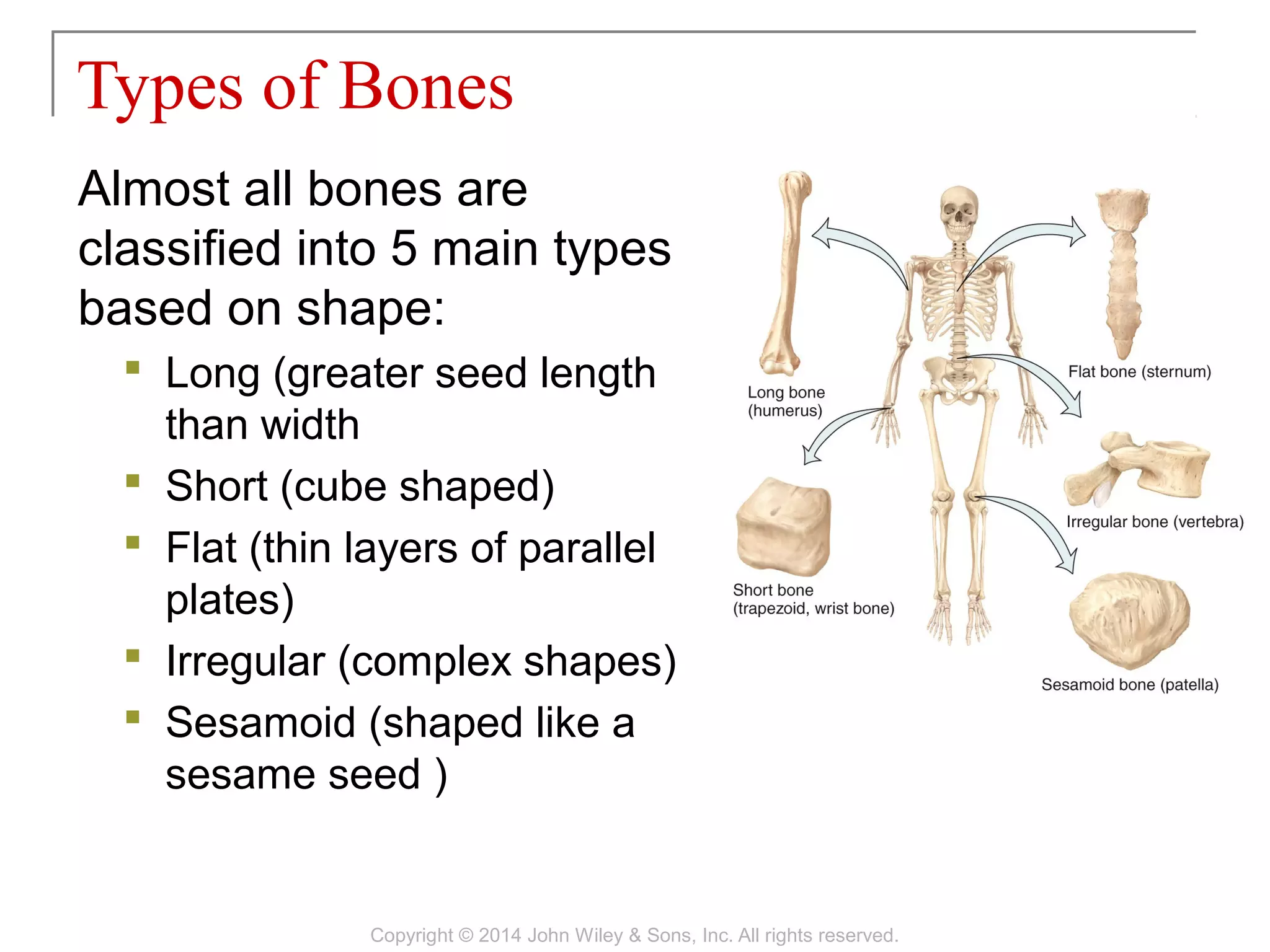
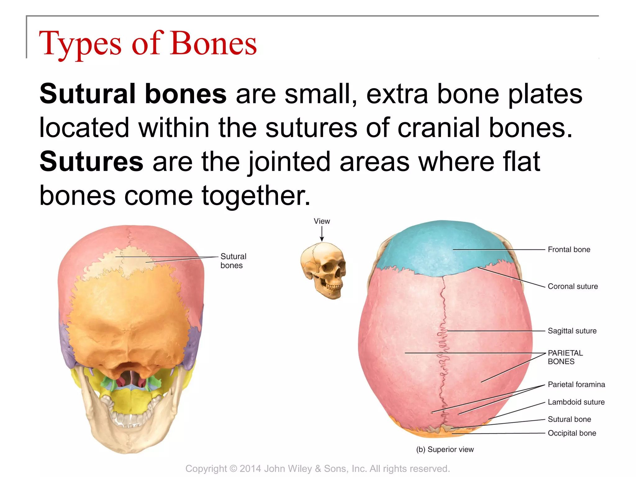

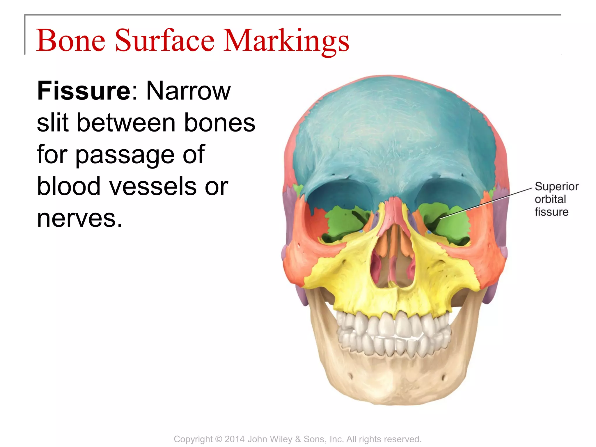
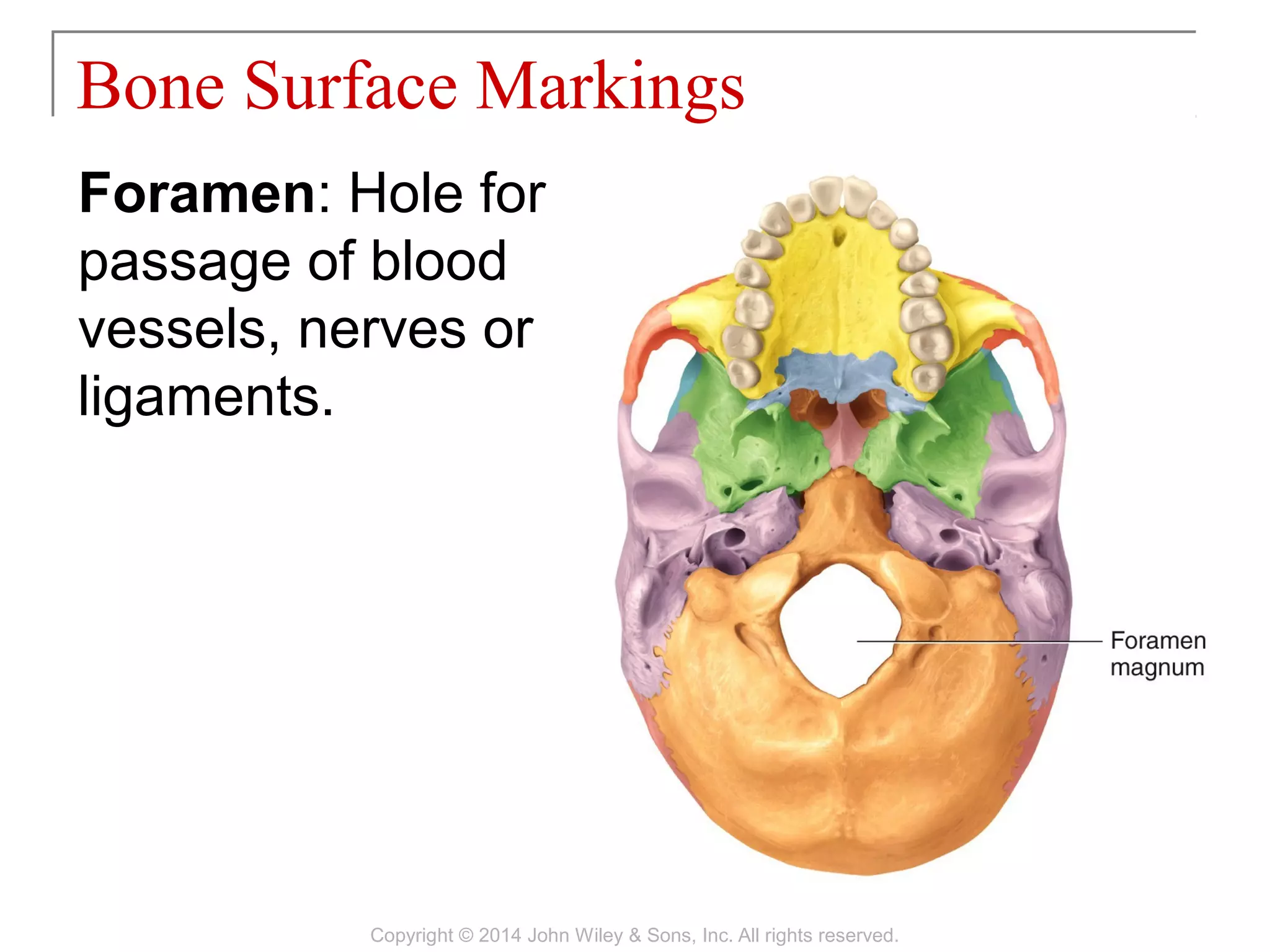
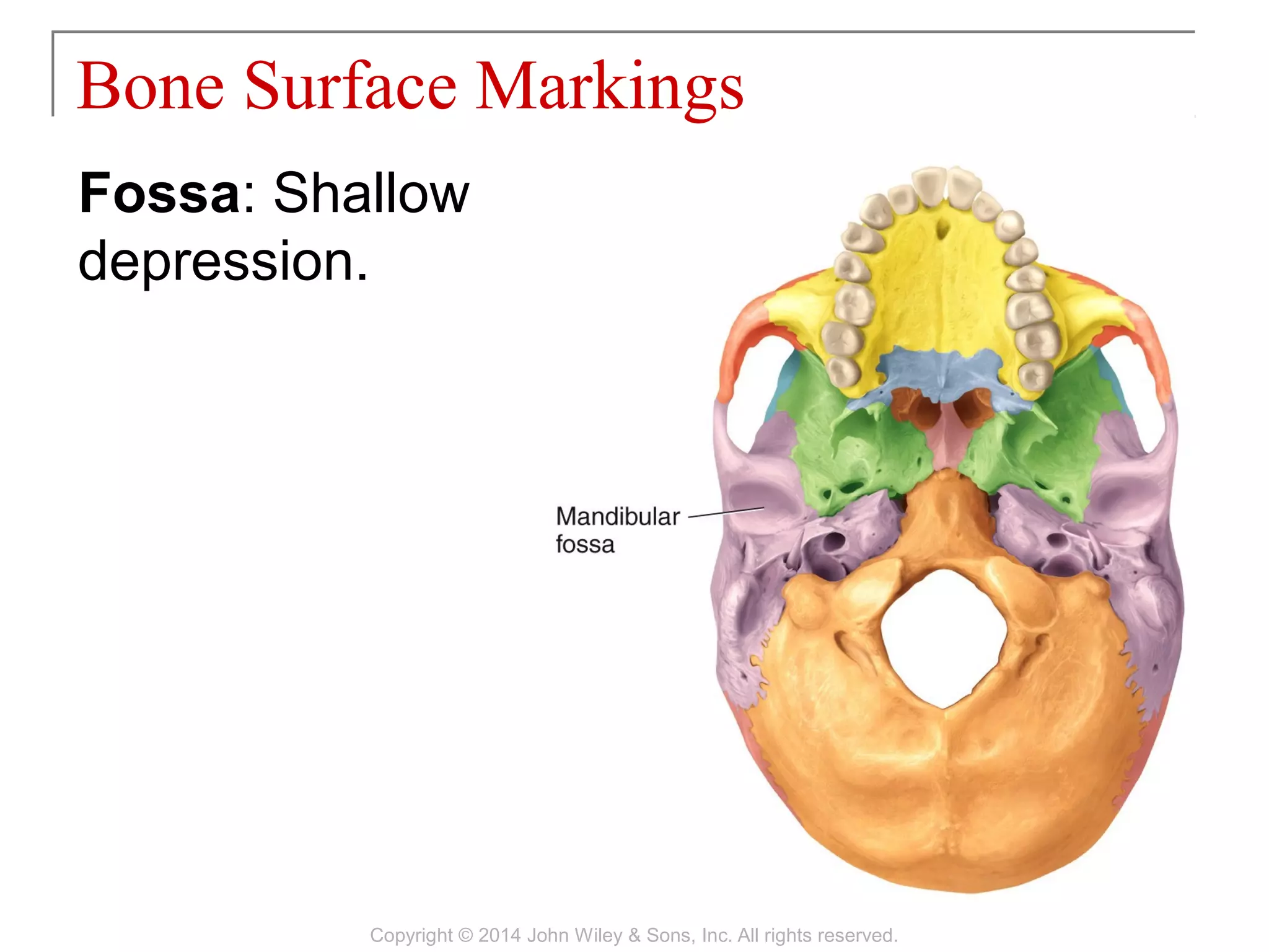
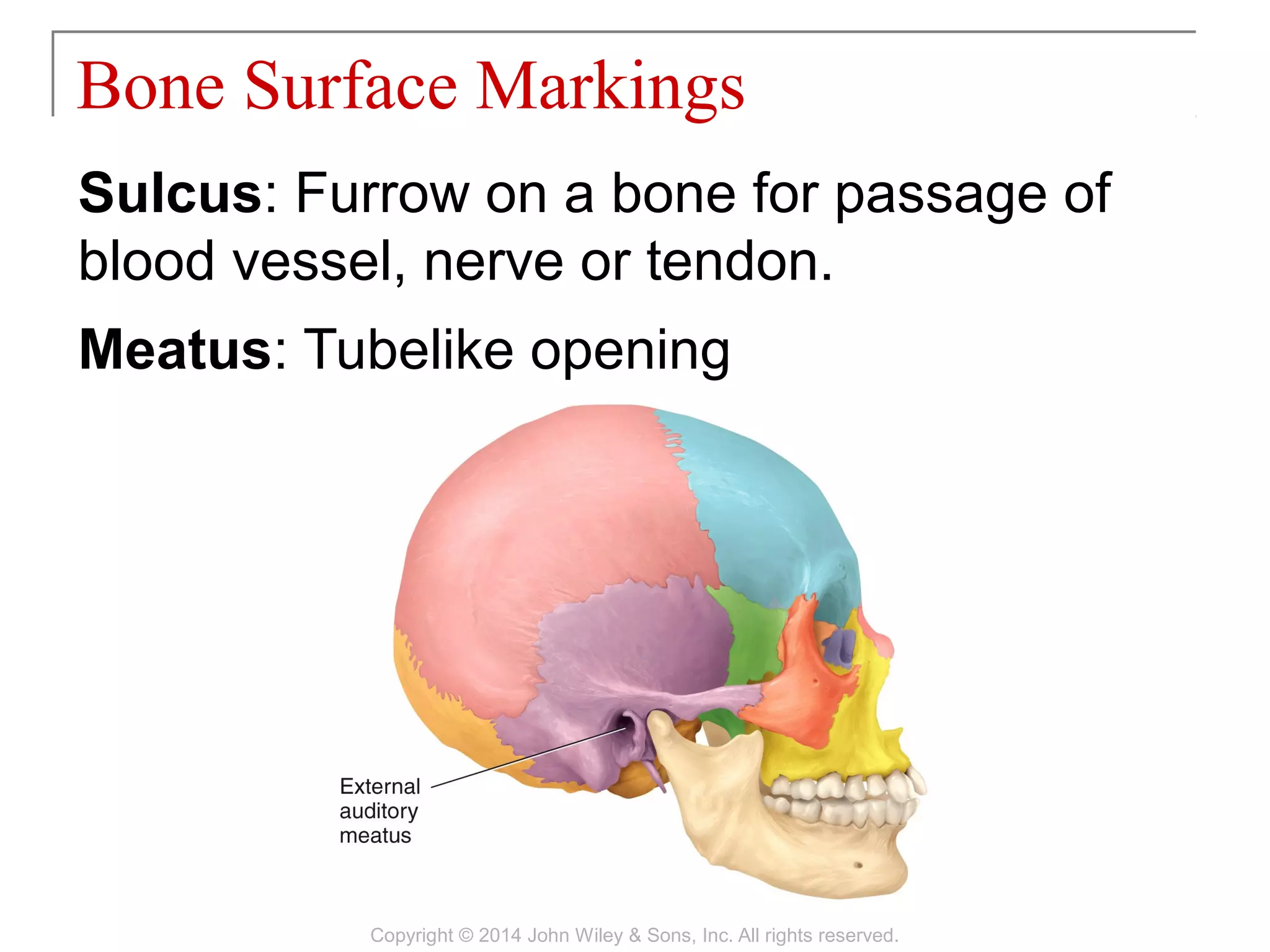
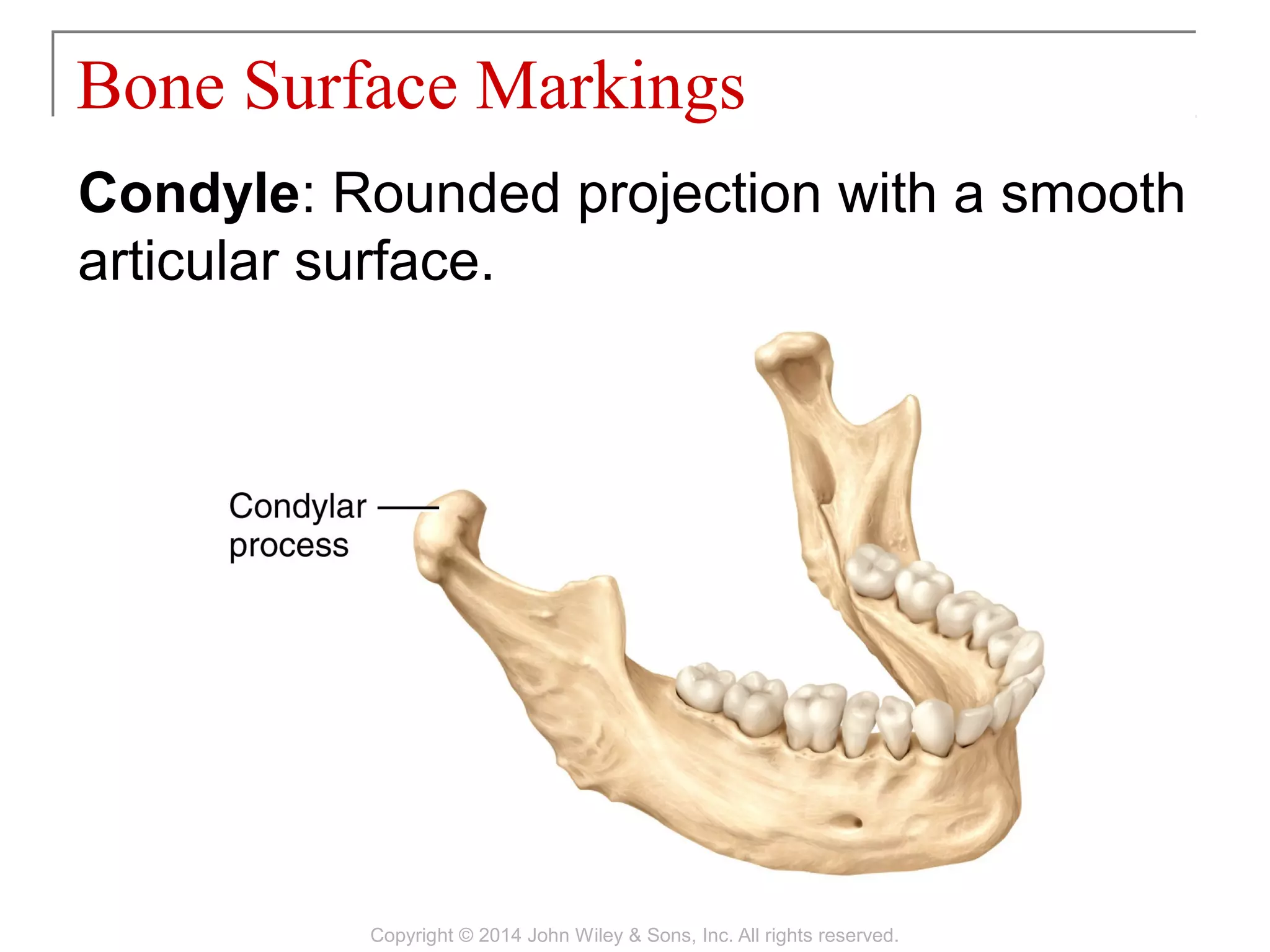
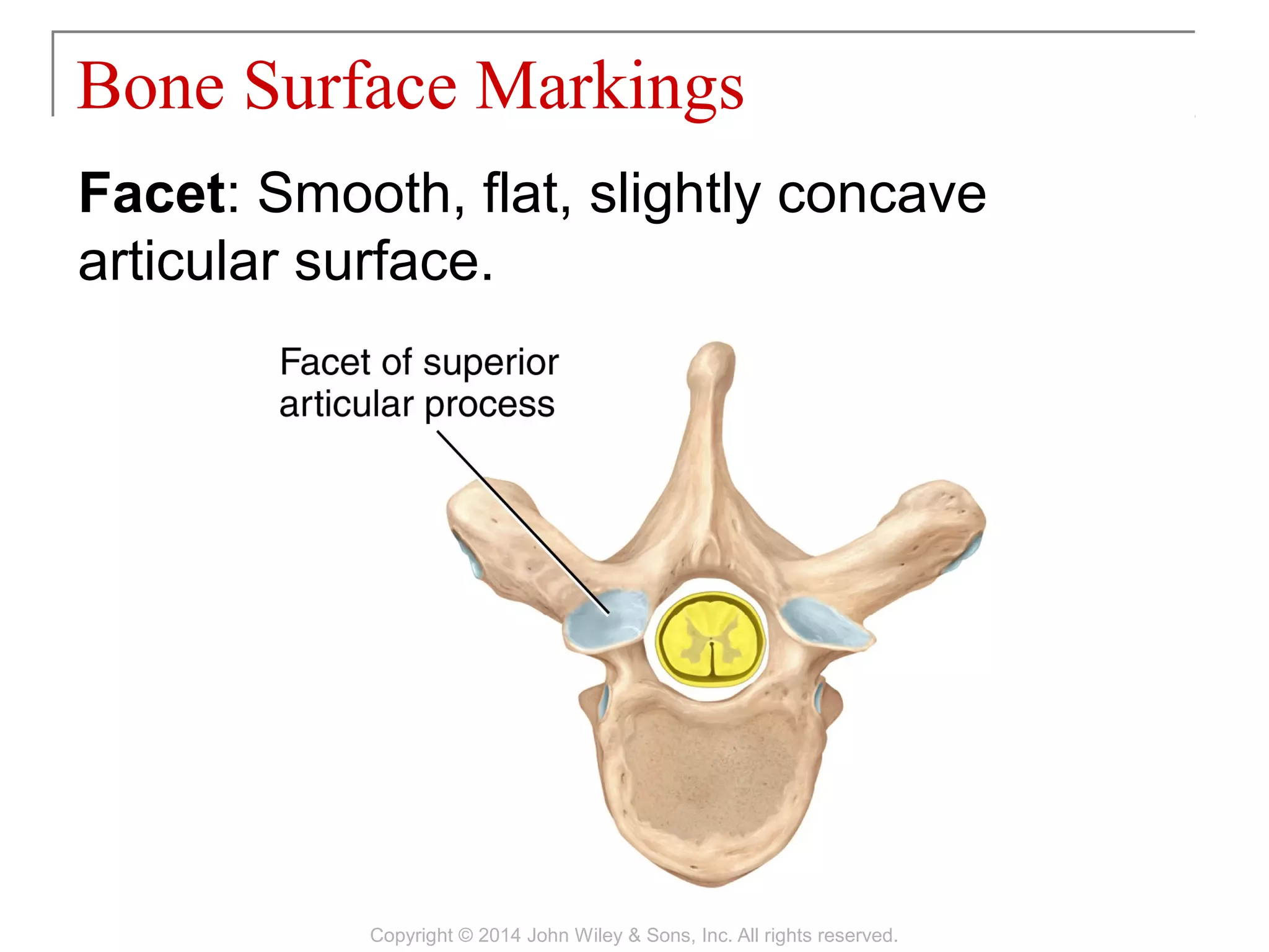
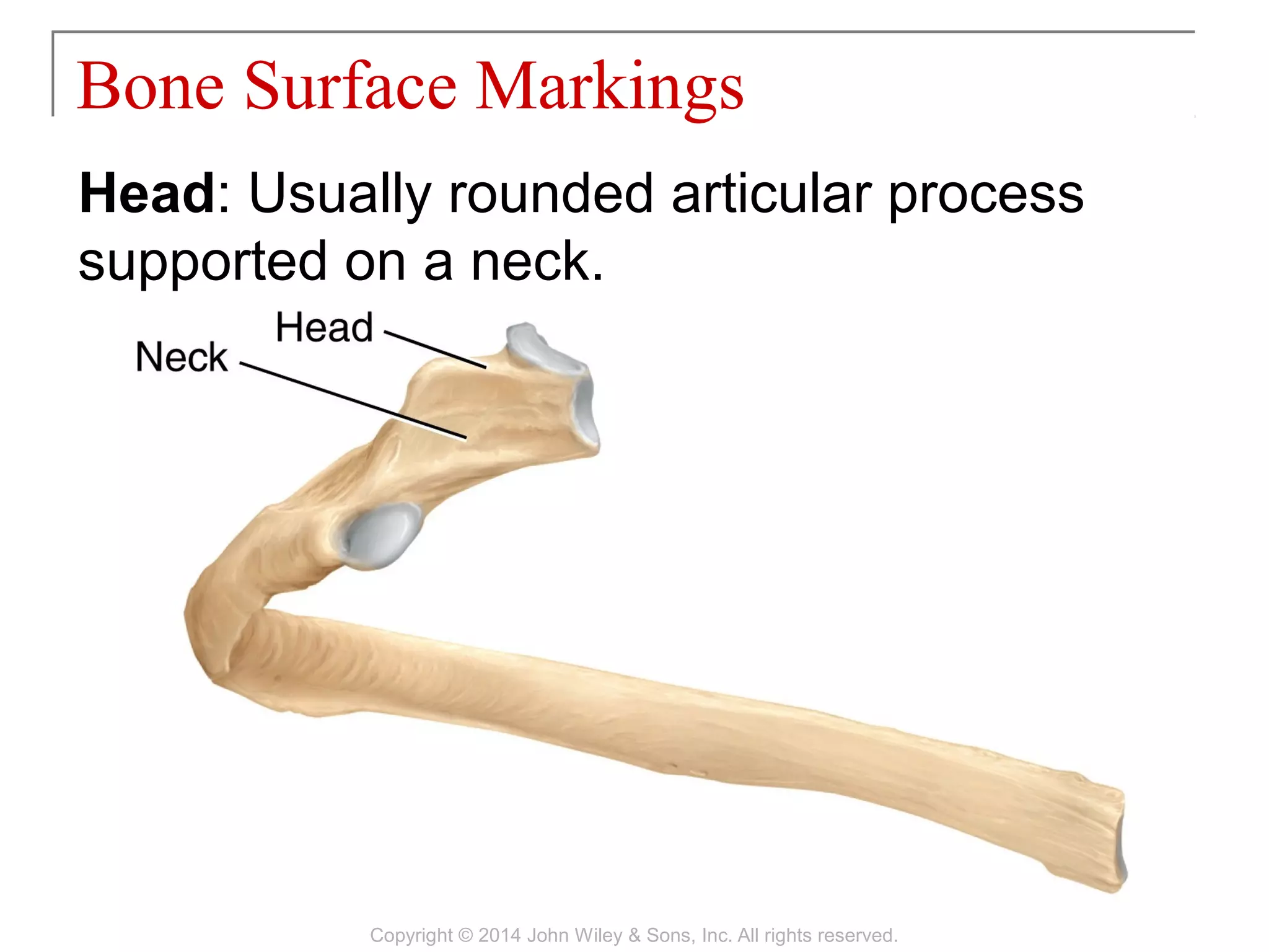

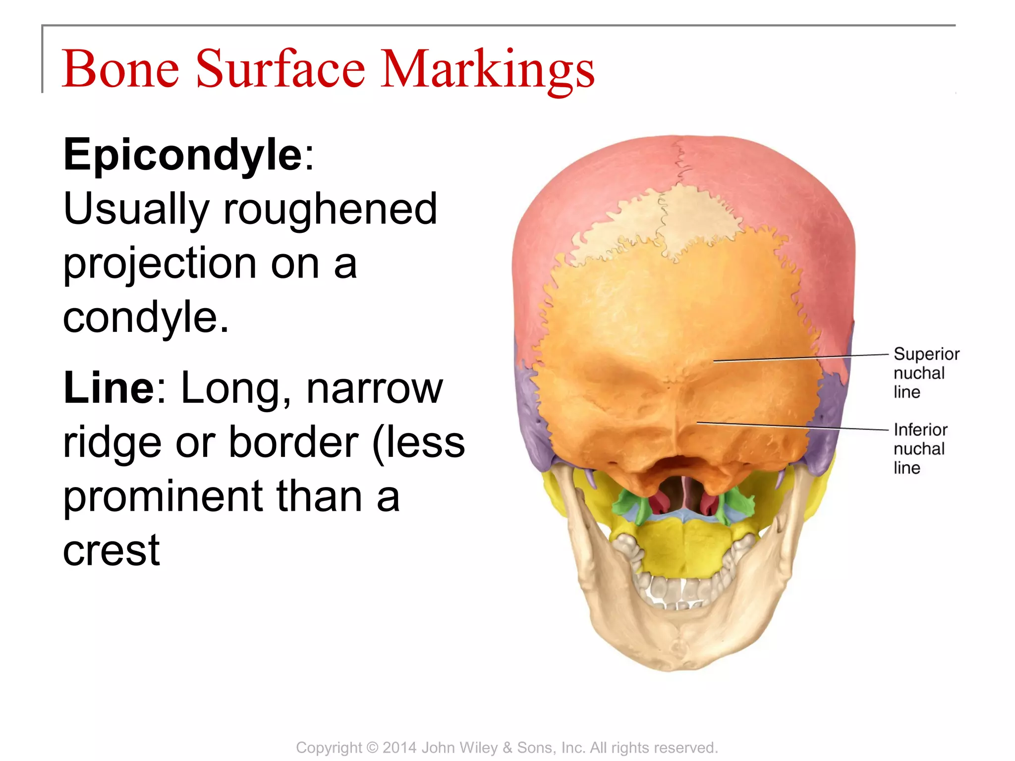
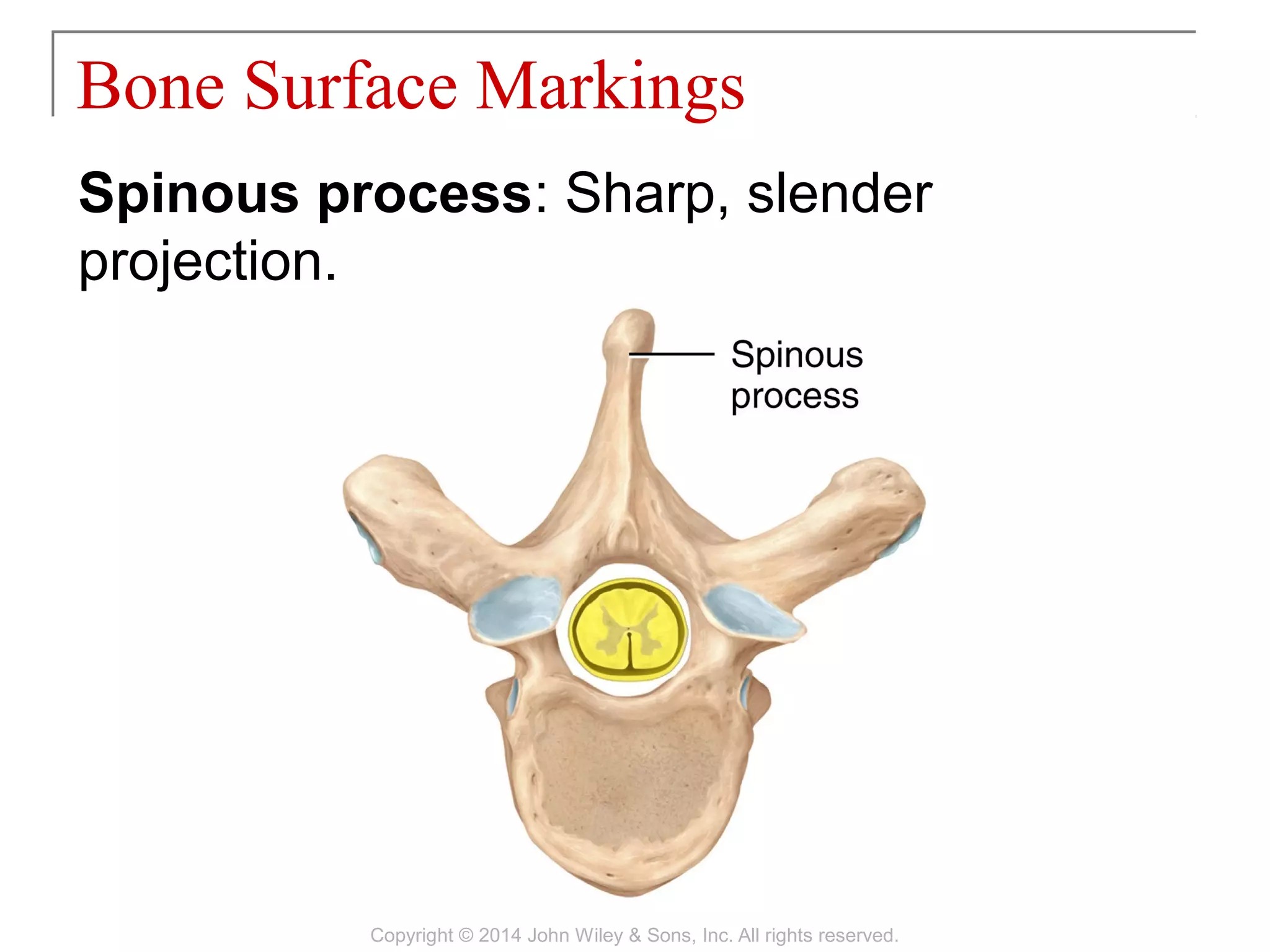
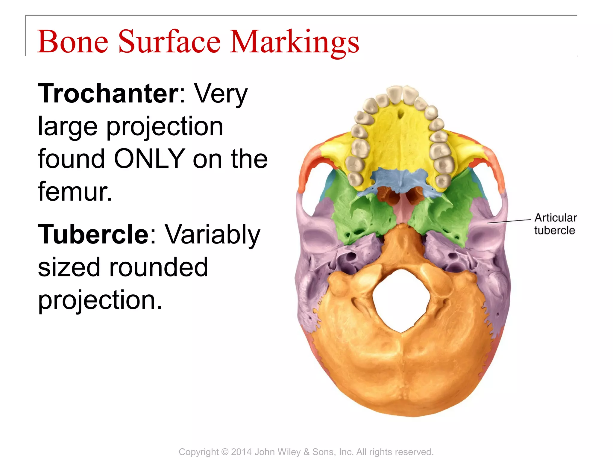
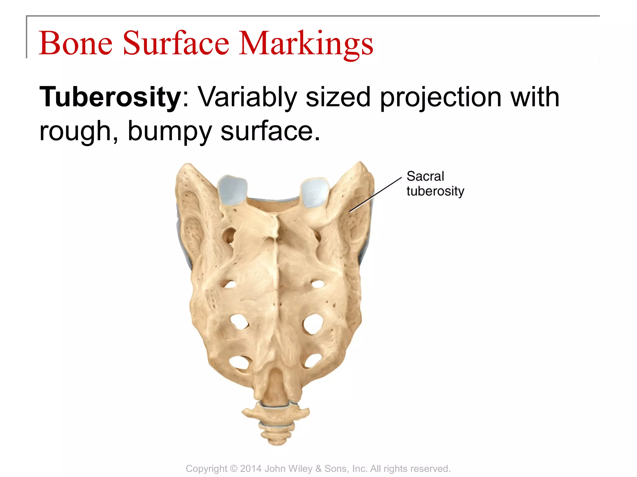
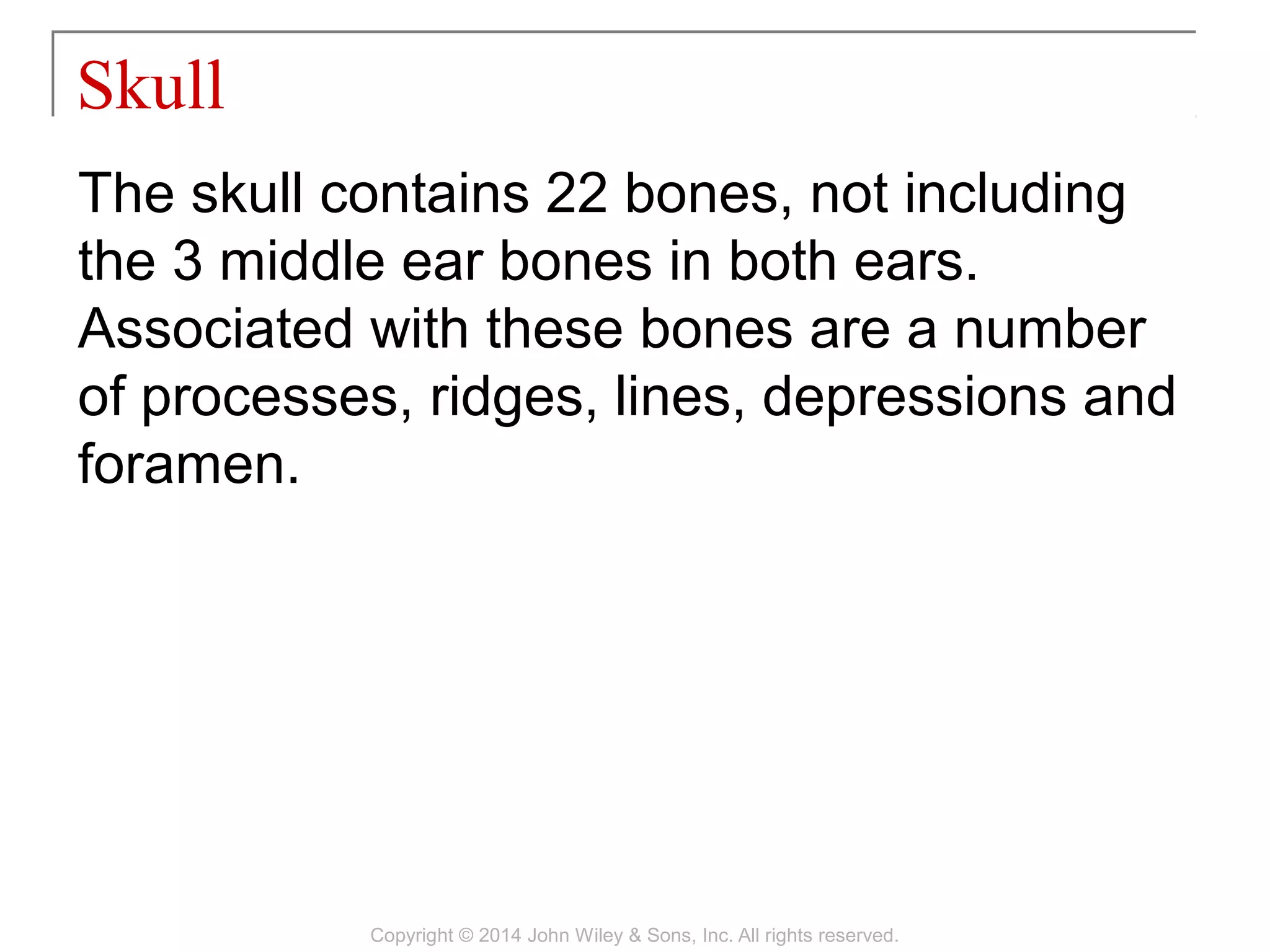
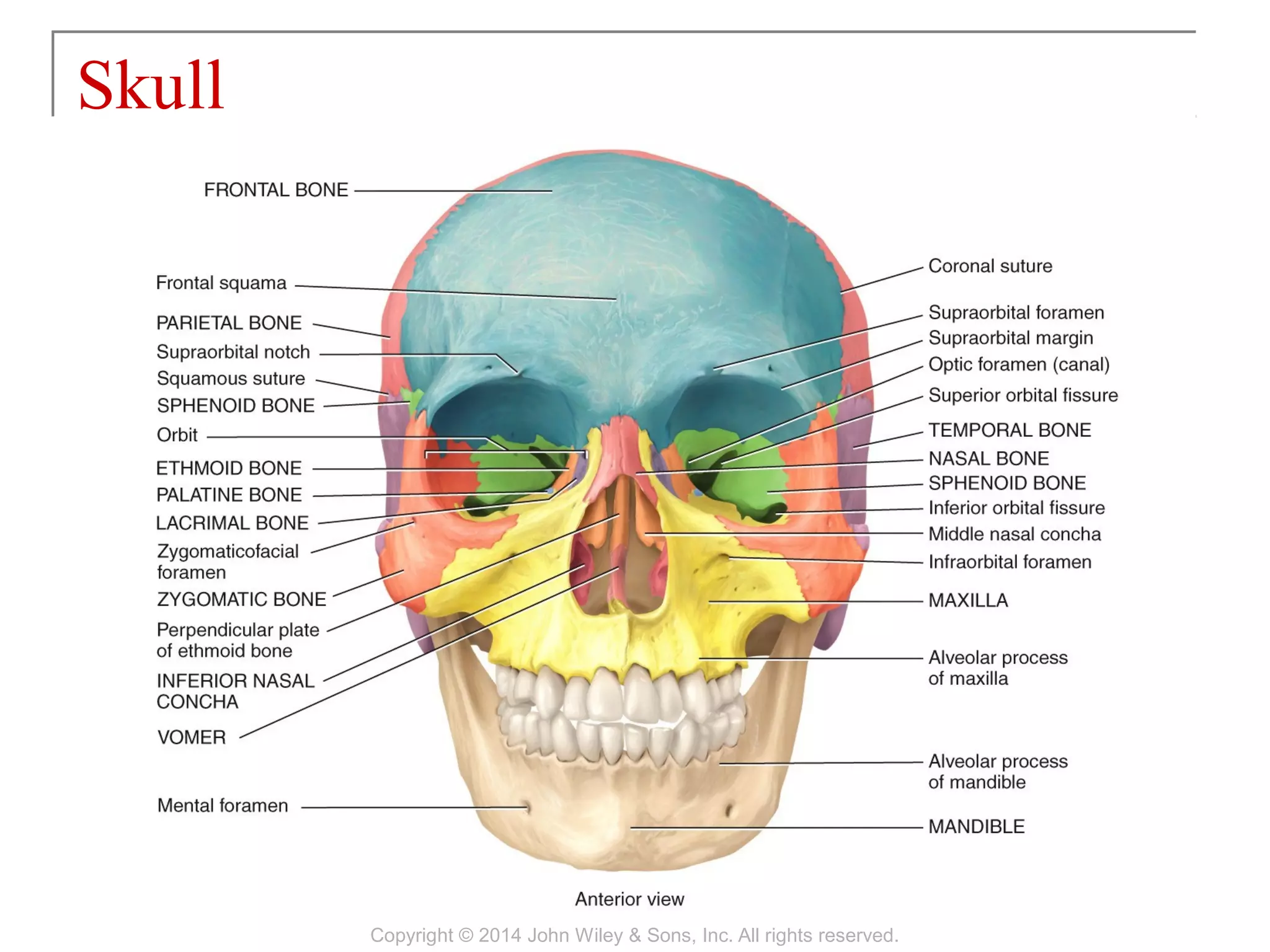

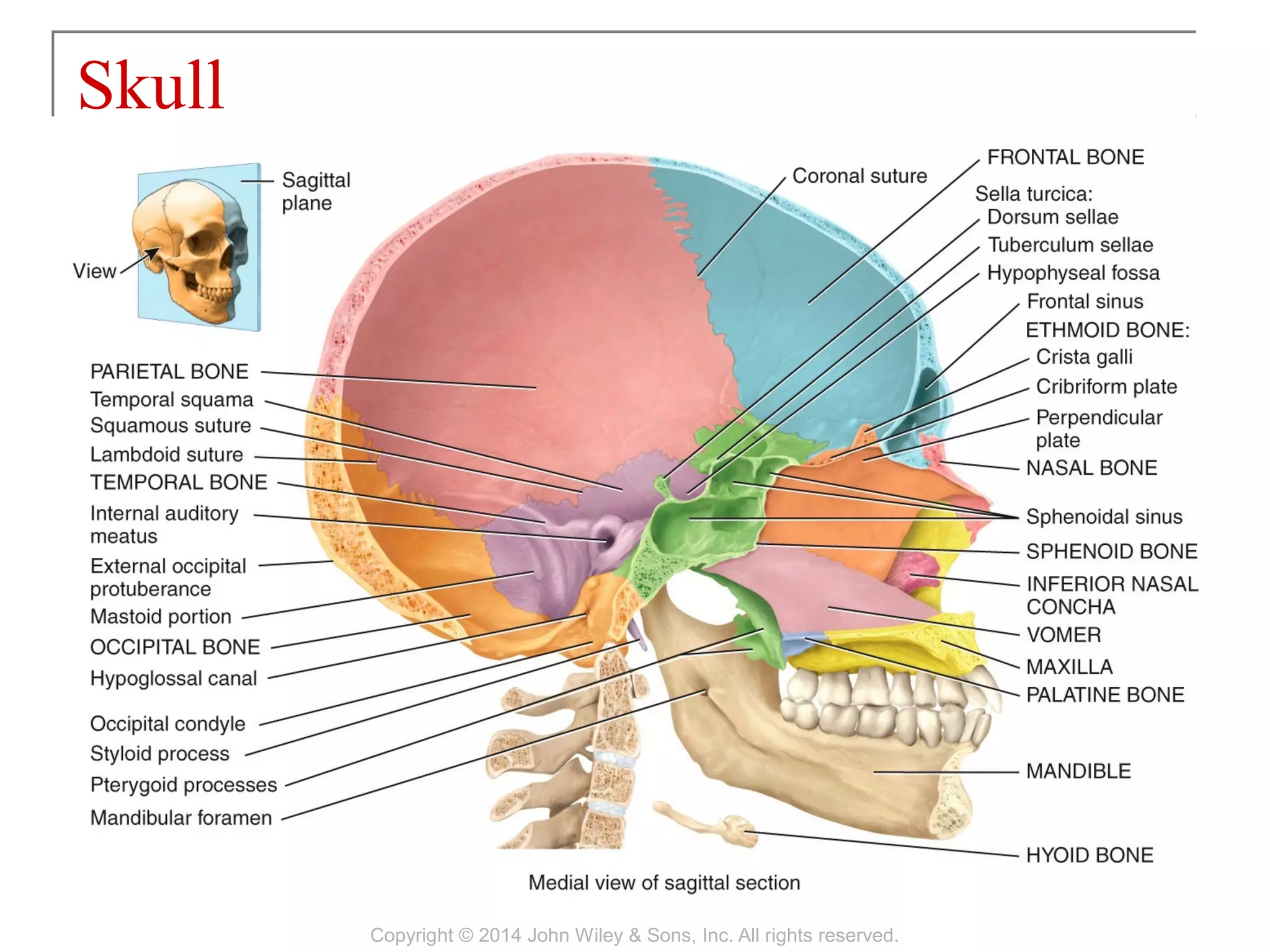
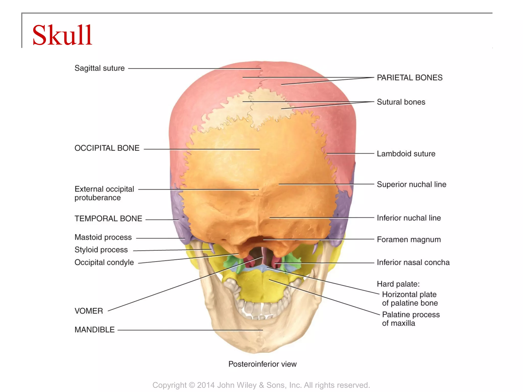
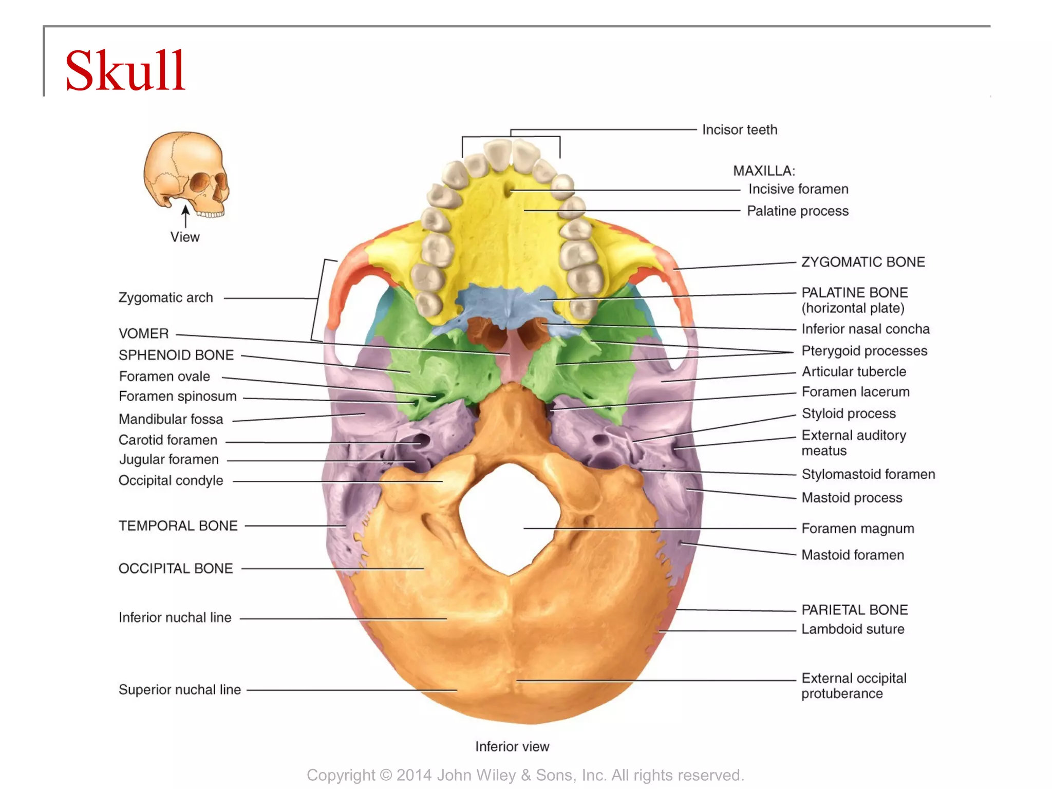
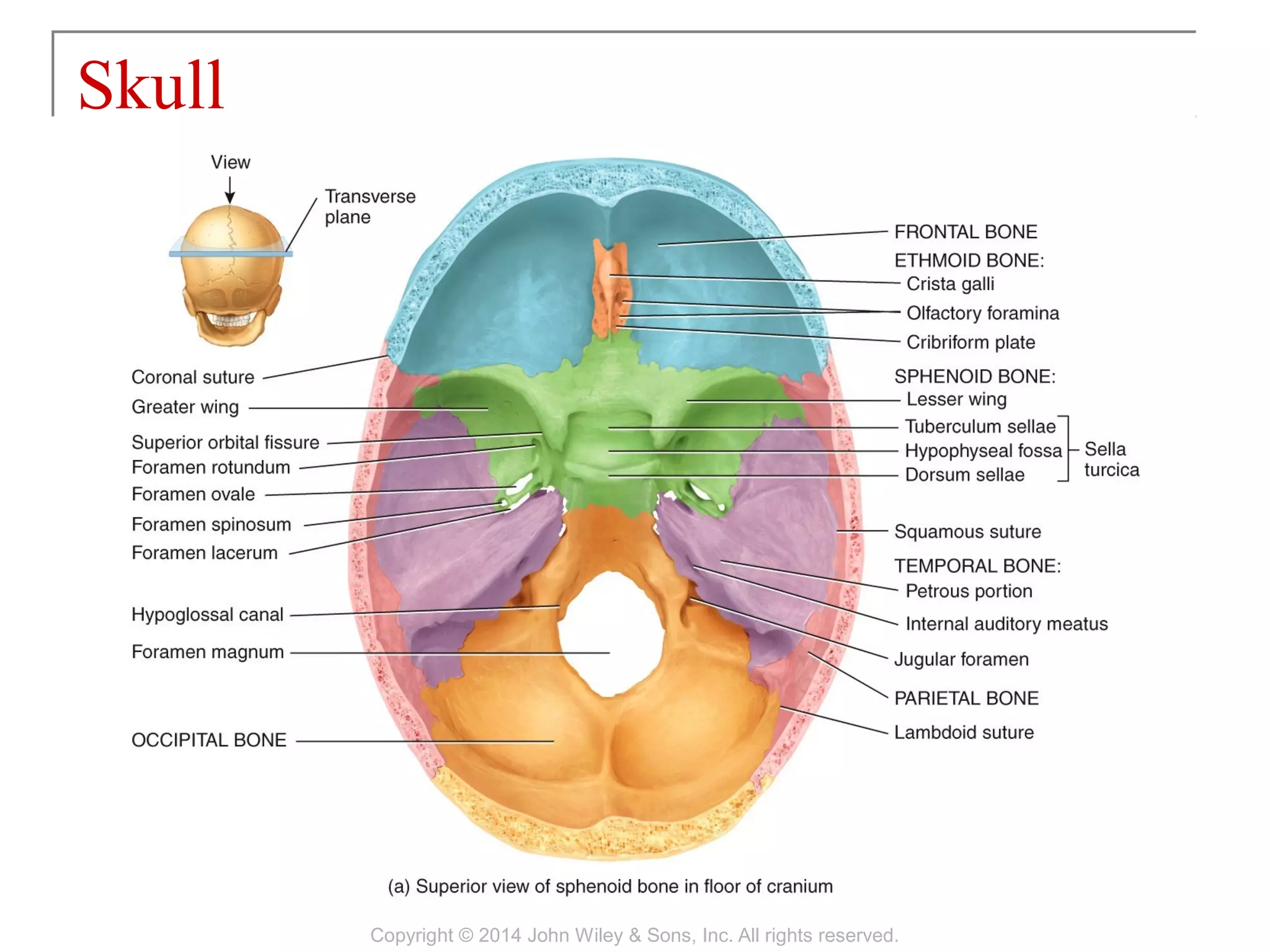


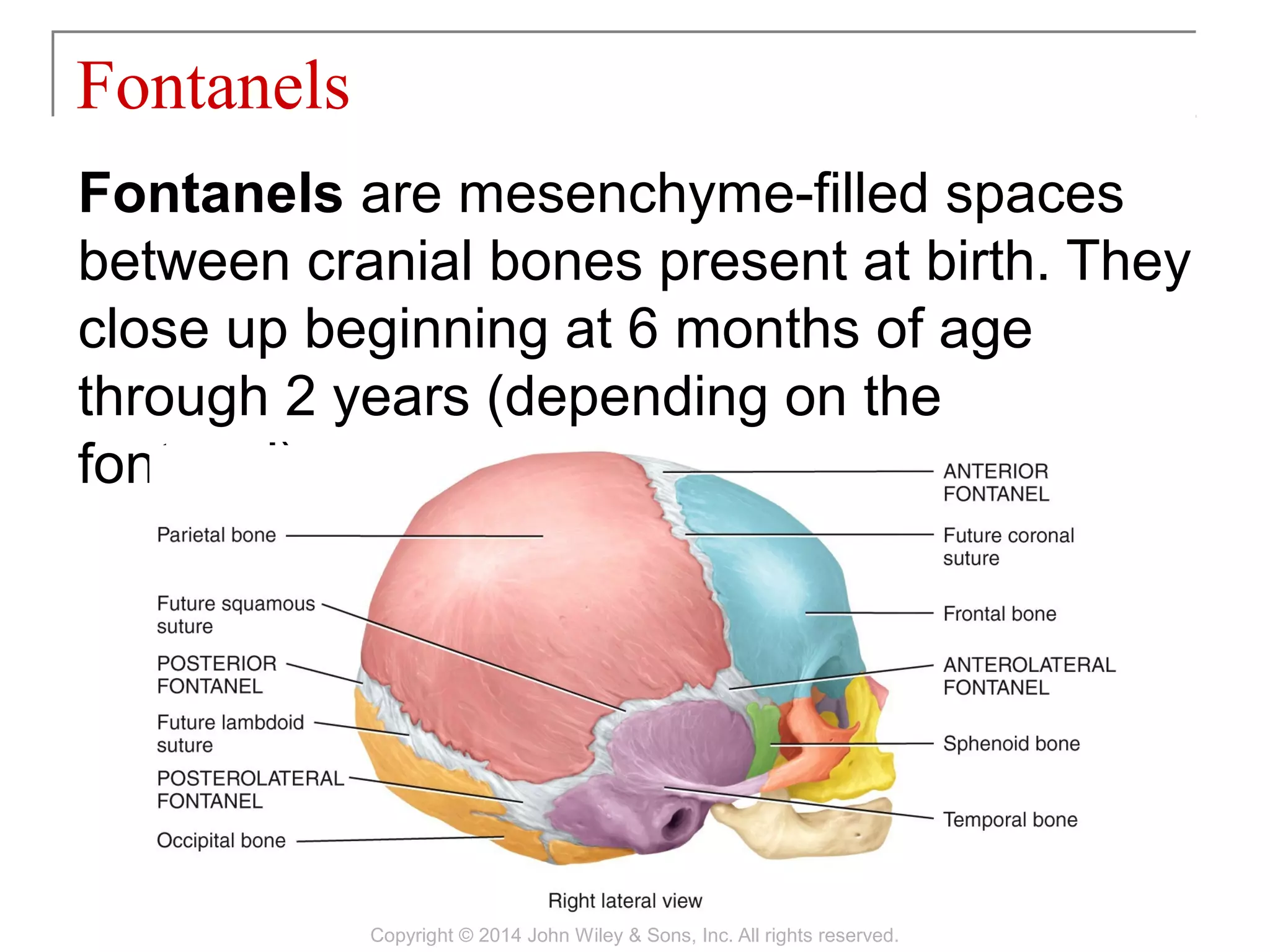
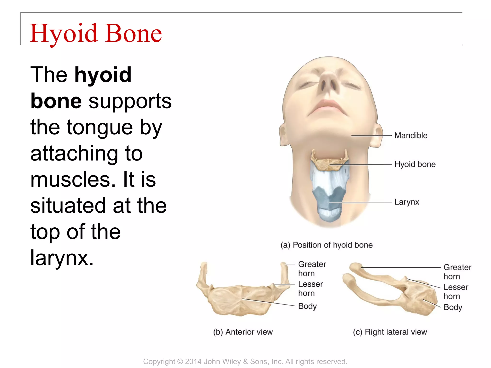


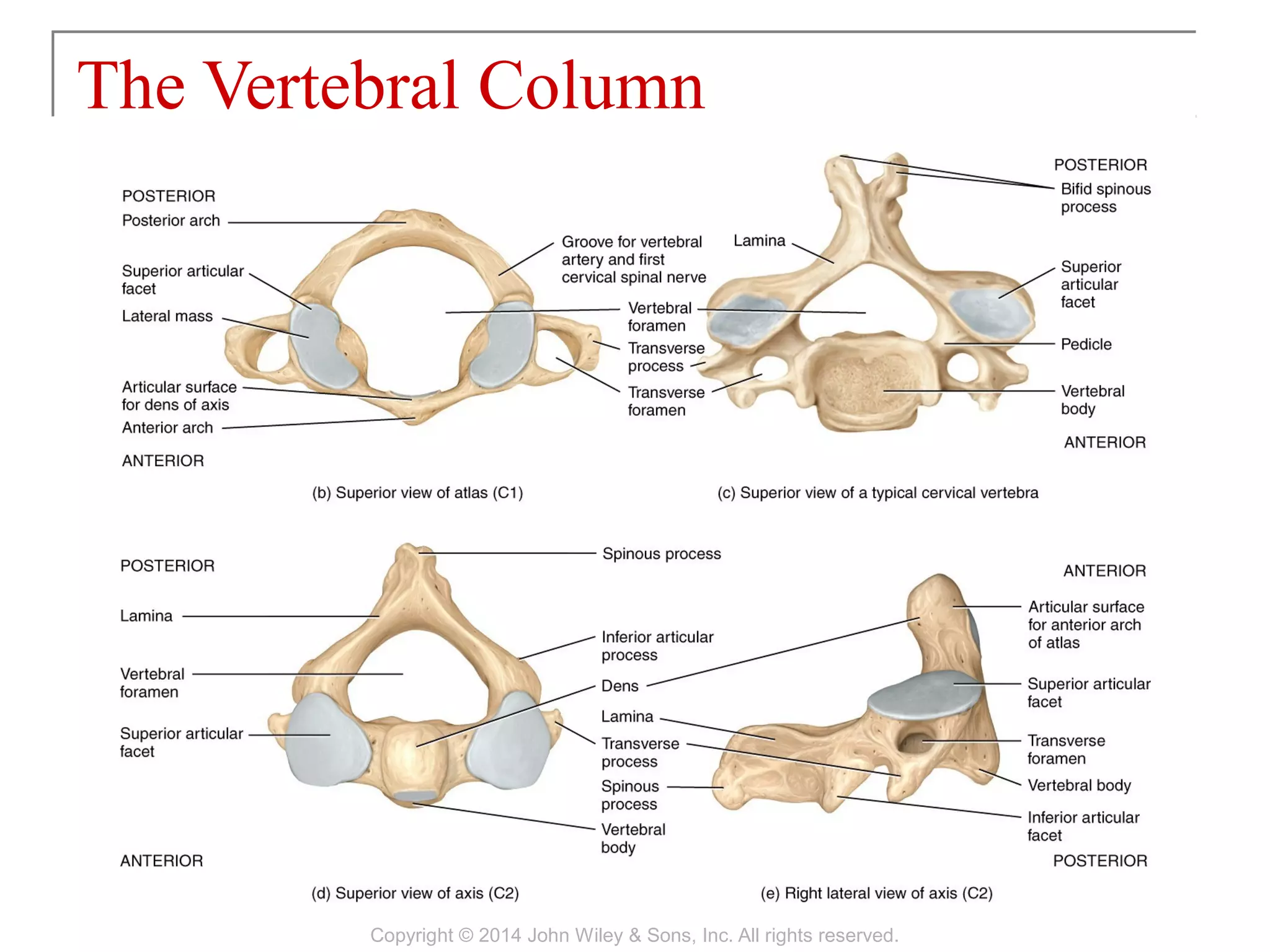
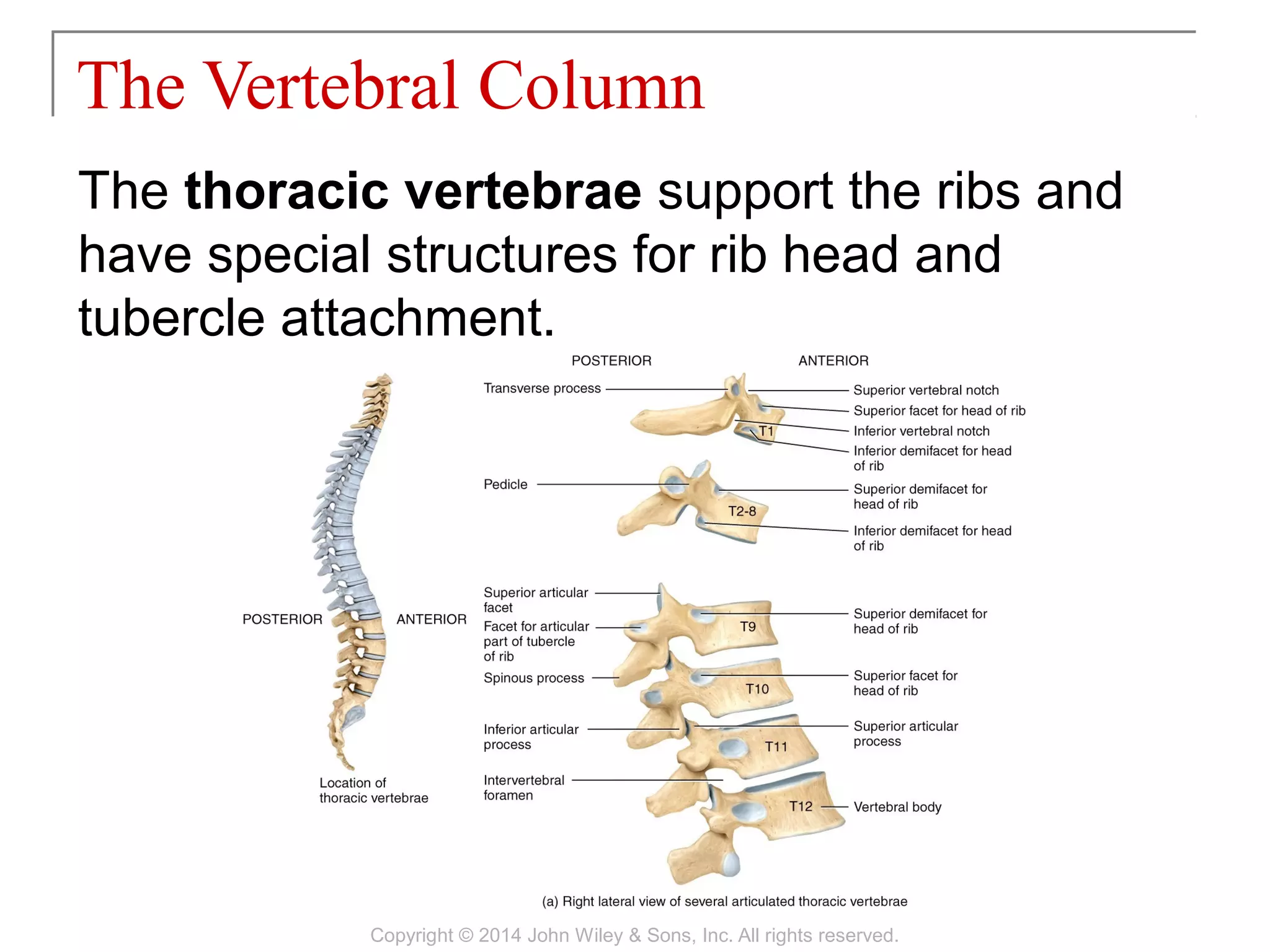
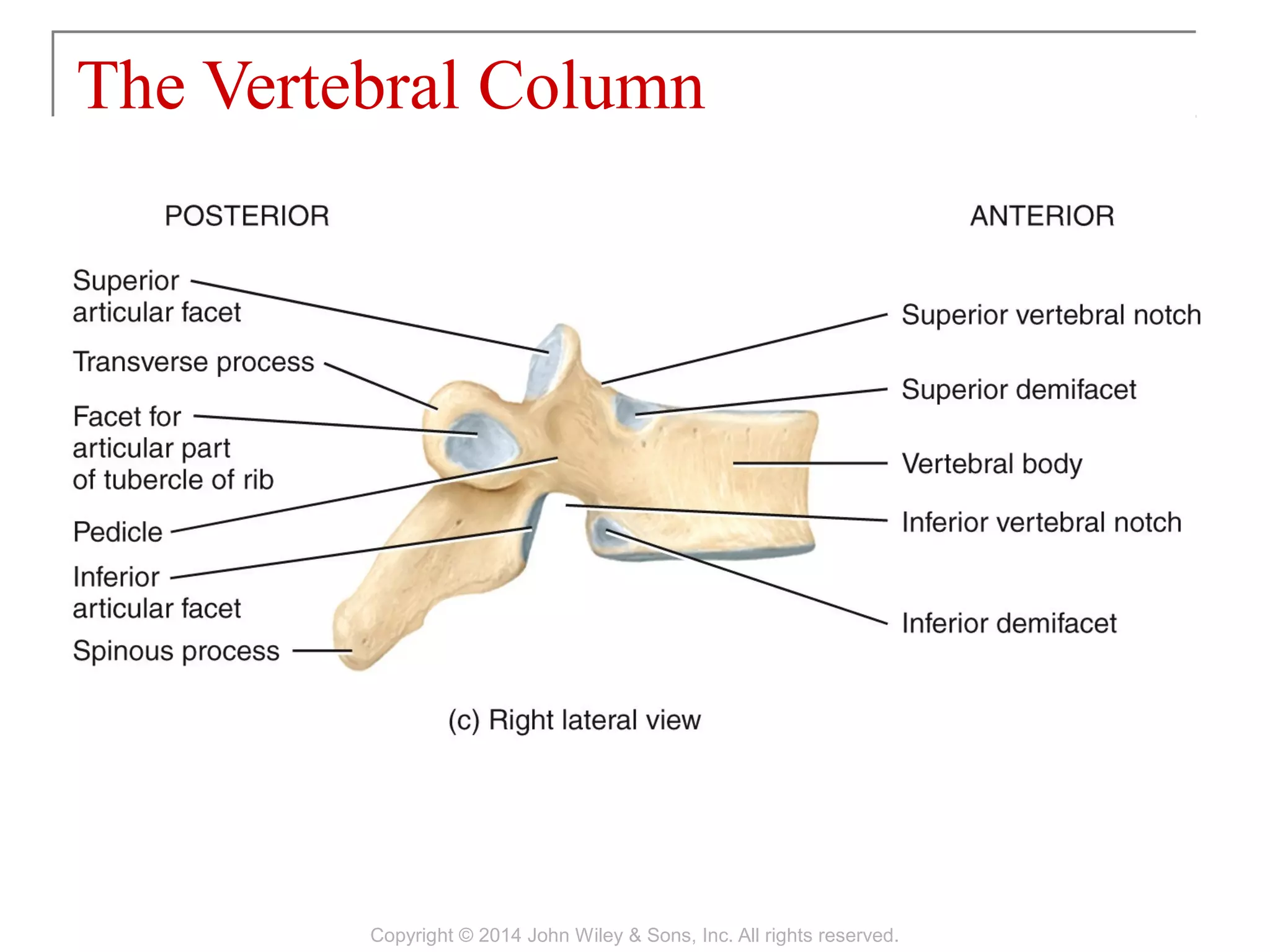


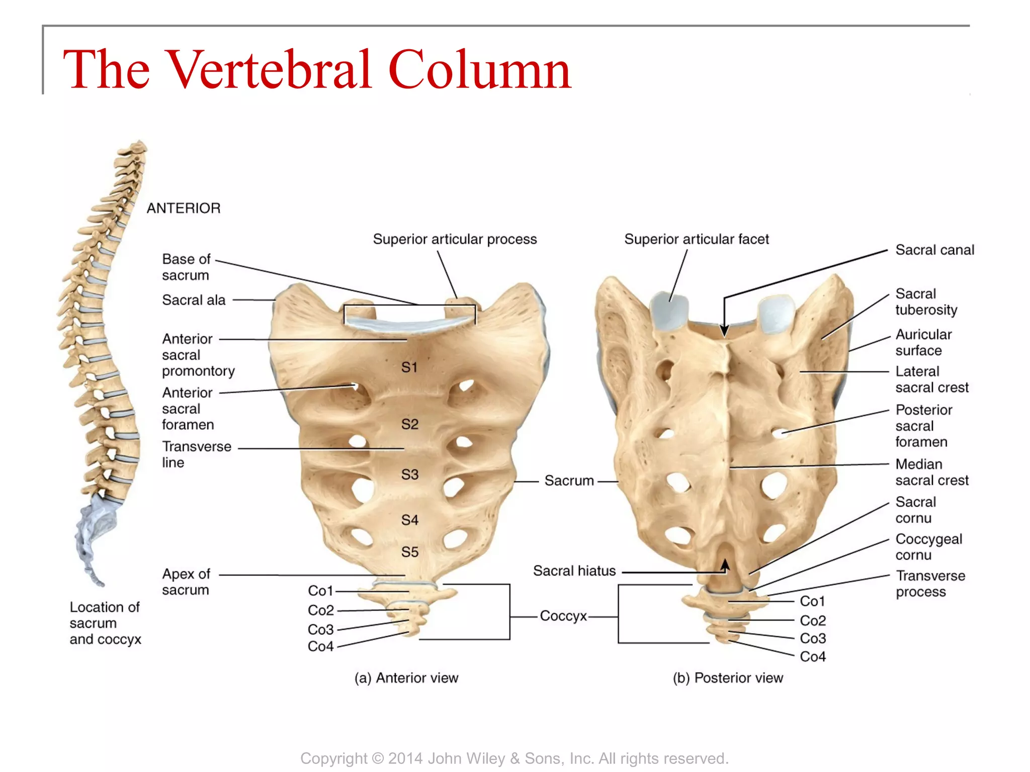
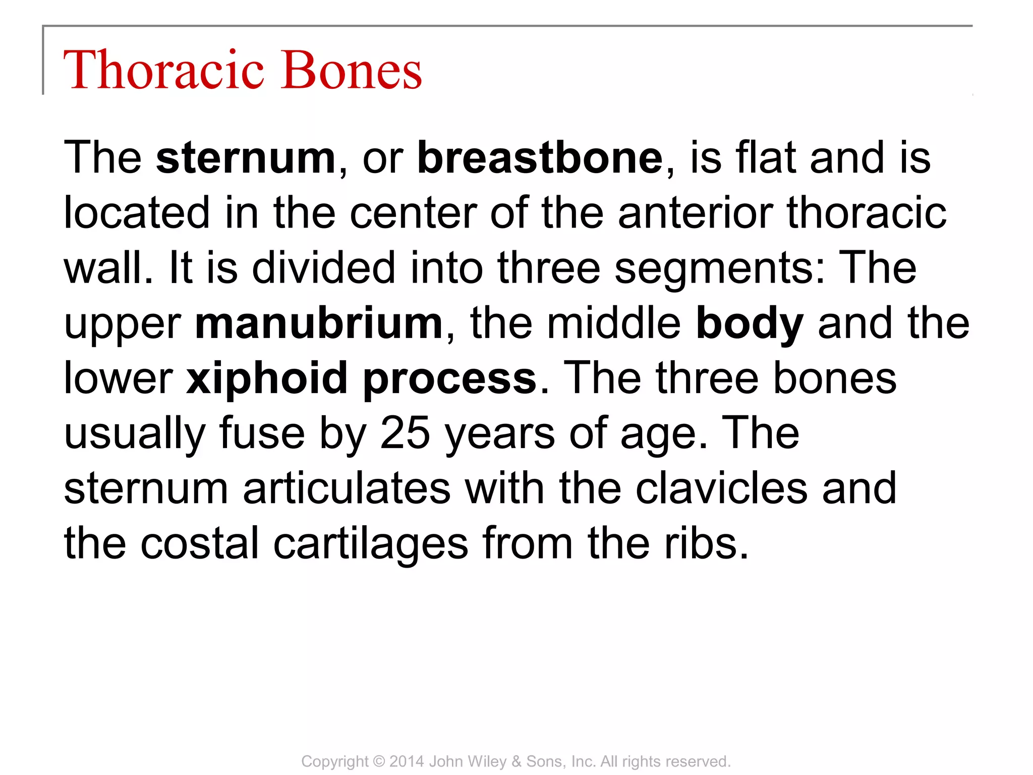
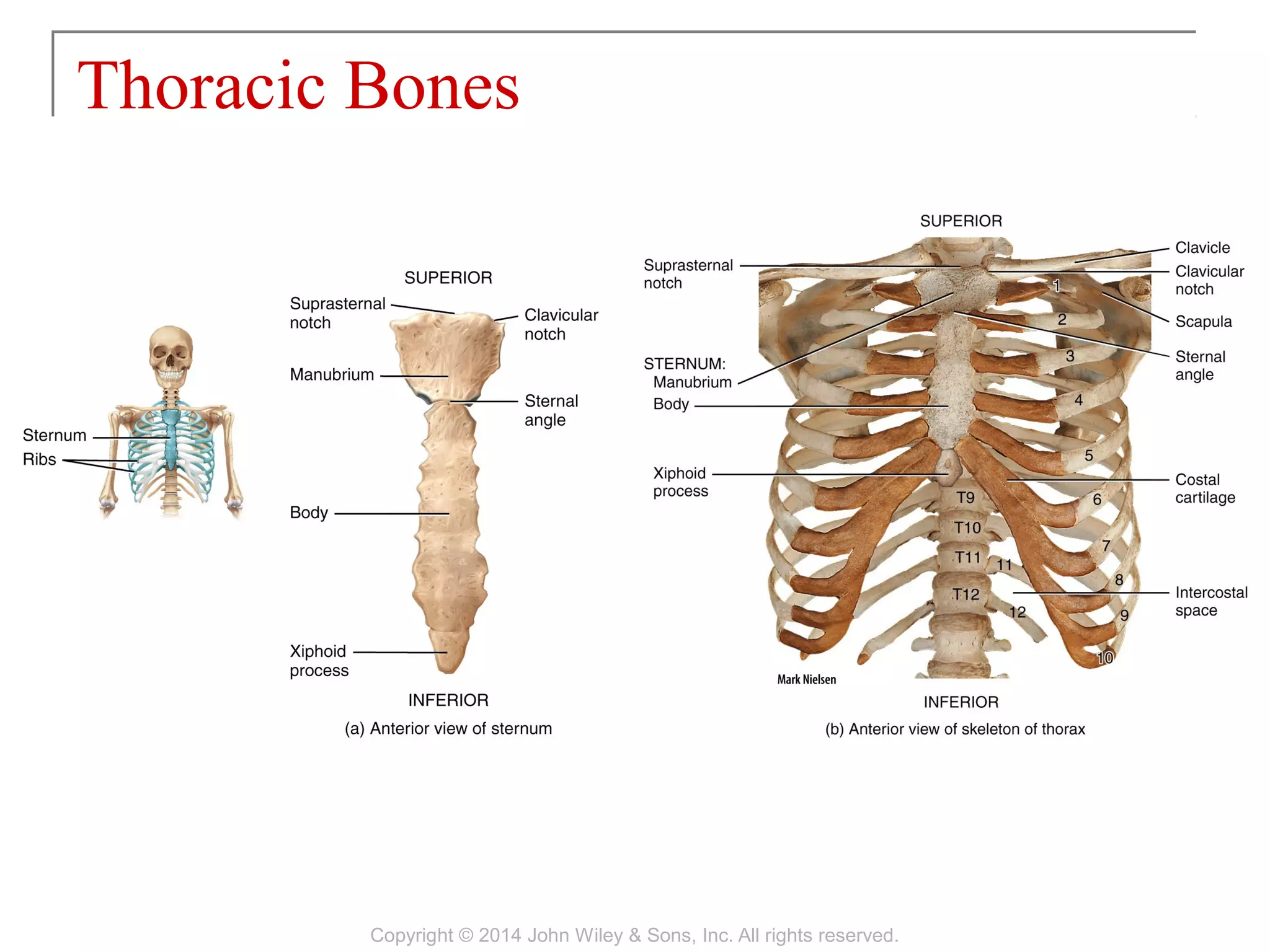

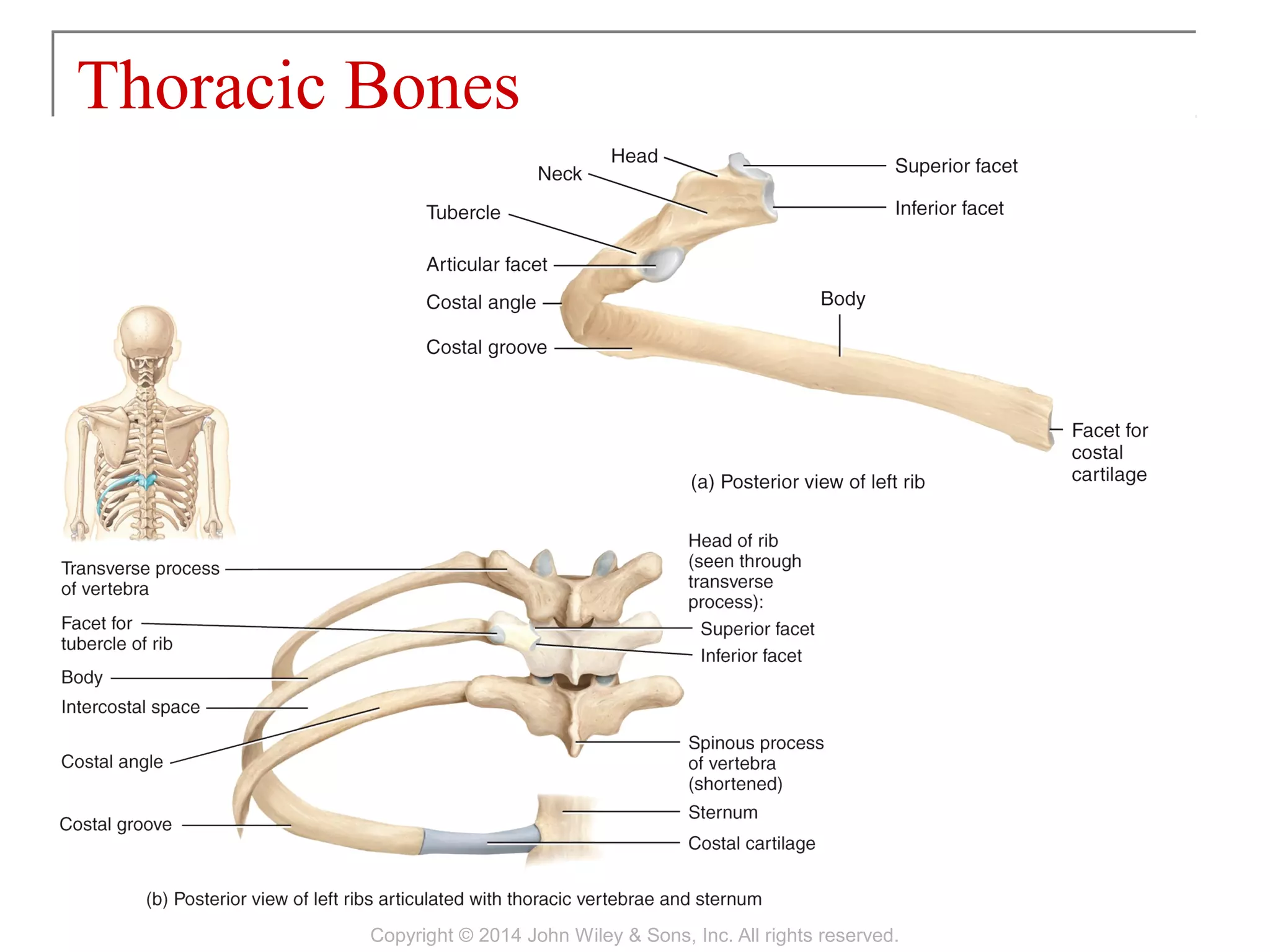
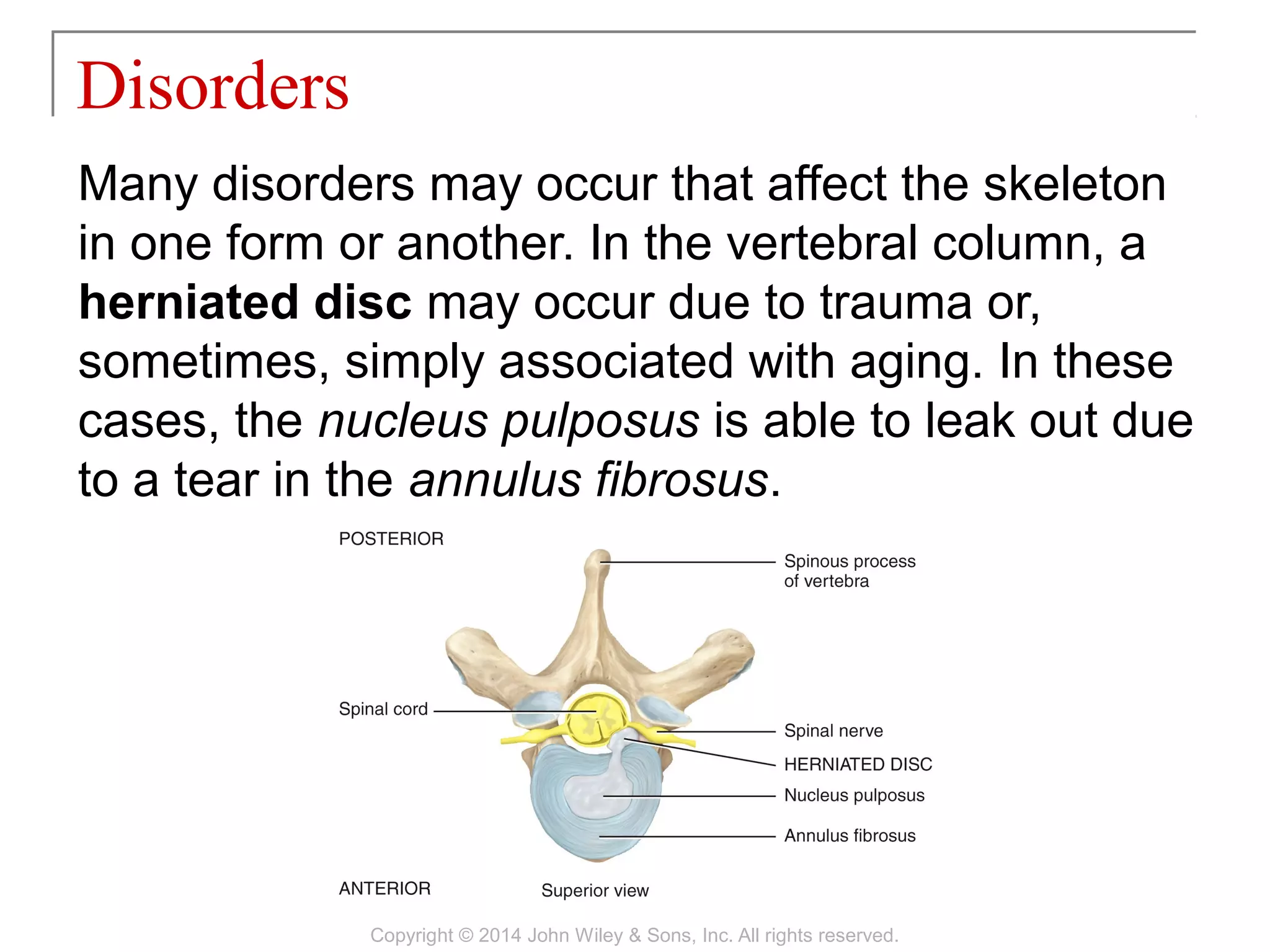


The skeletal system consists of 206 bones divided into the axial skeleton and appendicular skeleton. The axial skeleton includes the skull, vertebral column, ribs, and sternum. It forms the central axis of the body and protects the organs. Bones are classified based on their shape and have characteristic surface markings like depressions, openings, and processes. Common disorders include herniated discs, scoliosis, and spina bifida.
![08 [chapter 8 the skeletal system appendicular skeleton]](https://cdn.slidesharecdn.com/ss_thumbnails/08chapter8theskeletalsystem-appendicularskeleton-170828041008-thumbnail.jpg?width=640&height=640&fit=bounds)
![06 [chapter 6 the skeletal system bone tissue]](https://cdn.slidesharecdn.com/ss_thumbnails/06chapter6theskeletalsystem-bonetissue-170828035633-thumbnail.jpg?width=640&height=640&fit=bounds)
![09 [chapter 9 joints]](https://cdn.slidesharecdn.com/ss_thumbnails/09chapter9joints-170828041032-thumbnail.jpg?width=640&height=640&fit=bounds)
![13 [chapter 13 the spinal cord and spinal nerves]](https://cdn.slidesharecdn.com/ss_thumbnails/13chapter13thespinalcordandspinalnerves-170828040950-thumbnail.jpg?width=640&height=640&fit=bounds)
![11 [chapter 11 the muscular system]](https://cdn.slidesharecdn.com/ss_thumbnails/11chapter11themuscularsystem-170828041038-thumbnail.jpg?width=640&height=640&fit=bounds)
![14 [chapter 14 the brain and cranial nerves]](https://cdn.slidesharecdn.com/ss_thumbnails/14chapter14thebrainandcranialnerves-170828133437-thumbnail.jpg?width=640&height=640&fit=bounds)
![10 [chapter 10 muscular tissue]](https://cdn.slidesharecdn.com/ss_thumbnails/10chapter10musculartissue-170828040153-thumbnail.jpg?width=640&height=640&fit=bounds)
![05 [chapter 5 the integumentary system]](https://cdn.slidesharecdn.com/ss_thumbnails/05chapter5theintegumentarysystem-170828035624-thumbnail.jpg?width=640&height=640&fit=bounds)
![17 [chapter 17 the special senses]](https://cdn.slidesharecdn.com/ss_thumbnails/17chapter17thespecialsenses-170828041636-thumbnail.jpg?width=640&height=640&fit=bounds)

![01 [chapter 1 an introduction to the human body]](https://cdn.slidesharecdn.com/ss_thumbnails/01chapter1anintroductiontothehumanbody-170828035545-thumbnail.jpg?width=640&height=640&fit=bounds)
![11 [chapter 11 the muscular system][11e]](https://cdn.slidesharecdn.com/ss_thumbnails/11chapter11themuscularsystem11e-170828040427-thumbnail.jpg?width=640&height=640&fit=bounds)

![22 [chapter 22 the lymphatic system and immunity]](https://cdn.slidesharecdn.com/ss_thumbnails/22chapter22thelymphaticsystemandimmunity-170828153258-thumbnail.jpg?width=640&height=640&fit=bounds)
![19 [chapter 19 the cardiovascular system the blood]](https://cdn.slidesharecdn.com/ss_thumbnails/19chapter19thecardiovascularsystem-theblood-170828042033-thumbnail.jpg?width=640&height=640&fit=bounds)

![23 [chapter 23 the respiratory system]](https://cdn.slidesharecdn.com/ss_thumbnails/23chapter23therespiratorysystem-170828043650-thumbnail.jpg?width=640&height=640&fit=bounds)
![28 [chapter 28 the reproductive system]](https://cdn.slidesharecdn.com/ss_thumbnails/28chapter28thereproductivesystem-170828134050-thumbnail.jpg?width=640&height=640&fit=bounds)
![26 [chapter 26 the urinary system]](https://cdn.slidesharecdn.com/ss_thumbnails/26chapter26theurinarysystem-170828044011-thumbnail.jpg?width=640&height=640&fit=bounds)
![03 [chapter 3 the cellular level of organization]](https://cdn.slidesharecdn.com/ss_thumbnails/03chapter3thecellularleveloforganization-170828035521-thumbnail.jpg?width=640&height=640&fit=bounds)

![02 [chapter 2 the chemical level of organization]](https://cdn.slidesharecdn.com/ss_thumbnails/02chapter2thechemicalleveloforganization-170828035601-thumbnail.jpg?width=640&height=640&fit=bounds)
![15 [chapter 15 the autonomic nervous system]](https://cdn.slidesharecdn.com/ss_thumbnails/15chapter15theautonomicnervoussystem-170828041929-thumbnail.jpg?width=640&height=640&fit=bounds)
![16 [chapter 16 sensory, motor, and integrative systems]](https://cdn.slidesharecdn.com/ss_thumbnails/16chapter16sensorymotorandintegrativesystems-170828041940-thumbnail.jpg?width=640&height=640&fit=bounds)
![04 [chapter 4 the tissue level of organization][11e]](https://cdn.slidesharecdn.com/ss_thumbnails/04chapter4thetissueleveloforganization11e-170828035609-thumbnail.jpg?width=640&height=640&fit=bounds)
![18 [chapter 18 the endocrine system]](https://cdn.slidesharecdn.com/ss_thumbnails/18chapter18theendocrinesystem-170828042016-thumbnail.jpg?width=640&height=640&fit=bounds)
![21 [chapter 21 the cardiovascular system blood vessels and hemodynamics][11e]](https://cdn.slidesharecdn.com/ss_thumbnails/21chapter21thecardiovascularsystem-bloodvesselsandhemodynamics11e-170828043342-thumbnail.jpg?width=640&height=640&fit=bounds)
![12 [chapter 12 nervous tissue]](https://cdn.slidesharecdn.com/ss_thumbnails/12chapter12nervoustissue-170828041102-thumbnail.jpg?width=640&height=640&fit=bounds)


