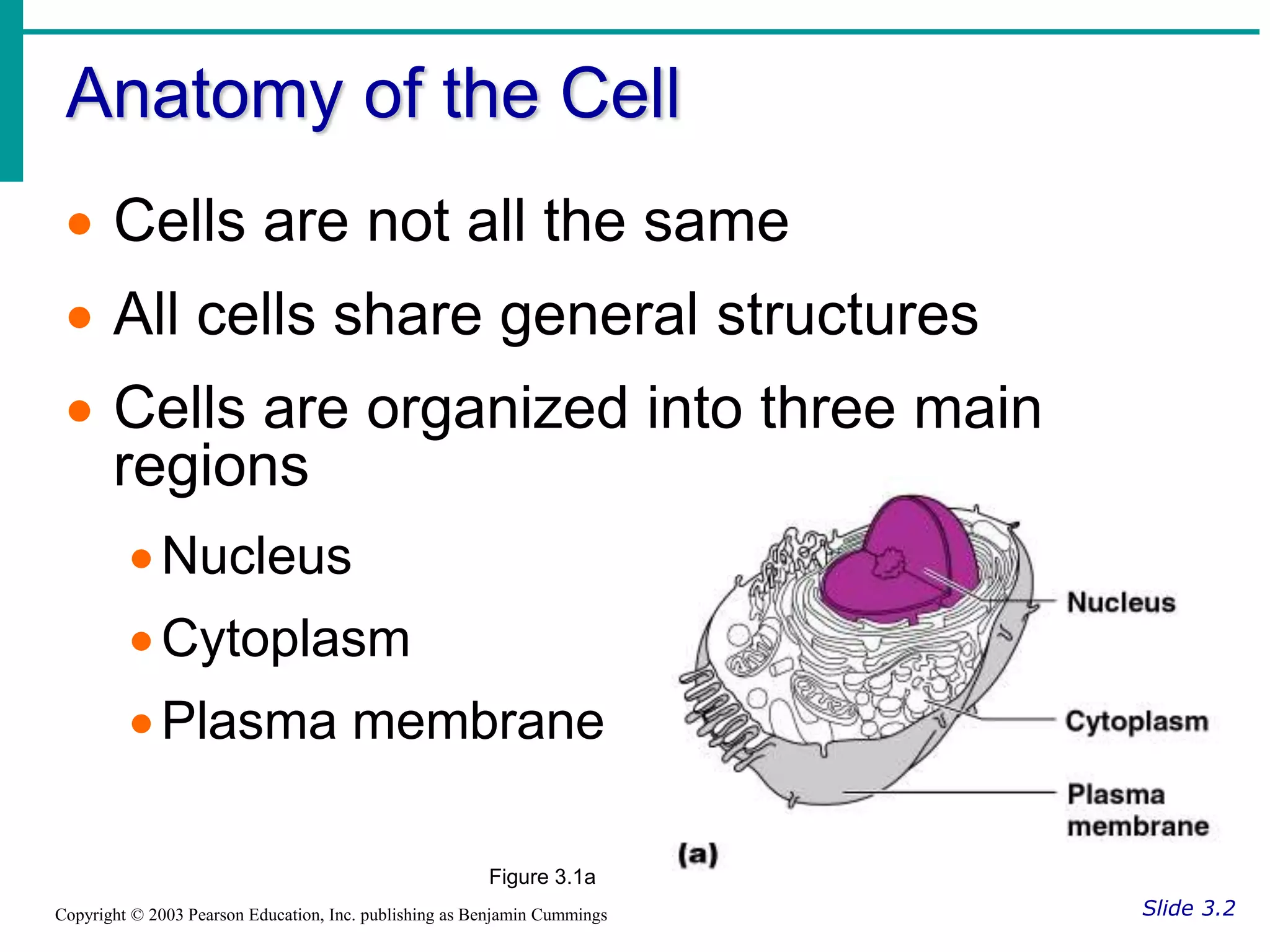
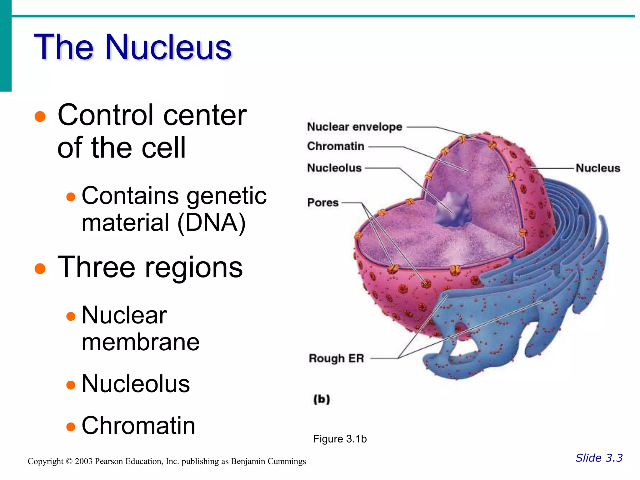
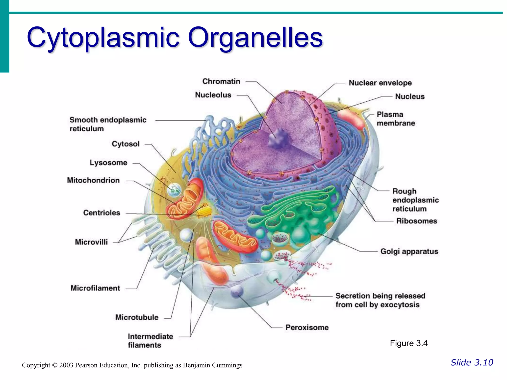
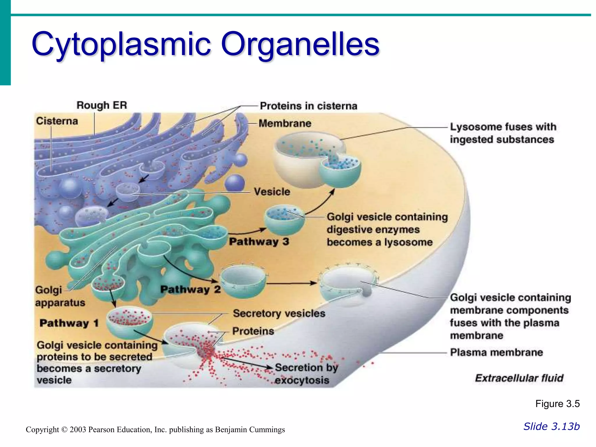
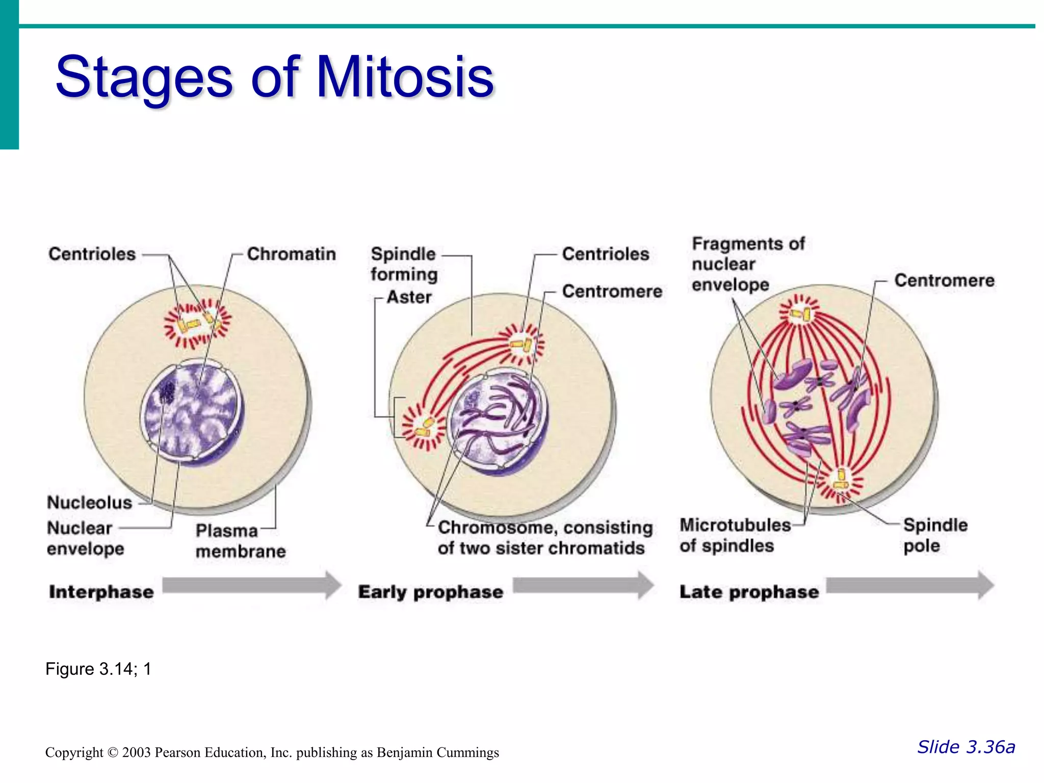
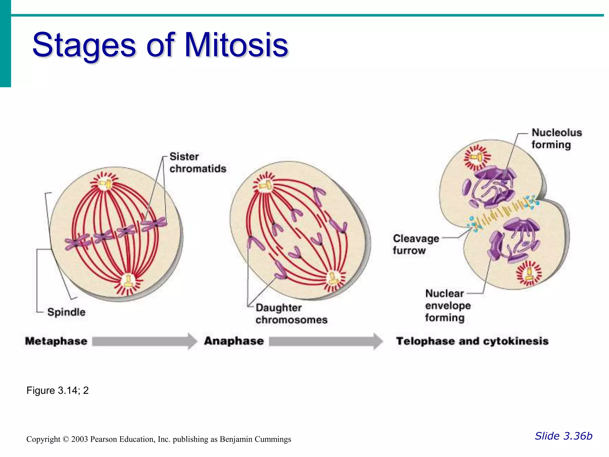
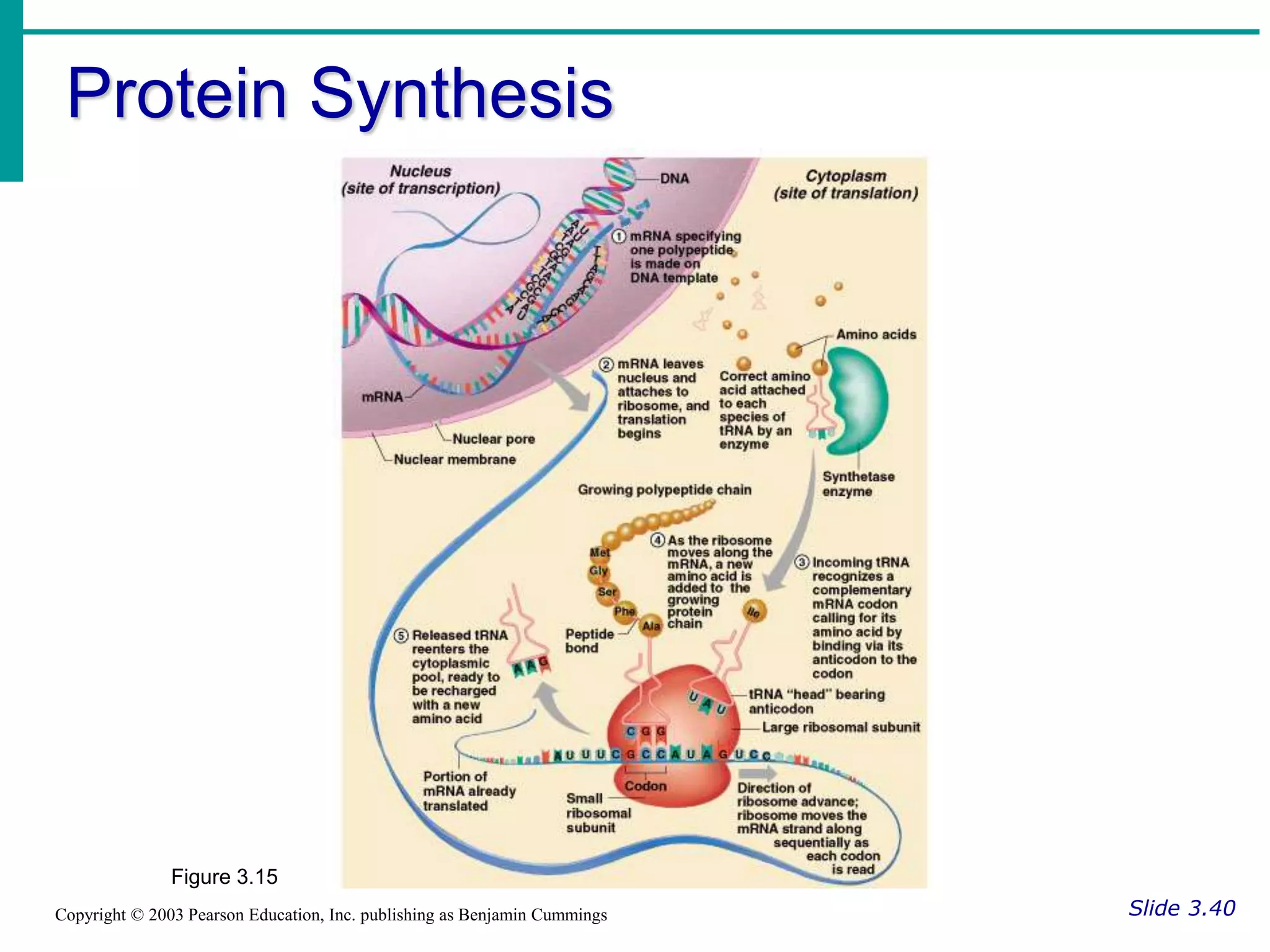
Essentials of Human Anatomy & Physiology
Copyright © 2003 Pearson Education, Inc. publishing as Benjamin Cummings
Slides 3.1 – 3.19
Seventh Edition
Elaine N. Marieb
Chapter 3
Cells and Tissues
Lecture Slides in PowerPoint by Jerry L. Cook
Cells and Tissues
Slide 3.1
Copyright © 2003 Pearson Education, Inc. publishing as Benjamin Cummings
 Carry out all chemical activities needed
to sustain life
 Cells are the building blocks of all living
things
 Tissues are groups of cells that are
similar in structure and function
Anatomy of the Cell
Slide 3.2
Copyright © 2003 Pearson Education, Inc. publishing as Benjamin Cummings
 Cells are not all the same
 All cells share general structures
 Cells are organized into three main
regions
Nucleus
Cytoplasm
Plasma membrane
Figure 3.1a
The Nucleus
Slide 3.3
Copyright © 2003 Pearson Education, Inc. publishing as Benjamin Cummings
 Control center
of the cell
 Contains genetic
material (DNA)
 Three regions
 Nuclear
membrane
 Nucleolus
 Chromatin Figure 3.1b
Nuclear Membrane
Slide 3.4
Copyright © 2003 Pearson Education, Inc. publishing as Benjamin Cummings
 Barrier of nucleus
 Consists of a double phospholipid
membrane
 Contain nuclear pores that allow for
exchange of material with the rest of the
cell
Nucleoli
Slide 3.5
Copyright © 2003 Pearson Education, Inc. publishing as Benjamin Cummings
 Nucleus contains one or more nucleoli
 Sites of ribosome production
 Ribosomes then migrate to the
cytoplasm through nuclear pores
Chromatin
Slide 3.6
Copyright © 2003 Pearson Education, Inc. publishing as Benjamin Cummings
 Composed of DNA and protein
 Scattered throughout the nucleus
 Chromatin condenses to form
chromosomes when the cell divides
Plasma Membrane
Slide 3.7a
Copyright © 2003 Pearson Education, Inc. publishing as Benjamin Cummings
 Barrier for cell contents
 Double phospholipid layer
Hydrophilic heads
Hydrophobic tails
 Other materials in plasma membrane
Protein
Cholesterol
Glycoproteins
Plasma Membrane
Slide 3.7b
Copyright © 2003 Pearson Education, Inc. publishing as Benjamin Cummings
Figure 3.2
Plasma Membrane Specializations
Slide 3.8a
Copyright © 2003 Pearson Education, Inc. publishing as Benjamin Cummings
 Microvilli
Finger-like
projections that
increase surface
area for absorption
Figure 3.3
Plasma Membrane Specializations
Slide 3.8b
Copyright © 2003 Pearson Education, Inc. publishing as Benjamin Cummings
 Membrane
junctions
Tight junctions
Desmosomes
Gap junctions
Figure 3.3
Cytoplasm
Slide 3.9
Copyright © 2003 Pearson Education, Inc. publishing as Benjamin Cummings
 Material outside the nucleus and inside
the plasma membrane
Cytosol
Fluid that suspends other elements
Organelles
Metabolic machinery of the cell
Inclusions
Non-functioning units
Cytoplasmic Organelles
Slide 3.10
Copyright © 2003 Pearson Education, Inc. publishing as Benjamin Cummings
Figure 3.4
Cytoplasmic Organelles
Slide 3.11
Copyright © 2003 Pearson Education, Inc. publishing as Benjamin Cummings
 Ribosomes
Made of protein and RNA
Sites of protein synthesis
Found at two locations
Free in the cytoplasm
Attached to rough endoplasmic reticulum
Cytoplasmic Organelles
Slide 3.12
Copyright © 2003 Pearson Education, Inc. publishing as Benjamin Cummings
 Endoplasmic reticulum (ER)
 Fluid-filled tubules for carrying substances
 Two types of ER
 Rough Endoplasmic Reticulum
 Studded with ribosomes
 Site where building materials of cellular
membrane are formed
 Smooth Endoplasmic Reticulum
 Functions in cholesterol synthesis and
breakdown, fat metabolism, and detoxification
of drugs
Cytoplasmic Organelles
Slide 3.13a
Copyright © 2003 Pearson Education, Inc. publishing as Benjamin Cummings
 Golgi apparatus
Modifies and packages proteins
Produces different types of packages
Secretory vesicles
Cell membrane components
Lysosomes
Cytoplasmic Organelles
Slide 3.13b
Copyright © 2003 Pearson Education, Inc. publishing as Benjamin Cummings
Figure 3.5
Cytoplasmic Organelles
Slide 3.14
Copyright © 2003 Pearson Education, Inc. publishing as Benjamin Cummings
 Lysosomes
Contain enzymes that digest nonusable
materials within the cell
 Peroxisomes
Membranous sacs of oxidase enzymes
Detoxify harmful substances
Break down free radicals
(highly reactive chemicals)
Replicate by pinching in half
Cytoplasmic Organelles
Slide 3.15
Copyright © 2003 Pearson Education, Inc. publishing as Benjamin Cummings
 Mitochondria
 “Powerhouses” of the cell
 Change shape continuously
 Carry out reactions where oxygen is used
to break down food
 Provides ATP for cellular energy
Cytoplasmic Organelles
Slide 3.16a
Copyright © 2003 Pearson Education, Inc. publishing as Benjamin Cummings
 Cytoskeleton
Network of protein structures that extend
throughout the cytoplasm
Provides the cell with an internal framework
Cytoplasmic Organelles
Slide 3.16b
Copyright © 2003 Pearson Education, Inc. publishing as Benjamin Cummings
 Cytoskeleton
Three different types
 Microfilaments
 Intermediate
filaments
 Microtubules
Figure 3.6
Cytoplasmic Organelles
Slide 3.17
Copyright © 2003 Pearson Education, Inc. publishing as Benjamin Cummings
 Centrioles
Rod-shaped bodies made of microtubules
Direct formation of mitotic spindle during
cell division
Cellular Projections
Slide 3.18
Copyright © 2003 Pearson Education, Inc. publishing as Benjamin Cummings
 Not found in all cells
 Used for movement
Cilia moves materials across the cell
surface
Flagellum propels the cell
Cell Diversity
Slide 3.19a
Copyright © 2003 Pearson Education, Inc. publishing as Benjamin Cummings
Figure 3.7; 1, 2
Cell Diversity
Slide 3.19b
Copyright © 2003 Pearson Education, Inc. publishing as Benjamin Cummings
Figure 3.7; 3
Cell Diversity
Slide 3.19c
Copyright © 2003 Pearson Education, Inc. publishing as Benjamin Cummings
Figure 3.7; 4, 5
Cell Diversity
Slide 3.19d
Copyright © 2003 Pearson Education, Inc. publishing as Benjamin Cummings
Figure 3.7; 6, 7
Essentials of Human Anatomy & Physiology
Copyright © 2003 Pearson Education, Inc. publishing as Benjamin Cummings
Slides 3.20 – 3.37
Seventh Edition
Elaine N. Marieb
Chapter 3
Cells and Tissues
Lecture Slides in PowerPoint by Jerry L. Cook
Cellular Physiology:
Membrane Transport
Slide 3.20
Copyright © 2003 Pearson Education, Inc. publishing as Benjamin Cummings
 Membrane Transport – movement of
substance into and out of the cell
 Transport is by two basic methods
Passive transport
No energy is required
Active transport
The cell must provide metabolic energy
Solutions and Transport
Slide 3.21
Copyright © 2003 Pearson Education, Inc. publishing as Benjamin Cummings
 Solution – homogeneous mixture of two
or more components
Solvent – dissolving medium
Solutes – components in smaller quantities
within a solution
 Intracellular fluid – nucleoplasm and
cytosol
 Interstitial fluid – fluid on the exterior of
the cell
Selective Permeability
Slide 3.22
Copyright © 2003 Pearson Education, Inc. publishing as Benjamin Cummings
 The plasma membrane allows some
materials to pass while excluding others
 This permeability includes movement
into and out of the cell
Passive Transport Processes
Slide 3.23
Copyright © 2003 Pearson Education, Inc. publishing as Benjamin Cummings
 Diffusion
 Particles tend to distribute themselves evenly
within a solution
 Movement is
from high
concentration
to low
concentration,
or down a
concentration
gradient
Figure 3.8
Passive Transport Processes
Slide 3.24a
Copyright © 2003 Pearson Education, Inc. publishing as Benjamin Cummings
 Types of diffusion
Simple diffusion
Unassisted process
Solutes are lipid-soluble materials or
small enough to pass through membrane
pores
Passive Transport Processes
Slide 3.24b
Copyright © 2003 Pearson Education, Inc. publishing as Benjamin Cummings
 Types of diffusion
Osmosis – simple diffusion of water
Highly polar water easily crosses the
plasma membrane
Facilitated diffusion
Substances require a protein carrier for
passive transport
Diffusion through the Plasma
Membrane
Slide 3.25
Copyright © 2003 Pearson Education, Inc. publishing as Benjamin Cummings
Figure 3.9
Passive Transport Processes
Slide 3.26
Copyright © 2003 Pearson Education, Inc. publishing as Benjamin Cummings
 Filtration
Water and solutes are forced through a
membrane by fluid, or hydrostatic pressure
A pressure gradient must exist
Solute-containing fluid is pushed from a
high pressure area to a lower pressure
area
Active Transport Processes
Slide 3.27
Copyright © 2003 Pearson Education, Inc. publishing as Benjamin Cummings
 Transport substances that are unable to pass
by diffusion
 They may be too large
 They may not be able to dissolve in the fat core
of the membrane
 They may have to move against a concentration
gradient
 Two common forms of active transport
 Solute pumping
 Bulk transport
Active Transport Processes
Slide 3.28a
Copyright © 2003 Pearson Education, Inc. publishing as Benjamin Cummings
 Solute pumping
Amino acids, some sugars and ions are
transported by solute pumps
ATP energizes protein carriers, and in most
cases, moves substances against
concentration gradients
Active Transport Processes
Slide 3.28b
Copyright © 2003 Pearson Education, Inc. publishing as Benjamin Cummings
Figure 3.10
Active Transport Processes
Slide 3.29a
Copyright © 2003 Pearson Education, Inc. publishing as Benjamin Cummings
 Bulk transport
Exocytosis
 Moves materials out of the cell
 Material is carried in a membranous vesicle
 Vesicle migrates to plasma membrane
 Vesicle combines with plasma membrane
 Material is emptied to the outside
Active Transport Processes
Slide 3.29b
Copyright © 2003 Pearson Education, Inc. publishing as Benjamin Cummings
Figure 3.11
Active Transport Processes
Slide 3.30a
Copyright © 2003 Pearson Education, Inc. publishing as Benjamin Cummings
 Bulk transport
Endocytosis
Extracellular substances are engulfed by
being enclosed in a membranous
vescicle
Types of endocytosis
Phagocytosis – cell eating
Pinocytosis – cell drinking
Active Transport Processes
Slide 3.30b
Copyright © 2003 Pearson Education, Inc. publishing as Benjamin Cummings
Figure 3.12
Cell Life Cycle
Slide 3.31
Copyright © 2003 Pearson Education, Inc. publishing as Benjamin Cummings
 Cells have two major periods
Interphase
Cell grows
Cell carries on metabolic processes
Cell division
Cell replicates itself
Function is to produce more cells for
growth and repair processes
DNA Replication
Slide 3.32
Copyright © 2003 Pearson Education, Inc. publishing as Benjamin Cummings
 Genetic material
duplicated and
readies a cell for
division into two cells
 Occurs toward the
end of interphase
 DNA uncoils and
each side serves
as a template
Figure 3.13
Events of Cell Division
Slide 3.33
Copyright © 2003 Pearson Education, Inc. publishing as Benjamin Cummings
 Mitosis
Division of the nucleus
Results in the formation of two daughter
nuclei
 Cytokinesis
Division of the cytoplasm
Begins when mitosis is near completion
Results in the formation of two daughter
cells
Stages of Mitosis
Slide 3.34a
Copyright © 2003 Pearson Education, Inc. publishing as Benjamin Cummings
 Interphase
No cell division occurs
The cell carries out normal metabolic
activity and growth
 Prophase
First part of cell division
Centromeres migrate to the poles
Stages of Mitosis
Slide 3.34b
Copyright © 2003 Pearson Education, Inc. publishing as Benjamin Cummings
 Metaphase
Spindle from centromeres are attached to
chromosomes that are aligned in the center
of the cell
Stages of Mitosis
Slide 3.35
Copyright © 2003 Pearson Education, Inc. publishing as Benjamin Cummings
 Anaphase
Daughter chromosomes are pulled toward
the poles
The cell begins to elongate
 Telophase
Daughter nuclei begin forming
A cleavage furrow (for cell division) begins
to form
Stages of Mitosis
Slide 3.36a
Copyright © 2003 Pearson Education, Inc. publishing as Benjamin Cummings
Figure 3.14; 1
Stages of Mitosis
Slide 3.36b
Copyright © 2003 Pearson Education, Inc. publishing as Benjamin Cummings
Figure 3.14; 2
Protein Synthesis
Slide 3.37
Copyright © 2003 Pearson Education, Inc. publishing as Benjamin Cummings
 Gene – DNA segment that carries a
blueprint for building one protein
 Proteins have many functions
Building materials for cells
Act as enzymes (biological catalysts)
 RNA is essential for protein synthesis
Essentials of Human Anatomy & Physiology
Copyright © 2003 Pearson Education, Inc. publishing as Benjamin Cummings
Slides 3.38 – 3.54
Seventh Edition
Elaine N. Marieb
Chapter 3
Cells and Tissues
Lecture Slides in PowerPoint by Jerry L. Cook
Protein Synthesis
Slide 3.37
Copyright © 2003 Pearson Education, Inc. publishing as Benjamin Cummings
 Gene – DNA segment that carries a
blueprint for building one protein
 Proteins have many functions
Building materials for cells
Act as enzymes (biological catalysts)
 RNA is essential for protein synthesis
Role of RNA
Slide 3.38
Copyright © 2003 Pearson Education, Inc. publishing as Benjamin Cummings
 Transfer RNA (tRNA)
Transfers appropriate amino acids to the
ribosome for building the protein
 Ribosomal RNA (rRNA)
Helps form the ribosomes where proteins
are built
 Messanger RN
Carries the instructions for building a
protein from the nucleus to the ribosome
Transcription and Translation
Slide 3.39
Copyright © 2003 Pearson Education, Inc. publishing as Benjamin Cummings
 Transcription
Transfer of information from DNA’s base
sequence to the complimentary base
sequence of mRNA
 Translation
Base sequence of nucleic acid is translated
to an amino acid sequence
Amino acids are the building blocks of
proteins
Protein Synthesis
Slide 3.40
Copyright © 2003 Pearson Education, Inc. publishing as Benjamin Cummings
Figure 3.15
Body Tissues
Slide 3.41
Copyright © 2003 Pearson Education, Inc. publishing as Benjamin Cummings
 Cells are specialized for particular functions
 Tissues
Groups of cells with similar structure and
function
Four primary types
 Epithelium
 Connective tissue
 Nervous tissue
 Muscle
Epithelial Tissues
Slide 3.42
Copyright © 2003 Pearson Education, Inc. publishing as Benjamin Cummings
 Found in different areas
Body coverings
Body linings
Glandular tissue
 Functions
Protection
Absorption
Filtration
Secretion
Epithelium Characteristics
Slide 3.43
Copyright © 2003 Pearson Education, Inc. publishing as Benjamin Cummings
 Cells fit closely together
 Tissue layer always has one free
surface
 The lower surface is bound by a
basement membrane
 Avascular (have no blood supply)
 Regenerate easily if well nourished
Classification of Epithelium
Slide 3.44a
Copyright © 2003 Pearson Education, Inc. publishing as Benjamin Cummings
 Number of cell layers
Simple – one layer
Stratified – more than
one layer
Figure 3.16a
Classification of Epithelium
Slide 3.44b
Copyright © 2003 Pearson Education, Inc. publishing as Benjamin Cummings
 Shape of cells
Squamous – flattened
Cuboidal – cube-shaped
Columnar – column-like
Figure 3.16b
Simple Epithelium
Slide 3.45
Copyright © 2003 Pearson Education, Inc. publishing as Benjamin Cummings
 Simple squamous
Single layer of flat
cells
Usually forms
membranes
Lines body
cavities
Lines lungs and
capillaries Figure 3.17a
Simple Epithelium
Slide 3.46
Copyright © 2003 Pearson Education, Inc. publishing as Benjamin Cummings
 Simple cuboidal
Single layer of
cube-like cells
Common in
glands and their
ducts
Forms walls
of kidney tubules
Covers the
ovaries Figure 3.17b
Simple Epithelium
Slide 3.47
Copyright © 2003 Pearson Education, Inc. publishing as Benjamin Cummings
 Simple columnar
Single layer of tall
cells
Often includes
goblet cells, which
produce mucus
Lines digestive
tract
Figure 3.17c
Simple Epithelium
Slide 3.48
Copyright © 2003 Pearson Education, Inc. publishing as Benjamin Cummings
 Pseudostratified
 Single layer, but
some cells are
shorter than others
 Often looks like a
double cell layer
 Sometimes ciliated,
such as in the
respiratory tract
 May function in
absorption or
secretion
Figure 3.17d
Stratified Epithelium
Slide 3.49
Copyright © 2003 Pearson Education, Inc. publishing as Benjamin Cummings
 Stratified squamous
 Cells at the free edge
are flattened
 Found as a protective
covering where
friction is common
 Locations
 Skin
 Mouth
 Esophagus Figure 3.17e
Stratified Epithelium
Slide 3.50
Copyright © 2003 Pearson Education, Inc. publishing as Benjamin Cummings
 Stratified cuboidal
 Two layers of cuboidal cells
 Stratified columnar
 Surface cells are columnar, cells
underneath vary in size and shape
 Stratified cuboidal and columnar
 Rare in human body
 Found mainly in ducts of large glands
Stratified Epithelium
Slide 3.51
Copyright © 2003 Pearson Education, Inc. publishing as Benjamin Cummings
 Transitional
epithelium
Shape of cells
depends upon the
amount of stretching
Lines organs of the
urinary system
Figure 3.17f
Glandular Epithelium
Slide 3.52
Copyright © 2003 Pearson Education, Inc. publishing as Benjamin Cummings
 Gland – one or more cells that secretes a
particular product
 Two major gland types
 Endocrine gland
 Ductless
 Secretions are hormones
 Exocrine gland
 Empty through ducts to the epithelial surface
 Include sweat and oil glands
Connective Tissue
Slide 3.53
Copyright © 2003 Pearson Education, Inc. publishing as Benjamin Cummings
 Found everywhere in the body
 Includes the most abundant and widely
distributed tissues
 Functions
Binds body tissues together
Supports the body
Provides protection
Connective Tissue Characteristics
Slide 3.54
Copyright © 2003 Pearson Education, Inc. publishing as Benjamin Cummings
 Variations in blood supply
Some tissue types are well vascularized
Some have poor blood supply or are
avascular
 Extracellular matrix
Non-living material that surrounds living
cells
Essentials of Human Anatomy & Physiology
Copyright © 2003 Pearson Education, Inc. publishing as Benjamin Cummings
Slides 3.55 – 3.73
Seventh Edition
Elaine N. Marieb
Chapter 3
Cells and Tissues
Lecture Slides in PowerPoint by Jerry L. Cook
Extracellular Matrix
Slide 3.55
Copyright © 2003 Pearson Education, Inc. publishing as Benjamin Cummings
 Two main elements
Ground substance – mostly water along
with adhesion proteins and polysaccharide
molecules
Fibers
Produced by the cells
Three types
 Collagen fibers
 Elastic fibers
 Reticular fibers
Connective Tissue Types
Slide 3.56
Copyright © 2003 Pearson Education, Inc. publishing as Benjamin Cummings
 Bone (osseous tissue)
Composed of:
Bone cells in lacunae
(cavities)
Hard matrix of calcium
salts
Large numbers of
collagen fibers
Used to protect and
support the body Figure 3.18a
Connective Tissue Types
Slide 3.57
Copyright © 2003 Pearson Education, Inc. publishing as Benjamin Cummings
 Hyaline cartilage
Most common
cartilage
Composed of:
Abundant collagen
fibers
Rubbery matrix
Entire fetal skeleton
is hyaline cartilage Figure 3.18b
Connective Tissue Types
Slide 3.58a
Copyright © 2003 Pearson Education, Inc. publishing as Benjamin Cummings
 Elastic cartilage
Provides elasticity
Example: supports the external ear
Connective Tissue Types
Slide 3.58b
Copyright © 2003 Pearson Education, Inc. publishing as Benjamin Cummings
 Fibrocartilage
Highly compressible
Example: forms
cushion-like discs
between vertebrae
Figure 3.18c
Connective Tissue Types
Slide 3.59
Copyright © 2003 Pearson Education, Inc. publishing as Benjamin Cummings
 Dense connective
tissue
Main matrix element
is collagen fibers
Cells are fibroblasts
Examples
 Tendon – attach
muscle to bone
 Ligaments – attach
bone to bone Figure 3.18d
Connective Tissue Types
Slide 3.60
Copyright © 2003 Pearson Education, Inc. publishing as Benjamin Cummings
 Areolar connective
tissue
Most widely
distributed
connective tissue
Soft, pliable tissue
Contains all fiber
types
Can soak up excess
fluid Figure 3.18e
Connective Tissue Types
Slide 3.61
Copyright © 2003 Pearson Education, Inc. publishing as Benjamin Cummings
 Adipose tissue
Matrix is an areolar tissue in which fat
globules predominate
Many cells contain
large lipid deposits
Functions
 Insulates the body
 Protects some organs
 Serves as a site of
fuel storage Figure 3.18f
Connective Tissue Types
Slide 3.62
Copyright © 2003 Pearson Education, Inc. publishing as Benjamin Cummings
 Reticular
connective tissue
 Delicate network of
interwoven fibers
 Forms stroma
(internal supporting
network) of
lymphoid organs
 Lymph nodes
 Spleen
 Bone marrow Figure 3.18g
Connective Tissue Types
Slide 3.63
Copyright © 2003 Pearson Education, Inc. publishing as Benjamin Cummings
 Blood
Blood cells
surrounded by fluid
matrix
Fibers are visible
during clotting
Functions as the
transport vehicle
for materials
Figure 3.18h
Muscle Tissue
Slide 3.64
Copyright © 2003 Pearson Education, Inc. publishing as Benjamin Cummings
 Function is to produce movement
 Three types
Skeletal muscle
Cardiac muscle
Smooth muscle
Muscle Tissue Types
Slide 3.65
Copyright © 2003 Pearson Education, Inc. publishing as Benjamin Cummings
 Skeletal muscle
Can be controlled
voluntarily
Cells attach to
connective tissue
Cells are striated
Cells have more than
one nucleus
Figure 3.19b
Muscle Tissue Types
Slide 3.66
Copyright © 2003 Pearson Education, Inc. publishing as Benjamin Cummings
 Cardiac muscle
 Found only in the
heart
 Function is to pump
blood (involuntary)
 Cells attached to
other cardiac muscle
cells at intercalated
disks
 Cells are striated
 One nucleus per cell Figure 3.19c
Muscle Tissue Types
Slide 3.67
Copyright © 2003 Pearson Education, Inc. publishing as Benjamin Cummings
 Smooth muscle
Involuntary muscle
Surrounds hollow
organs
Attached to other
smooth muscle cells
No visible striations
One nucleus per cell Figure 3.19a
Nervous Tissue
Slide 3.68
Copyright © 2003 Pearson Education, Inc. publishing as Benjamin Cummings
 Neurons and
nerve support
cells
 Function is to
send impulses to
other areas of
the body
Irritability
Conductivity Figure 3.20
Tissue Repair
Slide 3.69
Copyright © 2003 Pearson Education, Inc. publishing as Benjamin Cummings
 Regeneration
Replacement of destroyed tissue by the
same kind of cells
 Fibrosis
Repair by dense fibrous connective tissue
(scar tissue)
 Determination of method
Type of tissue damaged
Severity of the injury
Events in Tissue Repair
Slide 3.70
Copyright © 2003 Pearson Education, Inc. publishing as Benjamin Cummings
 Capillaries become very permeable
Introduce clotting proteins
Wall off injured area
 Formation of granulation tissue
 Regeneration of surface epithelium
Regeneration of Tissues
Slide 3.71
Copyright © 2003 Pearson Education, Inc. publishing as Benjamin Cummings
 Tissues that regenerate easily
 Epithelial tissue
 Fibrous connective tissue and bone
 Tissues that regenerate poorly
 Skeletal muscle
 Tissues that are replaced largely with scar
tissue
 Cardiac muscle
 Nervous tissue within the brain and spinal cord
Developmental Aspects of Tissue
Slide 3.72
Copyright © 2003 Pearson Education, Inc. publishing as Benjamin Cummings
 Epithelial tissue arises from all three
primary germ layers
 Muscle and connective tissue arise from
the mesoderm
 Nervous tissue arises from the
ectoderm
 With old age there is a decrease in
mass and viabililty in most tissues

Cells_and_Tissues.ppt

  • 1.
    Essentials of HumanAnatomy & Physiology Copyright © 2003 Pearson Education, Inc. publishing as Benjamin Cummings Slides 3.1 – 3.19 Seventh Edition Elaine N. Marieb Chapter 3 Cells and Tissues Lecture Slides in PowerPoint by Jerry L. Cook
  • 2.
    Cells and Tissues Slide3.1 Copyright © 2003 Pearson Education, Inc. publishing as Benjamin Cummings  Carry out all chemical activities needed to sustain life  Cells are the building blocks of all living things  Tissues are groups of cells that are similar in structure and function
  • 3.
    Anatomy of theCell Slide 3.2 Copyright © 2003 Pearson Education, Inc. publishing as Benjamin Cummings  Cells are not all the same  All cells share general structures  Cells are organized into three main regions Nucleus Cytoplasm Plasma membrane Figure 3.1a
  • 4.
    The Nucleus Slide 3.3 Copyright© 2003 Pearson Education, Inc. publishing as Benjamin Cummings  Control center of the cell  Contains genetic material (DNA)  Three regions  Nuclear membrane  Nucleolus  Chromatin Figure 3.1b
  • 5.
    Nuclear Membrane Slide 3.4 Copyright© 2003 Pearson Education, Inc. publishing as Benjamin Cummings  Barrier of nucleus  Consists of a double phospholipid membrane  Contain nuclear pores that allow for exchange of material with the rest of the cell
  • 6.
    Nucleoli Slide 3.5 Copyright ©2003 Pearson Education, Inc. publishing as Benjamin Cummings  Nucleus contains one or more nucleoli  Sites of ribosome production  Ribosomes then migrate to the cytoplasm through nuclear pores
  • 7.
    Chromatin Slide 3.6 Copyright ©2003 Pearson Education, Inc. publishing as Benjamin Cummings  Composed of DNA and protein  Scattered throughout the nucleus  Chromatin condenses to form chromosomes when the cell divides
  • 8.
    Plasma Membrane Slide 3.7a Copyright© 2003 Pearson Education, Inc. publishing as Benjamin Cummings  Barrier for cell contents  Double phospholipid layer Hydrophilic heads Hydrophobic tails  Other materials in plasma membrane Protein Cholesterol Glycoproteins
  • 9.
    Plasma Membrane Slide 3.7b Copyright© 2003 Pearson Education, Inc. publishing as Benjamin Cummings Figure 3.2
  • 10.
    Plasma Membrane Specializations Slide3.8a Copyright © 2003 Pearson Education, Inc. publishing as Benjamin Cummings  Microvilli Finger-like projections that increase surface area for absorption Figure 3.3
  • 11.
    Plasma Membrane Specializations Slide3.8b Copyright © 2003 Pearson Education, Inc. publishing as Benjamin Cummings  Membrane junctions Tight junctions Desmosomes Gap junctions Figure 3.3
  • 12.
    Cytoplasm Slide 3.9 Copyright ©2003 Pearson Education, Inc. publishing as Benjamin Cummings  Material outside the nucleus and inside the plasma membrane Cytosol Fluid that suspends other elements Organelles Metabolic machinery of the cell Inclusions Non-functioning units
  • 13.
    Cytoplasmic Organelles Slide 3.10 Copyright© 2003 Pearson Education, Inc. publishing as Benjamin Cummings Figure 3.4
  • 14.
    Cytoplasmic Organelles Slide 3.11 Copyright© 2003 Pearson Education, Inc. publishing as Benjamin Cummings  Ribosomes Made of protein and RNA Sites of protein synthesis Found at two locations Free in the cytoplasm Attached to rough endoplasmic reticulum
  • 15.
    Cytoplasmic Organelles Slide 3.12 Copyright© 2003 Pearson Education, Inc. publishing as Benjamin Cummings  Endoplasmic reticulum (ER)  Fluid-filled tubules for carrying substances  Two types of ER  Rough Endoplasmic Reticulum  Studded with ribosomes  Site where building materials of cellular membrane are formed  Smooth Endoplasmic Reticulum  Functions in cholesterol synthesis and breakdown, fat metabolism, and detoxification of drugs
  • 16.
    Cytoplasmic Organelles Slide 3.13a Copyright© 2003 Pearson Education, Inc. publishing as Benjamin Cummings  Golgi apparatus Modifies and packages proteins Produces different types of packages Secretory vesicles Cell membrane components Lysosomes
  • 17.
    Cytoplasmic Organelles Slide 3.13b Copyright© 2003 Pearson Education, Inc. publishing as Benjamin Cummings Figure 3.5
  • 18.
    Cytoplasmic Organelles Slide 3.14 Copyright© 2003 Pearson Education, Inc. publishing as Benjamin Cummings  Lysosomes Contain enzymes that digest nonusable materials within the cell  Peroxisomes Membranous sacs of oxidase enzymes Detoxify harmful substances Break down free radicals (highly reactive chemicals) Replicate by pinching in half
  • 19.
    Cytoplasmic Organelles Slide 3.15 Copyright© 2003 Pearson Education, Inc. publishing as Benjamin Cummings  Mitochondria  “Powerhouses” of the cell  Change shape continuously  Carry out reactions where oxygen is used to break down food  Provides ATP for cellular energy
  • 20.
    Cytoplasmic Organelles Slide 3.16a Copyright© 2003 Pearson Education, Inc. publishing as Benjamin Cummings  Cytoskeleton Network of protein structures that extend throughout the cytoplasm Provides the cell with an internal framework
  • 21.
    Cytoplasmic Organelles Slide 3.16b Copyright© 2003 Pearson Education, Inc. publishing as Benjamin Cummings  Cytoskeleton Three different types  Microfilaments  Intermediate filaments  Microtubules Figure 3.6
  • 22.
    Cytoplasmic Organelles Slide 3.17 Copyright© 2003 Pearson Education, Inc. publishing as Benjamin Cummings  Centrioles Rod-shaped bodies made of microtubules Direct formation of mitotic spindle during cell division
  • 23.
    Cellular Projections Slide 3.18 Copyright© 2003 Pearson Education, Inc. publishing as Benjamin Cummings  Not found in all cells  Used for movement Cilia moves materials across the cell surface Flagellum propels the cell
  • 24.
    Cell Diversity Slide 3.19a Copyright© 2003 Pearson Education, Inc. publishing as Benjamin Cummings Figure 3.7; 1, 2
  • 25.
    Cell Diversity Slide 3.19b Copyright© 2003 Pearson Education, Inc. publishing as Benjamin Cummings Figure 3.7; 3
  • 26.
    Cell Diversity Slide 3.19c Copyright© 2003 Pearson Education, Inc. publishing as Benjamin Cummings Figure 3.7; 4, 5
  • 27.
    Cell Diversity Slide 3.19d Copyright© 2003 Pearson Education, Inc. publishing as Benjamin Cummings Figure 3.7; 6, 7
  • 28.
    Essentials of HumanAnatomy & Physiology Copyright © 2003 Pearson Education, Inc. publishing as Benjamin Cummings Slides 3.20 – 3.37 Seventh Edition Elaine N. Marieb Chapter 3 Cells and Tissues Lecture Slides in PowerPoint by Jerry L. Cook
  • 29.
    Cellular Physiology: Membrane Transport Slide3.20 Copyright © 2003 Pearson Education, Inc. publishing as Benjamin Cummings  Membrane Transport – movement of substance into and out of the cell  Transport is by two basic methods Passive transport No energy is required Active transport The cell must provide metabolic energy
  • 30.
    Solutions and Transport Slide3.21 Copyright © 2003 Pearson Education, Inc. publishing as Benjamin Cummings  Solution – homogeneous mixture of two or more components Solvent – dissolving medium Solutes – components in smaller quantities within a solution  Intracellular fluid – nucleoplasm and cytosol  Interstitial fluid – fluid on the exterior of the cell
  • 31.
    Selective Permeability Slide 3.22 Copyright© 2003 Pearson Education, Inc. publishing as Benjamin Cummings  The plasma membrane allows some materials to pass while excluding others  This permeability includes movement into and out of the cell
  • 32.
    Passive Transport Processes Slide3.23 Copyright © 2003 Pearson Education, Inc. publishing as Benjamin Cummings  Diffusion  Particles tend to distribute themselves evenly within a solution  Movement is from high concentration to low concentration, or down a concentration gradient Figure 3.8
  • 33.
    Passive Transport Processes Slide3.24a Copyright © 2003 Pearson Education, Inc. publishing as Benjamin Cummings  Types of diffusion Simple diffusion Unassisted process Solutes are lipid-soluble materials or small enough to pass through membrane pores
  • 34.
    Passive Transport Processes Slide3.24b Copyright © 2003 Pearson Education, Inc. publishing as Benjamin Cummings  Types of diffusion Osmosis – simple diffusion of water Highly polar water easily crosses the plasma membrane Facilitated diffusion Substances require a protein carrier for passive transport
  • 35.
    Diffusion through thePlasma Membrane Slide 3.25 Copyright © 2003 Pearson Education, Inc. publishing as Benjamin Cummings Figure 3.9
  • 36.
    Passive Transport Processes Slide3.26 Copyright © 2003 Pearson Education, Inc. publishing as Benjamin Cummings  Filtration Water and solutes are forced through a membrane by fluid, or hydrostatic pressure A pressure gradient must exist Solute-containing fluid is pushed from a high pressure area to a lower pressure area
  • 37.
    Active Transport Processes Slide3.27 Copyright © 2003 Pearson Education, Inc. publishing as Benjamin Cummings  Transport substances that are unable to pass by diffusion  They may be too large  They may not be able to dissolve in the fat core of the membrane  They may have to move against a concentration gradient  Two common forms of active transport  Solute pumping  Bulk transport
  • 38.
    Active Transport Processes Slide3.28a Copyright © 2003 Pearson Education, Inc. publishing as Benjamin Cummings  Solute pumping Amino acids, some sugars and ions are transported by solute pumps ATP energizes protein carriers, and in most cases, moves substances against concentration gradients
  • 39.
    Active Transport Processes Slide3.28b Copyright © 2003 Pearson Education, Inc. publishing as Benjamin Cummings Figure 3.10
  • 40.
    Active Transport Processes Slide3.29a Copyright © 2003 Pearson Education, Inc. publishing as Benjamin Cummings  Bulk transport Exocytosis  Moves materials out of the cell  Material is carried in a membranous vesicle  Vesicle migrates to plasma membrane  Vesicle combines with plasma membrane  Material is emptied to the outside
  • 41.
    Active Transport Processes Slide3.29b Copyright © 2003 Pearson Education, Inc. publishing as Benjamin Cummings Figure 3.11
  • 42.
    Active Transport Processes Slide3.30a Copyright © 2003 Pearson Education, Inc. publishing as Benjamin Cummings  Bulk transport Endocytosis Extracellular substances are engulfed by being enclosed in a membranous vescicle Types of endocytosis Phagocytosis – cell eating Pinocytosis – cell drinking
  • 43.
    Active Transport Processes Slide3.30b Copyright © 2003 Pearson Education, Inc. publishing as Benjamin Cummings Figure 3.12
  • 44.
    Cell Life Cycle Slide3.31 Copyright © 2003 Pearson Education, Inc. publishing as Benjamin Cummings  Cells have two major periods Interphase Cell grows Cell carries on metabolic processes Cell division Cell replicates itself Function is to produce more cells for growth and repair processes
  • 45.
    DNA Replication Slide 3.32 Copyright© 2003 Pearson Education, Inc. publishing as Benjamin Cummings  Genetic material duplicated and readies a cell for division into two cells  Occurs toward the end of interphase  DNA uncoils and each side serves as a template Figure 3.13
  • 46.
    Events of CellDivision Slide 3.33 Copyright © 2003 Pearson Education, Inc. publishing as Benjamin Cummings  Mitosis Division of the nucleus Results in the formation of two daughter nuclei  Cytokinesis Division of the cytoplasm Begins when mitosis is near completion Results in the formation of two daughter cells
  • 47.
    Stages of Mitosis Slide3.34a Copyright © 2003 Pearson Education, Inc. publishing as Benjamin Cummings  Interphase No cell division occurs The cell carries out normal metabolic activity and growth  Prophase First part of cell division Centromeres migrate to the poles
  • 48.
    Stages of Mitosis Slide3.34b Copyright © 2003 Pearson Education, Inc. publishing as Benjamin Cummings  Metaphase Spindle from centromeres are attached to chromosomes that are aligned in the center of the cell
  • 49.
    Stages of Mitosis Slide3.35 Copyright © 2003 Pearson Education, Inc. publishing as Benjamin Cummings  Anaphase Daughter chromosomes are pulled toward the poles The cell begins to elongate  Telophase Daughter nuclei begin forming A cleavage furrow (for cell division) begins to form
  • 50.
    Stages of Mitosis Slide3.36a Copyright © 2003 Pearson Education, Inc. publishing as Benjamin Cummings Figure 3.14; 1
  • 51.
    Stages of Mitosis Slide3.36b Copyright © 2003 Pearson Education, Inc. publishing as Benjamin Cummings Figure 3.14; 2
  • 52.
    Protein Synthesis Slide 3.37 Copyright© 2003 Pearson Education, Inc. publishing as Benjamin Cummings  Gene – DNA segment that carries a blueprint for building one protein  Proteins have many functions Building materials for cells Act as enzymes (biological catalysts)  RNA is essential for protein synthesis
  • 53.
    Essentials of HumanAnatomy & Physiology Copyright © 2003 Pearson Education, Inc. publishing as Benjamin Cummings Slides 3.38 – 3.54 Seventh Edition Elaine N. Marieb Chapter 3 Cells and Tissues Lecture Slides in PowerPoint by Jerry L. Cook
  • 54.
    Protein Synthesis Slide 3.37 Copyright© 2003 Pearson Education, Inc. publishing as Benjamin Cummings  Gene – DNA segment that carries a blueprint for building one protein  Proteins have many functions Building materials for cells Act as enzymes (biological catalysts)  RNA is essential for protein synthesis
  • 55.
    Role of RNA Slide3.38 Copyright © 2003 Pearson Education, Inc. publishing as Benjamin Cummings  Transfer RNA (tRNA) Transfers appropriate amino acids to the ribosome for building the protein  Ribosomal RNA (rRNA) Helps form the ribosomes where proteins are built  Messanger RN Carries the instructions for building a protein from the nucleus to the ribosome
  • 56.
    Transcription and Translation Slide3.39 Copyright © 2003 Pearson Education, Inc. publishing as Benjamin Cummings  Transcription Transfer of information from DNA’s base sequence to the complimentary base sequence of mRNA  Translation Base sequence of nucleic acid is translated to an amino acid sequence Amino acids are the building blocks of proteins
  • 57.
    Protein Synthesis Slide 3.40 Copyright© 2003 Pearson Education, Inc. publishing as Benjamin Cummings Figure 3.15
  • 58.
    Body Tissues Slide 3.41 Copyright© 2003 Pearson Education, Inc. publishing as Benjamin Cummings  Cells are specialized for particular functions  Tissues Groups of cells with similar structure and function Four primary types  Epithelium  Connective tissue  Nervous tissue  Muscle
  • 59.
    Epithelial Tissues Slide 3.42 Copyright© 2003 Pearson Education, Inc. publishing as Benjamin Cummings  Found in different areas Body coverings Body linings Glandular tissue  Functions Protection Absorption Filtration Secretion
  • 60.
    Epithelium Characteristics Slide 3.43 Copyright© 2003 Pearson Education, Inc. publishing as Benjamin Cummings  Cells fit closely together  Tissue layer always has one free surface  The lower surface is bound by a basement membrane  Avascular (have no blood supply)  Regenerate easily if well nourished
  • 61.
    Classification of Epithelium Slide3.44a Copyright © 2003 Pearson Education, Inc. publishing as Benjamin Cummings  Number of cell layers Simple – one layer Stratified – more than one layer Figure 3.16a
  • 62.
    Classification of Epithelium Slide3.44b Copyright © 2003 Pearson Education, Inc. publishing as Benjamin Cummings  Shape of cells Squamous – flattened Cuboidal – cube-shaped Columnar – column-like Figure 3.16b
  • 63.
    Simple Epithelium Slide 3.45 Copyright© 2003 Pearson Education, Inc. publishing as Benjamin Cummings  Simple squamous Single layer of flat cells Usually forms membranes Lines body cavities Lines lungs and capillaries Figure 3.17a
  • 64.
    Simple Epithelium Slide 3.46 Copyright© 2003 Pearson Education, Inc. publishing as Benjamin Cummings  Simple cuboidal Single layer of cube-like cells Common in glands and their ducts Forms walls of kidney tubules Covers the ovaries Figure 3.17b
  • 65.
    Simple Epithelium Slide 3.47 Copyright© 2003 Pearson Education, Inc. publishing as Benjamin Cummings  Simple columnar Single layer of tall cells Often includes goblet cells, which produce mucus Lines digestive tract Figure 3.17c
  • 66.
    Simple Epithelium Slide 3.48 Copyright© 2003 Pearson Education, Inc. publishing as Benjamin Cummings  Pseudostratified  Single layer, but some cells are shorter than others  Often looks like a double cell layer  Sometimes ciliated, such as in the respiratory tract  May function in absorption or secretion Figure 3.17d
  • 67.
    Stratified Epithelium Slide 3.49 Copyright© 2003 Pearson Education, Inc. publishing as Benjamin Cummings  Stratified squamous  Cells at the free edge are flattened  Found as a protective covering where friction is common  Locations  Skin  Mouth  Esophagus Figure 3.17e
  • 68.
    Stratified Epithelium Slide 3.50 Copyright© 2003 Pearson Education, Inc. publishing as Benjamin Cummings  Stratified cuboidal  Two layers of cuboidal cells  Stratified columnar  Surface cells are columnar, cells underneath vary in size and shape  Stratified cuboidal and columnar  Rare in human body  Found mainly in ducts of large glands
  • 69.
    Stratified Epithelium Slide 3.51 Copyright© 2003 Pearson Education, Inc. publishing as Benjamin Cummings  Transitional epithelium Shape of cells depends upon the amount of stretching Lines organs of the urinary system Figure 3.17f
  • 70.
    Glandular Epithelium Slide 3.52 Copyright© 2003 Pearson Education, Inc. publishing as Benjamin Cummings  Gland – one or more cells that secretes a particular product  Two major gland types  Endocrine gland  Ductless  Secretions are hormones  Exocrine gland  Empty through ducts to the epithelial surface  Include sweat and oil glands
  • 71.
    Connective Tissue Slide 3.53 Copyright© 2003 Pearson Education, Inc. publishing as Benjamin Cummings  Found everywhere in the body  Includes the most abundant and widely distributed tissues  Functions Binds body tissues together Supports the body Provides protection
  • 72.
    Connective Tissue Characteristics Slide3.54 Copyright © 2003 Pearson Education, Inc. publishing as Benjamin Cummings  Variations in blood supply Some tissue types are well vascularized Some have poor blood supply or are avascular  Extracellular matrix Non-living material that surrounds living cells
  • 73.
    Essentials of HumanAnatomy & Physiology Copyright © 2003 Pearson Education, Inc. publishing as Benjamin Cummings Slides 3.55 – 3.73 Seventh Edition Elaine N. Marieb Chapter 3 Cells and Tissues Lecture Slides in PowerPoint by Jerry L. Cook
  • 74.
    Extracellular Matrix Slide 3.55 Copyright© 2003 Pearson Education, Inc. publishing as Benjamin Cummings  Two main elements Ground substance – mostly water along with adhesion proteins and polysaccharide molecules Fibers Produced by the cells Three types  Collagen fibers  Elastic fibers  Reticular fibers
  • 75.
    Connective Tissue Types Slide3.56 Copyright © 2003 Pearson Education, Inc. publishing as Benjamin Cummings  Bone (osseous tissue) Composed of: Bone cells in lacunae (cavities) Hard matrix of calcium salts Large numbers of collagen fibers Used to protect and support the body Figure 3.18a
  • 76.
    Connective Tissue Types Slide3.57 Copyright © 2003 Pearson Education, Inc. publishing as Benjamin Cummings  Hyaline cartilage Most common cartilage Composed of: Abundant collagen fibers Rubbery matrix Entire fetal skeleton is hyaline cartilage Figure 3.18b
  • 77.
    Connective Tissue Types Slide3.58a Copyright © 2003 Pearson Education, Inc. publishing as Benjamin Cummings  Elastic cartilage Provides elasticity Example: supports the external ear
  • 78.
    Connective Tissue Types Slide3.58b Copyright © 2003 Pearson Education, Inc. publishing as Benjamin Cummings  Fibrocartilage Highly compressible Example: forms cushion-like discs between vertebrae Figure 3.18c
  • 79.
    Connective Tissue Types Slide3.59 Copyright © 2003 Pearson Education, Inc. publishing as Benjamin Cummings  Dense connective tissue Main matrix element is collagen fibers Cells are fibroblasts Examples  Tendon – attach muscle to bone  Ligaments – attach bone to bone Figure 3.18d
  • 80.
    Connective Tissue Types Slide3.60 Copyright © 2003 Pearson Education, Inc. publishing as Benjamin Cummings  Areolar connective tissue Most widely distributed connective tissue Soft, pliable tissue Contains all fiber types Can soak up excess fluid Figure 3.18e
  • 81.
    Connective Tissue Types Slide3.61 Copyright © 2003 Pearson Education, Inc. publishing as Benjamin Cummings  Adipose tissue Matrix is an areolar tissue in which fat globules predominate Many cells contain large lipid deposits Functions  Insulates the body  Protects some organs  Serves as a site of fuel storage Figure 3.18f
  • 82.
    Connective Tissue Types Slide3.62 Copyright © 2003 Pearson Education, Inc. publishing as Benjamin Cummings  Reticular connective tissue  Delicate network of interwoven fibers  Forms stroma (internal supporting network) of lymphoid organs  Lymph nodes  Spleen  Bone marrow Figure 3.18g
  • 83.
    Connective Tissue Types Slide3.63 Copyright © 2003 Pearson Education, Inc. publishing as Benjamin Cummings  Blood Blood cells surrounded by fluid matrix Fibers are visible during clotting Functions as the transport vehicle for materials Figure 3.18h
  • 84.
    Muscle Tissue Slide 3.64 Copyright© 2003 Pearson Education, Inc. publishing as Benjamin Cummings  Function is to produce movement  Three types Skeletal muscle Cardiac muscle Smooth muscle
  • 85.
    Muscle Tissue Types Slide3.65 Copyright © 2003 Pearson Education, Inc. publishing as Benjamin Cummings  Skeletal muscle Can be controlled voluntarily Cells attach to connective tissue Cells are striated Cells have more than one nucleus Figure 3.19b
  • 86.
    Muscle Tissue Types Slide3.66 Copyright © 2003 Pearson Education, Inc. publishing as Benjamin Cummings  Cardiac muscle  Found only in the heart  Function is to pump blood (involuntary)  Cells attached to other cardiac muscle cells at intercalated disks  Cells are striated  One nucleus per cell Figure 3.19c
  • 87.
    Muscle Tissue Types Slide3.67 Copyright © 2003 Pearson Education, Inc. publishing as Benjamin Cummings  Smooth muscle Involuntary muscle Surrounds hollow organs Attached to other smooth muscle cells No visible striations One nucleus per cell Figure 3.19a
  • 88.
    Nervous Tissue Slide 3.68 Copyright© 2003 Pearson Education, Inc. publishing as Benjamin Cummings  Neurons and nerve support cells  Function is to send impulses to other areas of the body Irritability Conductivity Figure 3.20
  • 89.
    Tissue Repair Slide 3.69 Copyright© 2003 Pearson Education, Inc. publishing as Benjamin Cummings  Regeneration Replacement of destroyed tissue by the same kind of cells  Fibrosis Repair by dense fibrous connective tissue (scar tissue)  Determination of method Type of tissue damaged Severity of the injury
  • 90.
    Events in TissueRepair Slide 3.70 Copyright © 2003 Pearson Education, Inc. publishing as Benjamin Cummings  Capillaries become very permeable Introduce clotting proteins Wall off injured area  Formation of granulation tissue  Regeneration of surface epithelium
  • 91.
    Regeneration of Tissues Slide3.71 Copyright © 2003 Pearson Education, Inc. publishing as Benjamin Cummings  Tissues that regenerate easily  Epithelial tissue  Fibrous connective tissue and bone  Tissues that regenerate poorly  Skeletal muscle  Tissues that are replaced largely with scar tissue  Cardiac muscle  Nervous tissue within the brain and spinal cord
  • 92.
    Developmental Aspects ofTissue Slide 3.72 Copyright © 2003 Pearson Education, Inc. publishing as Benjamin Cummings  Epithelial tissue arises from all three primary germ layers  Muscle and connective tissue arise from the mesoderm  Nervous tissue arises from the ectoderm  With old age there is a decrease in mass and viabililty in most tissues