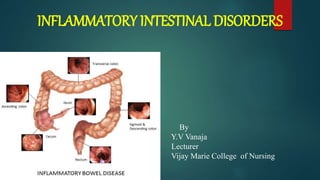
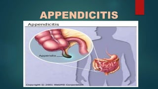
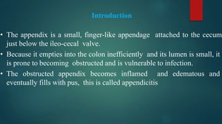
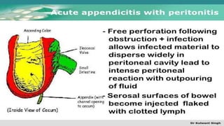
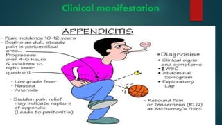
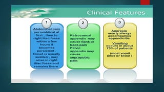
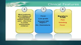
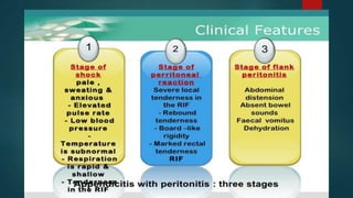
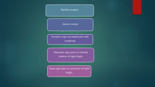
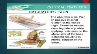
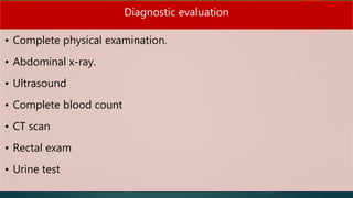
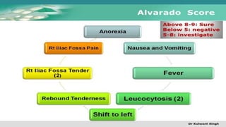
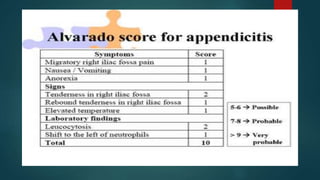
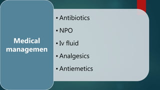
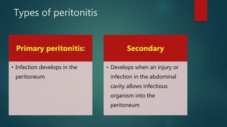
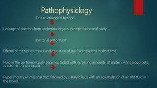
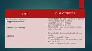
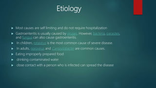
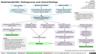
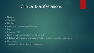
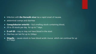
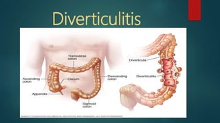
SlideShare a Scribd company logo
INFLAMMATORY INTESTINAL DISORDERS
By
Y.V Vanaja
Lecturer
Vijay Marie College of Nursing
A part from the lower GI tract is susceptible to acute inflammation
caused by bacterial, viral or fungal infection.
 Appendicitis
 Peritonitis
 Gastroenteritis
 Diverticulitis
APPENDICITIS
Introduction
• The appendix is a small, finger-like appendage attached to the cecum
just below the ileo-cecal valve.
• Because it empties into the colon inefficiently and its lumen is small, it
is prone to becoming obstructed and is vulnerable to infection.
• The obstructed appendix becomes inflamed and edematous and
eventually fills with pus, this is called appendicitis
Definition
 Appendicitis is an acute Inflammation of the vermiform appendix.
Inflammation occurs when the lumen of the appendix is obstructed,
leading to infection as bacteria invade the wall of the appendix
“ “Ignatavivcious”
• Appendicitis is an inflammation of the appendix, a narrow blind tube
that extends from the interior part of the cecum. “Lewis”
• the appendix becomes inflamed and edematous as a result of either
kinked or occluded by a fecalith, tumor or foreign body
“Brunner”
INCIDENCE
• Males are affected more than females.
• Teenagers more frequently than adults.
• The highest incidence is in those between the ages
of 10 and 30years
Types of appendicitis
 Lumen of the appendix is obstructed as a result of fecaliths
 Less common causes are – malignant tumors
 Trauma to the abdomen
 Intramural thickening caused by hyper
growth of lymphoid tissue
 Venous engorgement
 Accumulation of mucus and bacteria
Pathophysiology
Obstruction can result from food matter, adhesion, or lymphoid hyperplasia
Mucosa continues to secrete fluid.
Increase in the intraluminal pressure and restricting the blood flow.
End result is perforation and spillage of infected appendiceal contents into the
peritoneum
as inflammation continues, serosa and adjacent structures become inflammed
 Resulting in the generalized or upper abdominal pain that becomes localized in the
RLQ
 eventually the inflamed appendix fills with pus
 If rapid process may result in peritonitis
Clinical manifestation
Painful urination
Severe cramps
Dunphy’s sign (increased pain with
coughing).
Obturator sign( pain on internal
rotation of right thigh)
Psoas sign (pain on extension of right
thigh).
Complications
 Perforation
 Peritonitis
 Abscess
Diagnostic evaluation
• Complete physical examination.
• Abdominal x-ray.
• Ultrasound
• Complete blood count
• CT scan
• Rectal exam
• Urine test
• Antibiotics
• NPO
• Iv fluid
• Analgesics
• Antiemetics
Medical
managemen
Surgical management
 laparoscopic Appendectomy
 A new procedure is known as Natural orifice transluminal endoscopic
surgery , does not require an external skin incision
 Laparotomy is an open surgical approach with a larger abdominal incision for complicated or
atypical appendicitis or peritonitis
PERITONOTIS
Introduction:
The peritoneum is the serous membrane forming the lining of the abdominal
cavity
Peritonitis is an inflammation of the peritoneum, the tissue that lines the inner
wall of the abdomen and covers and supports most of abdominal organs.
Peritonitis is usually caused by infection from bacteria or fungi.
Definition:
Peritonitis is an inflammation of the peritoneum, the serous membrane lining the abdominal cavity and
covering the viscera.
“Brunner”
Peritonitis is a life threatening, acute inflammation of the visceral/ parietal peritoneum and endothelial
lining of the abdominal cavity.
“ Ignatavicious”
Types of peritonitis
Primary peritonitis:
• Infection develops in the
peritoneum
Secondary
• Develops when an injury or
infection in the abdominal
cavity allows infectious
organism into the
peritoneum
Risk factors
Riskfactors for primary peritonitis:
 Liver disease (cirrhosis)
 Fluid in the abdomen
 Weakened immune system
 Pelvic inflammatory disease
Risk factors for secondary peritonitis include:
 Appendicitis (inflammation of the appendix)
 Stomach ulcers,
 Twisted intestine,
 Pancreatitis
 Inflammatory bowel disease,
 Injury caused by an operation.
 Peritoneal dialysis,
 Trauma.
I- Infected peritonitis:
 1) Generalized Infected peritonitis:
Perforation of the part of the gastrointestinal tract is the most
common cause of peritonitis
2) Systemic or localized infections (such as tuberculosis)
may rarely have a peritoneal localisation.
II –Non Infected Peritonitis
 Leakage of sterile body fluids into the peritoneum such as blood gastric juice
Pathophysiology
Due to etiological factors
Leakage of contents from abdominal organs into the abdominal cavity
Bacterial perforation
Edema of the tissues results and exudation of the fluid develops in short time
Fluid in the peritoneal cavity becomes turbid with increasing amounts of protein, white blood cells,
cellular debris and blood
Hyper motility of intestinal tract followed by paralytic ileus with an accumulation of air and fluid in
the bowel
Clinical manifestations:
 Abdominal pain
 Tenderness over the involved area
 Rebound tenderness
 Muscular rigidity and spasm
 Abdominal distention
 Fever
 Tachycardia
 Tachypnea
 Nausea, vomiting
 Altered bowel disease
 Muscular rigidity
 Peristalsis slows or stops in response to severe peritoneal
inflammation and the lumen of bowel becomes distended with
gas and fluid
 Respiratory problems can occur as a result of increased
abdominal pressure against the diaphragm from intestinal
distention and fluid shifts to the peritoneal cavity.
Diagnostic Evaluation:
 History collection
 Physical Examination
 CBC
 Serum electrolytes
 Abdominal X-ray
 Abdominal paracentesis & culture of fluid
Management
Nonsurgical management
 IV fluids and broad spectrum antibiotics
 Monitor daily weight and I/O chart carefully
 NG tube decompresses the stomach and the intestine.
 Patient is NPO
 Monitor O2 saturation and Apply O2 as prescribed
 Administer analgesics
 Antiemetics
 Intestinal intubation and suction assist in relieving abdominal
distention and in promoting intestinal function
Surgical Management
 Exploratory Laparotomy – surgical opening into the abdomen
Laparoscopy is used to remove or repair the inflamed or perforated organ
 Colon resection with or without colostomy for a perforated diverticulum
 Before the incision is closed the surgeon irrigates the peritoneum with antibiotic
solutions
 Several catheters may be inserted to drain the cavity and provide a route for
irrigation after surgery.
GASTROENTERITIS
Introduction
 Gastroenteritis is a medical condition characterized by inflammation of
the gastrointestinal tract that involves both the stomach the small
intestine resulting in some combination of diarrhea, vomiting, and
abdominal pain and cramping. It usually happens because of infection
by a virus or bacteria.
 It affects mainly the small bowel and can be caused by either viral or
bacterial infections, which have similar manifestations.
Definitions:
 Gastroenteritis is an increase in the frequency and water content of
stools and/or vomiting as a result of inflammation of the mucous
membranes of the stomach and intestinal tract
“ Ignatavicious”
Gastroenteritis is an inflammation of the mucosa of the stomach and
small intestine “ Lewis”
Types of Gastroenteritis:
TYPE CHARACTERISTICS
Viral Gastroenteritis :
Epidemic viral:
Rotavirus & Norwalk virus
• Caused by many parvovirus –type organisms
transmitted by the fecal-oral route in food and water
• Incubation period is 10-51hrs
• Communicable during acute illness
o Transmitted by the fecal-oral route or by contact with
infected animals or infants
o Incubation period 1-10 days
o Rotavirus is most common in infants and young
children
o Norwalk virus affects young children and adults
TYPE CHARACTERISTICS
Bacterial Gastroenteritis:
Campylobacter enteritis:
Escherichia coli diarrhea
Shigellosis:
• Transmitted by the fecal- oral route or contact
with infected animals or infants
• Incubation period – 1-10 days
• Communicable for 2-7 weeks
 Transmitted by fecal contamination of food,
water, or fomites
• Transmitted by direct and indirect fecal- oral
routes
• Incubation period 1-7 days
• Communicable during the acute illness to 4
after the illness
• Humans possibly carries for months
Etiology
 Most causes are self limiting and do not require hospitalization
 Gastroenteritis is usually caused by viruses. However, bacteria, parasites,
and fungus can also cause gastroenteritis.
 In children, rotavirus is the most common cause of severe disease.
 In adults, norovirus and Campylobacter are common causes.
 Eating improperly prepared food
 drinking contaminated water
 close contact with a person who is infected can spread the disease
Clinical Manifestations
 Nausea
 Vomiting
 Diarrhea
 Abdominal cramping and distention
 Fever
 Increased WBC
 Blood or mucus in the stool may be present
 In Patient with epidemic viral gastroenteritis – myalgia, headache, and malaise
 Weakness
 cardiac dysrhythmias due to hypokalemia
 Infection with the Norwalk virus has a rapid onset of nausea,
 abdominal cramps and diarrhea
 Campylobacter enteritis – foul smelling stools containing blood,
 20 to 30 stools per day for up to 7 days.
 E-coli GE – may or may not have blood in the stool.
 Diarrhea can last for up to 10days
 Shigella - causes stools to have blood acidic mucus which can continue for up
to 5 days
Diagnosis
 Stool culture
 serum glucose levels
 CBP
 Serum electrolytes
Management
 Monitoring of intake and output
 Fluid replacement
 IV fluids such as half strength normal saline to replace sodiulm lost in
vomitus
 Obtain weight and orthostatic B.P
 Until vomitings has ceased the patient should be on NPO status
 Skin care -
Drugs:
 Antiperistaltic agents an initial dose of loperamide
(Imodium) 4mg
 Antibiotics eg – ciprofloxacin, levofloxacin, azithromycin
 If GE due to shigellosis, antiinfective agents such as
trimethoprim/sulfamethoxazole
Diverticulitis
Introduction
Diverticula are small, bulging pouches that can form in the
lining of your digestive system. They are found most often in
the lower part of the large intestine (colon). Diverticula are
common, especially after age 40, and seldom cause problems.
Sometimes, however, one or more of the pouches become
inflamed or infected. That condition is known as diverticulitis .
Definitions:
Diverticulitis is the inflammation of one or more diverticula
“Ignatavicious”
Diverticulitis results when food and bacteria retained in
diverticulum produce infection and inflammation that can
impede drainage and lead to perforation or abscess formation.
“ Brunner”
Risk factors
 Congenital predisposition id suspected when the disorder occurs in those
younger than 40 yrs of age.
 Aging: The incidence of diverticulitis increases with age.
 Obesity.
 Smoking.
 Lack of exercise.
 Diet high in animal fat and low in fiber.
 Certain medications Several drugs are associated with an increased risk of
diverticulitis, including steroids, opioids and nonsteroidal anti-inflammatory
drugs, such as ibuprofen (Advil, Motrin IB, others) and naproxen sodium
(Aleve).
Causes
 Diverticula usually develop when naturally weak
places in your colon give way under pressure. This
causes marble-sized pouches to protrude through
the colon wall.
 High intraluminal pressure.
 Diet low in fiber
 Retained undigested food in diverticula
Pathophysiology
Due to High Intraluminal pressure
The muscle of the colon hypertrophies, thickens, and becomes rigid, and herniation of the mucosa and
sub mucosa through the colon wall
Decreased muscle strength in the colon wall
If undigested food or bacteria become trapped in a diverticulum, however blood supply to that area is
reduced
Bacteria invade the diverticulum
A diverticulum can become obstructed and then inflamed if the obstruction continues
 Which can perforate and develop a local abscess
 A perforated diverticulum can progress to an intra abdominal perforation with peritonitis.
 Inflammation can also result in fistulas to other organs, such as the bladder and the vagina
Clinical Manifestations:
 Signs of acute diverticulosis are bowel irregularity
 intervals of diarrhea
 Abrupt crampy pain in the left lower quadrant of the abdomen .
If diverticulitis is suspected
 Low-grade fever
 Nausea
 Abdominal pain
 Constipation
 Bleeding from the rectum
 Tenderness over the abdominal palpation
 Elevated temperature with chills
 Tachycardia
Diagnosis
 History collection
 Physical examination
 CBP
 Stool test for occult blood
 Urine analysis may show a few RBC
 X ray of the intestinal tract barium contrast
 Barium enema shows the diverticula of the large intestine
 Abdominal X ray too identify free air and fluid indicating perforation
 CT scan
 Abdominal Ultrasonography
 Colonoscopy 4 to 8 wks after acute phase– if rectal bleeding
Management
Nonsurgical management
 A combination of drug and no nutrition therapy with rest is used to decrease the
inflammation.
 Broad- spectrum antimicrobial drugs, such as metronidazole plus trimethoprim/
sulfamethoxazole or ciprofloxacin
 Opioid analgesics – such as morphine sulfate.
 IV fluids to correct Dehydration & IV drug therapy
 antispasmodics such as propantheline bromide and oxyphencyclimine
 Assess the patient on ongoing basis for manifestations of fluid and electrolyte
imbalance.
 Restriction of high fiber diet because it may cause irritation
 NPO
 NG tube - if nausea, vomiting or abdominal distention is severe.
 When inflammation is resolved and bowel function returns to normal, fiber
containing diet is introduced gradually
Surgical management
 Emergency surgery if peritonitis, bowel resection, or pelvic abscess is present.
 Colon resection with or without colostomy
 Some patients may have colostomy closure and anastomosis after the bowel
has been allowed to rest for 3 to 6 months
 The patient may have one of two surgical approaches
 Conventional open approach or
 Minimally invasive surgery via a laparoscopy
Postoperative care
 The patient may have drain in place at the abdominal incision site for
several days
 The stoma may be covered with a petroleum gauze dressing because the
colostomy does not drain for about 2 days or colostomy bag may be
placed over the stoma
 If the stoma is visible monitor for color and integrity
 The may be NPO with an NGT until peristalsis returns if open surgery is
performed
 A tight seal around the stoma is essential to avoid contact feces with the
skin
 Colostomy care
Inflammatory intestinal disorders

More Related Content

What's hot

Acute peritonitis
Acute peritonitisAcute peritonitis
Acute peritonitis
Yuvaraj Karthick
 
Abdominal Tuberculosis
Abdominal TuberculosisAbdominal Tuberculosis
Abdominal Tuberculosis
GunJee Gj
 
Abdominal tb (dr masood tareen)
Abdominal tb (dr masood tareen)Abdominal tb (dr masood tareen)
Abdominal tb (dr masood tareen)
Masood Tareen
 
PERITONEAL TUBERCULOSIS & TUBERCULOUS MESENTERIC LYMPHADENITIS
PERITONEAL TUBERCULOSIS & TUBERCULOUS MESENTERIC LYMPHADENITISPERITONEAL TUBERCULOSIS & TUBERCULOUS MESENTERIC LYMPHADENITIS
PERITONEAL TUBERCULOSIS & TUBERCULOUS MESENTERIC LYMPHADENITIS
Praveen RK
 
Acute Suppurative Peritonitis
Acute Suppurative PeritonitisAcute Suppurative Peritonitis
Acute Suppurative PeritonitisDeep Deep
 
ABDOMINAL TUBERCULOSIS
ABDOMINAL TUBERCULOSISABDOMINAL TUBERCULOSIS
ABDOMINAL TUBERCULOSIS
akjlm10
 
Abdominal tuberculosis
Abdominal tuberculosisAbdominal tuberculosis
Abdominal tuberculosis
Shilpa Bhaskar
 
Abdiminal tuberculosis
Abdiminal tuberculosisAbdiminal tuberculosis
Abdiminal tuberculosis
Thorlikonda Sasidhar
 
GIT Tb
GIT Tb GIT Tb
GIT Tb
RMLIMS
 
Peritonitis
PeritonitisPeritonitis
Peritonitis
Ayesha Batool
 
Abdominal tb
Abdominal tbAbdominal tb
Abdominal tb
Vrishit Saraswat
 
Abdominal tuberculosis
Abdominal tuberculosisAbdominal tuberculosis
Abdominal tuberculosis
gku1990
 
Abdominal tuberculosis
Abdominal tuberculosisAbdominal tuberculosis
Abdominal tuberculosis
Abdominal tuberculosisAbdominal tuberculosis
Abdominal tuberculosisBPKIHS
 
IMAGING IN ABDOMINAL TUBERCULOSIS
IMAGING IN ABDOMINAL TUBERCULOSISIMAGING IN ABDOMINAL TUBERCULOSIS
IMAGING IN ABDOMINAL TUBERCULOSIS
Navni Garg
 
Abdominal tuberculosis
Abdominal tuberculosisAbdominal tuberculosis
Abdominal tuberculosisJohn Peter
 
Tb
TbTb
Peritonitis
PeritonitisPeritonitis
Peritonitis
abhishekmehta149
 

What's hot (20)

Acute peritonitis
Acute peritonitisAcute peritonitis
Acute peritonitis
 
Abdominal Tuberculosis
Abdominal TuberculosisAbdominal Tuberculosis
Abdominal Tuberculosis
 
Abdominal tb (dr masood tareen)
Abdominal tb (dr masood tareen)Abdominal tb (dr masood tareen)
Abdominal tb (dr masood tareen)
 
PERITONEAL TUBERCULOSIS & TUBERCULOUS MESENTERIC LYMPHADENITIS
PERITONEAL TUBERCULOSIS & TUBERCULOUS MESENTERIC LYMPHADENITISPERITONEAL TUBERCULOSIS & TUBERCULOUS MESENTERIC LYMPHADENITIS
PERITONEAL TUBERCULOSIS & TUBERCULOUS MESENTERIC LYMPHADENITIS
 
Acute Suppurative Peritonitis
Acute Suppurative PeritonitisAcute Suppurative Peritonitis
Acute Suppurative Peritonitis
 
ABDOMINAL TUBERCULOSIS
ABDOMINAL TUBERCULOSISABDOMINAL TUBERCULOSIS
ABDOMINAL TUBERCULOSIS
 
Abdominal tuberculosis
Abdominal tuberculosisAbdominal tuberculosis
Abdominal tuberculosis
 
Abdiminal tuberculosis
Abdiminal tuberculosisAbdiminal tuberculosis
Abdiminal tuberculosis
 
GIT Tb
GIT Tb GIT Tb
GIT Tb
 
Peritonitis
PeritonitisPeritonitis
Peritonitis
 
Abdominal tb
Abdominal tbAbdominal tb
Abdominal tb
 
Abdominal tb
Abdominal tbAbdominal tb
Abdominal tb
 
Abdominal tuberculosis
Abdominal tuberculosisAbdominal tuberculosis
Abdominal tuberculosis
 
Abdominal tuberculosis
Abdominal tuberculosisAbdominal tuberculosis
Abdominal tuberculosis
 
Abdominal tuberculosis
Abdominal tuberculosisAbdominal tuberculosis
Abdominal tuberculosis
 
IMAGING IN ABDOMINAL TUBERCULOSIS
IMAGING IN ABDOMINAL TUBERCULOSISIMAGING IN ABDOMINAL TUBERCULOSIS
IMAGING IN ABDOMINAL TUBERCULOSIS
 
Abdominal tuberculosis
Abdominal tuberculosisAbdominal tuberculosis
Abdominal tuberculosis
 
Tb
TbTb
Tb
 
Peritonitis (lecture mogilevec e.v
Peritonitis (lecture mogilevec e.vPeritonitis (lecture mogilevec e.v
Peritonitis (lecture mogilevec e.v
 
Peritonitis
PeritonitisPeritonitis
Peritonitis
 

Similar to Inflammatory intestinal disorders

Peritonitis.pptx
Peritonitis.pptxPeritonitis.pptx
Peritonitis.pptx
Melba Shaya Sweety
 
Appendicitis
AppendicitisAppendicitis
Appendicitis
vanajayarrlagadda
 
Peritonitis.ppt
Peritonitis.pptPeritonitis.ppt
Peritonitis.ppt
YohelioPriawanSibu
 
Common pediatric surgical conditions 2 By Dr Hatem El Gohary
Common pediatric surgical conditions 2 By Dr Hatem El GoharyCommon pediatric surgical conditions 2 By Dr Hatem El Gohary
Common pediatric surgical conditions 2 By Dr Hatem El GoharyHatem Elgohary
 
APPENDICITIS power point presentations.pptx
APPENDICITIS power point presentations.pptxAPPENDICITIS power point presentations.pptx
APPENDICITIS power point presentations.pptx
IbrahimKargbo13
 
acuteperitonitis-161018013220 (1).pdf
acuteperitonitis-161018013220 (1).pdfacuteperitonitis-161018013220 (1).pdf
acuteperitonitis-161018013220 (1).pdf
ssuserc27f73
 
3.peritonitis
3.peritonitis3.peritonitis
3.peritonitis
ChristopherJohn67
 
Seminar presentation on Appendicitis.pptx
Seminar presentation on Appendicitis.pptxSeminar presentation on Appendicitis.pptx
Seminar presentation on Appendicitis.pptx
DaudaUsman6
 
DIVERTICULAR DISEASE BYBHUMIKA.G.THAKOR
DIVERTICULAR DISEASE  BYBHUMIKA.G.THAKORDIVERTICULAR DISEASE  BYBHUMIKA.G.THAKOR
DIVERTICULAR DISEASE BYBHUMIKA.G.THAKOR
BhumikaThakor1
 
peritonium.pptx
peritonium.pptxperitonium.pptx
peritonium.pptx
arunabhasinha2
 
02. Purulent-inflammatory diseases of abdominal cavity_d38fe14abb13872ba8f006...
02. Purulent-inflammatory diseases of abdominal cavity_d38fe14abb13872ba8f006...02. Purulent-inflammatory diseases of abdominal cavity_d38fe14abb13872ba8f006...
02. Purulent-inflammatory diseases of abdominal cavity_d38fe14abb13872ba8f006...
ssuserca828a
 
11. Other Problems in Inflammatory Response.pptx
11. Other Problems in Inflammatory Response.pptx11. Other Problems in Inflammatory Response.pptx
11. Other Problems in Inflammatory Response.pptx
JRRolfNeuqelet
 
Urinary Tract Infection.pptx
Urinary Tract Infection.pptxUrinary Tract Infection.pptx
Urinary Tract Infection.pptx
Dr.Navaneethakrishnan S
 
Diverticulitis, Gastroenteritis and dehydration .pptx
Diverticulitis, Gastroenteritis and dehydration .pptxDiverticulitis, Gastroenteritis and dehydration .pptx
Diverticulitis, Gastroenteritis and dehydration .pptx
christy199321
 
Diverticular disease of colon.pptx
Diverticular disease of colon.pptxDiverticular disease of colon.pptx
Diverticular disease of colon.pptx
Pradeep Pande
 
Abdominal Tuberculosis
Abdominal TuberculosisAbdominal Tuberculosis
Abdominal Tuberculosis
Prateek Kumar
 
Ulcerative intestine
Ulcerative  intestineUlcerative  intestine
Ulcerative intestineSaurav Singh
 
Peptic Ulcer complications By Abdullah Farooqi GM20-148.pptx
Peptic Ulcer complications By Abdullah Farooqi GM20-148.pptxPeptic Ulcer complications By Abdullah Farooqi GM20-148.pptx
Peptic Ulcer complications By Abdullah Farooqi GM20-148.pptx
shiv847105
 
Diarrhea tutorial 2023 students copy.pptx
Diarrhea tutorial 2023 students copy.pptxDiarrhea tutorial 2023 students copy.pptx
Diarrhea tutorial 2023 students copy.pptx
MUHAMMADCHAUDHRY39
 

Similar to Inflammatory intestinal disorders (20)

Peritonitis.pptx
Peritonitis.pptxPeritonitis.pptx
Peritonitis.pptx
 
Appendicitis
AppendicitisAppendicitis
Appendicitis
 
Peritonitis.ppt
Peritonitis.pptPeritonitis.ppt
Peritonitis.ppt
 
Common pediatric surgical conditions 2 By Dr Hatem El Gohary
Common pediatric surgical conditions 2 By Dr Hatem El GoharyCommon pediatric surgical conditions 2 By Dr Hatem El Gohary
Common pediatric surgical conditions 2 By Dr Hatem El Gohary
 
APPENDICITIS power point presentations.pptx
APPENDICITIS power point presentations.pptxAPPENDICITIS power point presentations.pptx
APPENDICITIS power point presentations.pptx
 
acuteperitonitis-161018013220 (1).pdf
acuteperitonitis-161018013220 (1).pdfacuteperitonitis-161018013220 (1).pdf
acuteperitonitis-161018013220 (1).pdf
 
Peritonitis
PeritonitisPeritonitis
Peritonitis
 
3.peritonitis
3.peritonitis3.peritonitis
3.peritonitis
 
Seminar presentation on Appendicitis.pptx
Seminar presentation on Appendicitis.pptxSeminar presentation on Appendicitis.pptx
Seminar presentation on Appendicitis.pptx
 
DIVERTICULAR DISEASE BYBHUMIKA.G.THAKOR
DIVERTICULAR DISEASE  BYBHUMIKA.G.THAKORDIVERTICULAR DISEASE  BYBHUMIKA.G.THAKOR
DIVERTICULAR DISEASE BYBHUMIKA.G.THAKOR
 
peritonium.pptx
peritonium.pptxperitonium.pptx
peritonium.pptx
 
02. Purulent-inflammatory diseases of abdominal cavity_d38fe14abb13872ba8f006...
02. Purulent-inflammatory diseases of abdominal cavity_d38fe14abb13872ba8f006...02. Purulent-inflammatory diseases of abdominal cavity_d38fe14abb13872ba8f006...
02. Purulent-inflammatory diseases of abdominal cavity_d38fe14abb13872ba8f006...
 
11. Other Problems in Inflammatory Response.pptx
11. Other Problems in Inflammatory Response.pptx11. Other Problems in Inflammatory Response.pptx
11. Other Problems in Inflammatory Response.pptx
 
Urinary Tract Infection.pptx
Urinary Tract Infection.pptxUrinary Tract Infection.pptx
Urinary Tract Infection.pptx
 
Diverticulitis, Gastroenteritis and dehydration .pptx
Diverticulitis, Gastroenteritis and dehydration .pptxDiverticulitis, Gastroenteritis and dehydration .pptx
Diverticulitis, Gastroenteritis and dehydration .pptx
 
Diverticular disease of colon.pptx
Diverticular disease of colon.pptxDiverticular disease of colon.pptx
Diverticular disease of colon.pptx
 
Abdominal Tuberculosis
Abdominal TuberculosisAbdominal Tuberculosis
Abdominal Tuberculosis
 
Ulcerative intestine
Ulcerative  intestineUlcerative  intestine
Ulcerative intestine
 
Peptic Ulcer complications By Abdullah Farooqi GM20-148.pptx
Peptic Ulcer complications By Abdullah Farooqi GM20-148.pptxPeptic Ulcer complications By Abdullah Farooqi GM20-148.pptx
Peptic Ulcer complications By Abdullah Farooqi GM20-148.pptx
 
Diarrhea tutorial 2023 students copy.pptx
Diarrhea tutorial 2023 students copy.pptxDiarrhea tutorial 2023 students copy.pptx
Diarrhea tutorial 2023 students copy.pptx
 

More from vanajayarrlagadda

Cardiac cycle
Cardiac cycle Cardiac cycle
Cardiac cycle
vanajayarrlagadda
 
Blood groups ppt
Blood groups pptBlood groups ppt
Blood groups ppt
vanajayarrlagadda
 
Pancreatic disorders
Pancreatic disordersPancreatic disorders
Pancreatic disorders
vanajayarrlagadda
 
Mastoiditis
MastoiditisMastoiditis
Mastoiditis
vanajayarrlagadda
 
Otitis media
Otitis mediaOtitis media
Otitis media
vanajayarrlagadda
 
Pancreatic islets
Pancreatic isletsPancreatic islets
Pancreatic islets
vanajayarrlagadda
 
Cholecystitis
CholecystitisCholecystitis
Cholecystitis
vanajayarrlagadda
 
Adrenal glands
Adrenal glandsAdrenal glands
Adrenal glands
vanajayarrlagadda
 
Anatomy and physiology of thyroid gland
Anatomy and physiology of thyroid glandAnatomy and physiology of thyroid gland
Anatomy and physiology of thyroid gland
vanajayarrlagadda
 
Malabsorption syndrome
Malabsorption syndromeMalabsorption syndrome
Malabsorption syndrome
vanajayarrlagadda
 
Intestinal obstruction
Intestinal obstructionIntestinal obstruction
Intestinal obstruction
vanajayarrlagadda
 
Physiology of pituitary gland
Physiology of pituitary glandPhysiology of pituitary gland
Physiology of pituitary gland
vanajayarrlagadda
 
Anatomy of endocrine system
Anatomy of endocrine systemAnatomy of endocrine system
Anatomy of endocrine system
vanajayarrlagadda
 
Gustaation
GustaationGustaation
Gustaation
vanajayarrlagadda
 
Physiology of hearing
Physiology of hearingPhysiology of hearing
Physiology of hearing
vanajayarrlagadda
 
Nose
NoseNose
Skin
SkinSkin
Internal ear
Internal earInternal ear
Internal ear
vanajayarrlagadda
 
Fracture healing
Fracture healingFracture healing
Fracture healing
vanajayarrlagadda
 
Tissues
TissuesTissues

More from vanajayarrlagadda (20)

Cardiac cycle
Cardiac cycle Cardiac cycle
Cardiac cycle
 
Blood groups ppt
Blood groups pptBlood groups ppt
Blood groups ppt
 
Pancreatic disorders
Pancreatic disordersPancreatic disorders
Pancreatic disorders
 
Mastoiditis
MastoiditisMastoiditis
Mastoiditis
 
Otitis media
Otitis mediaOtitis media
Otitis media
 
Pancreatic islets
Pancreatic isletsPancreatic islets
Pancreatic islets
 
Cholecystitis
CholecystitisCholecystitis
Cholecystitis
 
Adrenal glands
Adrenal glandsAdrenal glands
Adrenal glands
 
Anatomy and physiology of thyroid gland
Anatomy and physiology of thyroid glandAnatomy and physiology of thyroid gland
Anatomy and physiology of thyroid gland
 
Malabsorption syndrome
Malabsorption syndromeMalabsorption syndrome
Malabsorption syndrome
 
Intestinal obstruction
Intestinal obstructionIntestinal obstruction
Intestinal obstruction
 
Physiology of pituitary gland
Physiology of pituitary glandPhysiology of pituitary gland
Physiology of pituitary gland
 
Anatomy of endocrine system
Anatomy of endocrine systemAnatomy of endocrine system
Anatomy of endocrine system
 
Gustaation
GustaationGustaation
Gustaation
 
Physiology of hearing
Physiology of hearingPhysiology of hearing
Physiology of hearing
 
Nose
NoseNose
Nose
 
Skin
SkinSkin
Skin
 
Internal ear
Internal earInternal ear
Internal ear
 
Fracture healing
Fracture healingFracture healing
Fracture healing
 
Tissues
TissuesTissues
Tissues
 

Recently uploaded

Phrasal Verbs.XXXXXXXXXXXXXXXXXXXXXXXXXX
Phrasal Verbs.XXXXXXXXXXXXXXXXXXXXXXXXXXPhrasal Verbs.XXXXXXXXXXXXXXXXXXXXXXXXXX
Phrasal Verbs.XXXXXXXXXXXXXXXXXXXXXXXXXX
MIRIAMSALINAS13
 
Introduction to AI for Nonprofits with Tapp Network
Introduction to AI for Nonprofits with Tapp NetworkIntroduction to AI for Nonprofits with Tapp Network
Introduction to AI for Nonprofits with Tapp Network
TechSoup
 
Mule 4.6 & Java 17 Upgrade | MuleSoft Mysore Meetup #46
Mule 4.6 & Java 17 Upgrade | MuleSoft Mysore Meetup #46Mule 4.6 & Java 17 Upgrade | MuleSoft Mysore Meetup #46
Mule 4.6 & Java 17 Upgrade | MuleSoft Mysore Meetup #46
MysoreMuleSoftMeetup
 
How to Make a Field invisible in Odoo 17
How to Make a Field invisible in Odoo 17How to Make a Field invisible in Odoo 17
How to Make a Field invisible in Odoo 17
Celine George
 
The approach at University of Liverpool.pptx
The approach at University of Liverpool.pptxThe approach at University of Liverpool.pptx
The approach at University of Liverpool.pptx
Jisc
 
A Strategic Approach: GenAI in Education
A Strategic Approach: GenAI in EducationA Strategic Approach: GenAI in Education
A Strategic Approach: GenAI in Education
Peter Windle
 
Language Across the Curriculm LAC B.Ed.
Language Across the  Curriculm LAC B.Ed.Language Across the  Curriculm LAC B.Ed.
Language Across the Curriculm LAC B.Ed.
Atul Kumar Singh
 
CLASS 11 CBSE B.St Project AIDS TO TRADE - INSURANCE
CLASS 11 CBSE B.St Project AIDS TO TRADE - INSURANCECLASS 11 CBSE B.St Project AIDS TO TRADE - INSURANCE
CLASS 11 CBSE B.St Project AIDS TO TRADE - INSURANCE
BhavyaRajput3
 
Thesis Statement for students diagnonsed withADHD.ppt
Thesis Statement for students diagnonsed withADHD.pptThesis Statement for students diagnonsed withADHD.ppt
Thesis Statement for students diagnonsed withADHD.ppt
EverAndrsGuerraGuerr
 
Lapbook sobre os Regimes Totalitários.pdf
Lapbook sobre os Regimes Totalitários.pdfLapbook sobre os Regimes Totalitários.pdf
Lapbook sobre os Regimes Totalitários.pdf
Jean Carlos Nunes Paixão
 
Polish students' mobility in the Czech Republic
Polish students' mobility in the Czech RepublicPolish students' mobility in the Czech Republic
Polish students' mobility in the Czech Republic
Anna Sz.
 
Unit 2- Research Aptitude (UGC NET Paper I).pdf
Unit 2- Research Aptitude (UGC NET Paper I).pdfUnit 2- Research Aptitude (UGC NET Paper I).pdf
Unit 2- Research Aptitude (UGC NET Paper I).pdf
Thiyagu K
 
The geography of Taylor Swift - some ideas
The geography of Taylor Swift - some ideasThe geography of Taylor Swift - some ideas
The geography of Taylor Swift - some ideas
GeoBlogs
 
Honest Reviews of Tim Han LMA Course Program.pptx
Honest Reviews of Tim Han LMA Course Program.pptxHonest Reviews of Tim Han LMA Course Program.pptx
Honest Reviews of Tim Han LMA Course Program.pptx
timhan337
 
Synthetic Fiber Construction in lab .pptx
Synthetic Fiber Construction in lab .pptxSynthetic Fiber Construction in lab .pptx
Synthetic Fiber Construction in lab .pptx
Pavel ( NSTU)
 
1.4 modern child centered education - mahatma gandhi-2.pptx
1.4 modern child centered education - mahatma gandhi-2.pptx1.4 modern child centered education - mahatma gandhi-2.pptx
1.4 modern child centered education - mahatma gandhi-2.pptx
JosvitaDsouza2
 
The French Revolution Class 9 Study Material pdf free download
The French Revolution Class 9 Study Material pdf free downloadThe French Revolution Class 9 Study Material pdf free download
The French Revolution Class 9 Study Material pdf free download
Vivekanand Anglo Vedic Academy
 
The Roman Empire A Historical Colossus.pdf
The Roman Empire A Historical Colossus.pdfThe Roman Empire A Historical Colossus.pdf
The Roman Empire A Historical Colossus.pdf
kaushalkr1407
 
The Accursed House by Émile Gaboriau.pptx
The Accursed House by Émile Gaboriau.pptxThe Accursed House by Émile Gaboriau.pptx
The Accursed House by Émile Gaboriau.pptx
DhatriParmar
 
Guidance_and_Counselling.pdf B.Ed. 4th Semester
Guidance_and_Counselling.pdf B.Ed. 4th SemesterGuidance_and_Counselling.pdf B.Ed. 4th Semester
Guidance_and_Counselling.pdf B.Ed. 4th Semester
Atul Kumar Singh
 

Recently uploaded (20)

Phrasal Verbs.XXXXXXXXXXXXXXXXXXXXXXXXXX
Phrasal Verbs.XXXXXXXXXXXXXXXXXXXXXXXXXXPhrasal Verbs.XXXXXXXXXXXXXXXXXXXXXXXXXX
Phrasal Verbs.XXXXXXXXXXXXXXXXXXXXXXXXXX
 
Introduction to AI for Nonprofits with Tapp Network
Introduction to AI for Nonprofits with Tapp NetworkIntroduction to AI for Nonprofits with Tapp Network
Introduction to AI for Nonprofits with Tapp Network
 
Mule 4.6 & Java 17 Upgrade | MuleSoft Mysore Meetup #46
Mule 4.6 & Java 17 Upgrade | MuleSoft Mysore Meetup #46Mule 4.6 & Java 17 Upgrade | MuleSoft Mysore Meetup #46
Mule 4.6 & Java 17 Upgrade | MuleSoft Mysore Meetup #46
 
How to Make a Field invisible in Odoo 17
How to Make a Field invisible in Odoo 17How to Make a Field invisible in Odoo 17
How to Make a Field invisible in Odoo 17
 
The approach at University of Liverpool.pptx
The approach at University of Liverpool.pptxThe approach at University of Liverpool.pptx
The approach at University of Liverpool.pptx
 
A Strategic Approach: GenAI in Education
A Strategic Approach: GenAI in EducationA Strategic Approach: GenAI in Education
A Strategic Approach: GenAI in Education
 
Language Across the Curriculm LAC B.Ed.
Language Across the  Curriculm LAC B.Ed.Language Across the  Curriculm LAC B.Ed.
Language Across the Curriculm LAC B.Ed.
 
CLASS 11 CBSE B.St Project AIDS TO TRADE - INSURANCE
CLASS 11 CBSE B.St Project AIDS TO TRADE - INSURANCECLASS 11 CBSE B.St Project AIDS TO TRADE - INSURANCE
CLASS 11 CBSE B.St Project AIDS TO TRADE - INSURANCE
 
Thesis Statement for students diagnonsed withADHD.ppt
Thesis Statement for students diagnonsed withADHD.pptThesis Statement for students diagnonsed withADHD.ppt
Thesis Statement for students diagnonsed withADHD.ppt
 
Lapbook sobre os Regimes Totalitários.pdf
Lapbook sobre os Regimes Totalitários.pdfLapbook sobre os Regimes Totalitários.pdf
Lapbook sobre os Regimes Totalitários.pdf
 
Polish students' mobility in the Czech Republic
Polish students' mobility in the Czech RepublicPolish students' mobility in the Czech Republic
Polish students' mobility in the Czech Republic
 
Unit 2- Research Aptitude (UGC NET Paper I).pdf
Unit 2- Research Aptitude (UGC NET Paper I).pdfUnit 2- Research Aptitude (UGC NET Paper I).pdf
Unit 2- Research Aptitude (UGC NET Paper I).pdf
 
The geography of Taylor Swift - some ideas
The geography of Taylor Swift - some ideasThe geography of Taylor Swift - some ideas
The geography of Taylor Swift - some ideas
 
Honest Reviews of Tim Han LMA Course Program.pptx
Honest Reviews of Tim Han LMA Course Program.pptxHonest Reviews of Tim Han LMA Course Program.pptx
Honest Reviews of Tim Han LMA Course Program.pptx
 
Synthetic Fiber Construction in lab .pptx
Synthetic Fiber Construction in lab .pptxSynthetic Fiber Construction in lab .pptx
Synthetic Fiber Construction in lab .pptx
 
1.4 modern child centered education - mahatma gandhi-2.pptx
1.4 modern child centered education - mahatma gandhi-2.pptx1.4 modern child centered education - mahatma gandhi-2.pptx
1.4 modern child centered education - mahatma gandhi-2.pptx
 
The French Revolution Class 9 Study Material pdf free download
The French Revolution Class 9 Study Material pdf free downloadThe French Revolution Class 9 Study Material pdf free download
The French Revolution Class 9 Study Material pdf free download
 
The Roman Empire A Historical Colossus.pdf
The Roman Empire A Historical Colossus.pdfThe Roman Empire A Historical Colossus.pdf
The Roman Empire A Historical Colossus.pdf
 
The Accursed House by Émile Gaboriau.pptx
The Accursed House by Émile Gaboriau.pptxThe Accursed House by Émile Gaboriau.pptx
The Accursed House by Émile Gaboriau.pptx
 
Guidance_and_Counselling.pdf B.Ed. 4th Semester
Guidance_and_Counselling.pdf B.Ed. 4th SemesterGuidance_and_Counselling.pdf B.Ed. 4th Semester
Guidance_and_Counselling.pdf B.Ed. 4th Semester
 

Inflammatory intestinal disorders

  • 1. INFLAMMATORY INTESTINAL DISORDERS By Y.V Vanaja Lecturer Vijay Marie College of Nursing
  • 2. A part from the lower GI tract is susceptible to acute inflammation caused by bacterial, viral or fungal infection.  Appendicitis  Peritonitis  Gastroenteritis  Diverticulitis
  • 4. Introduction • The appendix is a small, finger-like appendage attached to the cecum just below the ileo-cecal valve. • Because it empties into the colon inefficiently and its lumen is small, it is prone to becoming obstructed and is vulnerable to infection. • The obstructed appendix becomes inflamed and edematous and eventually fills with pus, this is called appendicitis
  • 5. Definition  Appendicitis is an acute Inflammation of the vermiform appendix. Inflammation occurs when the lumen of the appendix is obstructed, leading to infection as bacteria invade the wall of the appendix “ “Ignatavivcious” • Appendicitis is an inflammation of the appendix, a narrow blind tube that extends from the interior part of the cecum. “Lewis” • the appendix becomes inflamed and edematous as a result of either kinked or occluded by a fecalith, tumor or foreign body “Brunner”
  • 6. INCIDENCE • Males are affected more than females. • Teenagers more frequently than adults. • The highest incidence is in those between the ages of 10 and 30years
  • 8.
  • 9.
  • 10.
  • 11.
  • 12.  Lumen of the appendix is obstructed as a result of fecaliths  Less common causes are – malignant tumors  Trauma to the abdomen  Intramural thickening caused by hyper growth of lymphoid tissue  Venous engorgement  Accumulation of mucus and bacteria
  • 13. Pathophysiology Obstruction can result from food matter, adhesion, or lymphoid hyperplasia Mucosa continues to secrete fluid. Increase in the intraluminal pressure and restricting the blood flow. End result is perforation and spillage of infected appendiceal contents into the peritoneum as inflammation continues, serosa and adjacent structures become inflammed
  • 14.  Resulting in the generalized or upper abdominal pain that becomes localized in the RLQ  eventually the inflamed appendix fills with pus  If rapid process may result in peritonitis
  • 16.
  • 17.
  • 18.
  • 19. Painful urination Severe cramps Dunphy’s sign (increased pain with coughing). Obturator sign( pain on internal rotation of right thigh) Psoas sign (pain on extension of right thigh).
  • 21.
  • 22.
  • 23.
  • 24.
  • 25.
  • 26. Diagnostic evaluation • Complete physical examination. • Abdominal x-ray. • Ultrasound • Complete blood count • CT scan • Rectal exam • Urine test
  • 27.
  • 28.
  • 29. • Antibiotics • NPO • Iv fluid • Analgesics • Antiemetics Medical managemen
  • 30. Surgical management  laparoscopic Appendectomy  A new procedure is known as Natural orifice transluminal endoscopic surgery , does not require an external skin incision
  • 31.
  • 32.  Laparotomy is an open surgical approach with a larger abdominal incision for complicated or atypical appendicitis or peritonitis
  • 34. Introduction: The peritoneum is the serous membrane forming the lining of the abdominal cavity Peritonitis is an inflammation of the peritoneum, the tissue that lines the inner wall of the abdomen and covers and supports most of abdominal organs. Peritonitis is usually caused by infection from bacteria or fungi.
  • 35. Definition: Peritonitis is an inflammation of the peritoneum, the serous membrane lining the abdominal cavity and covering the viscera. “Brunner” Peritonitis is a life threatening, acute inflammation of the visceral/ parietal peritoneum and endothelial lining of the abdominal cavity. “ Ignatavicious”
  • 36. Types of peritonitis Primary peritonitis: • Infection develops in the peritoneum Secondary • Develops when an injury or infection in the abdominal cavity allows infectious organism into the peritoneum
  • 37. Risk factors Riskfactors for primary peritonitis:  Liver disease (cirrhosis)  Fluid in the abdomen  Weakened immune system  Pelvic inflammatory disease Risk factors for secondary peritonitis include:  Appendicitis (inflammation of the appendix)  Stomach ulcers,  Twisted intestine,  Pancreatitis  Inflammatory bowel disease,  Injury caused by an operation.  Peritoneal dialysis,  Trauma.
  • 38. I- Infected peritonitis:  1) Generalized Infected peritonitis: Perforation of the part of the gastrointestinal tract is the most common cause of peritonitis 2) Systemic or localized infections (such as tuberculosis) may rarely have a peritoneal localisation. II –Non Infected Peritonitis  Leakage of sterile body fluids into the peritoneum such as blood gastric juice
  • 39. Pathophysiology Due to etiological factors Leakage of contents from abdominal organs into the abdominal cavity Bacterial perforation Edema of the tissues results and exudation of the fluid develops in short time Fluid in the peritoneal cavity becomes turbid with increasing amounts of protein, white blood cells, cellular debris and blood Hyper motility of intestinal tract followed by paralytic ileus with an accumulation of air and fluid in the bowel
  • 40. Clinical manifestations:  Abdominal pain  Tenderness over the involved area  Rebound tenderness  Muscular rigidity and spasm  Abdominal distention  Fever  Tachycardia  Tachypnea  Nausea, vomiting  Altered bowel disease
  • 41.  Muscular rigidity  Peristalsis slows or stops in response to severe peritoneal inflammation and the lumen of bowel becomes distended with gas and fluid  Respiratory problems can occur as a result of increased abdominal pressure against the diaphragm from intestinal distention and fluid shifts to the peritoneal cavity.
  • 42. Diagnostic Evaluation:  History collection  Physical Examination  CBC  Serum electrolytes  Abdominal X-ray  Abdominal paracentesis & culture of fluid
  • 43. Management Nonsurgical management  IV fluids and broad spectrum antibiotics  Monitor daily weight and I/O chart carefully  NG tube decompresses the stomach and the intestine.  Patient is NPO  Monitor O2 saturation and Apply O2 as prescribed  Administer analgesics  Antiemetics  Intestinal intubation and suction assist in relieving abdominal distention and in promoting intestinal function
  • 44. Surgical Management  Exploratory Laparotomy – surgical opening into the abdomen Laparoscopy is used to remove or repair the inflamed or perforated organ  Colon resection with or without colostomy for a perforated diverticulum  Before the incision is closed the surgeon irrigates the peritoneum with antibiotic solutions  Several catheters may be inserted to drain the cavity and provide a route for irrigation after surgery.
  • 46. Introduction  Gastroenteritis is a medical condition characterized by inflammation of the gastrointestinal tract that involves both the stomach the small intestine resulting in some combination of diarrhea, vomiting, and abdominal pain and cramping. It usually happens because of infection by a virus or bacteria.  It affects mainly the small bowel and can be caused by either viral or bacterial infections, which have similar manifestations.
  • 47. Definitions:  Gastroenteritis is an increase in the frequency and water content of stools and/or vomiting as a result of inflammation of the mucous membranes of the stomach and intestinal tract “ Ignatavicious” Gastroenteritis is an inflammation of the mucosa of the stomach and small intestine “ Lewis”
  • 48. Types of Gastroenteritis: TYPE CHARACTERISTICS Viral Gastroenteritis : Epidemic viral: Rotavirus & Norwalk virus • Caused by many parvovirus –type organisms transmitted by the fecal-oral route in food and water • Incubation period is 10-51hrs • Communicable during acute illness o Transmitted by the fecal-oral route or by contact with infected animals or infants o Incubation period 1-10 days o Rotavirus is most common in infants and young children o Norwalk virus affects young children and adults
  • 49. TYPE CHARACTERISTICS Bacterial Gastroenteritis: Campylobacter enteritis: Escherichia coli diarrhea Shigellosis: • Transmitted by the fecal- oral route or contact with infected animals or infants • Incubation period – 1-10 days • Communicable for 2-7 weeks  Transmitted by fecal contamination of food, water, or fomites • Transmitted by direct and indirect fecal- oral routes • Incubation period 1-7 days • Communicable during the acute illness to 4 after the illness • Humans possibly carries for months
  • 50. Etiology  Most causes are self limiting and do not require hospitalization  Gastroenteritis is usually caused by viruses. However, bacteria, parasites, and fungus can also cause gastroenteritis.  In children, rotavirus is the most common cause of severe disease.  In adults, norovirus and Campylobacter are common causes.  Eating improperly prepared food  drinking contaminated water  close contact with a person who is infected can spread the disease
  • 51.
  • 52. Clinical Manifestations  Nausea  Vomiting  Diarrhea  Abdominal cramping and distention  Fever  Increased WBC  Blood or mucus in the stool may be present  In Patient with epidemic viral gastroenteritis – myalgia, headache, and malaise  Weakness  cardiac dysrhythmias due to hypokalemia
  • 53.  Infection with the Norwalk virus has a rapid onset of nausea,  abdominal cramps and diarrhea  Campylobacter enteritis – foul smelling stools containing blood,  20 to 30 stools per day for up to 7 days.  E-coli GE – may or may not have blood in the stool.  Diarrhea can last for up to 10days  Shigella - causes stools to have blood acidic mucus which can continue for up to 5 days
  • 54. Diagnosis  Stool culture  serum glucose levels  CBP  Serum electrolytes
  • 55. Management  Monitoring of intake and output  Fluid replacement  IV fluids such as half strength normal saline to replace sodiulm lost in vomitus  Obtain weight and orthostatic B.P  Until vomitings has ceased the patient should be on NPO status  Skin care -
  • 56. Drugs:  Antiperistaltic agents an initial dose of loperamide (Imodium) 4mg  Antibiotics eg – ciprofloxacin, levofloxacin, azithromycin  If GE due to shigellosis, antiinfective agents such as trimethoprim/sulfamethoxazole
  • 58. Introduction Diverticula are small, bulging pouches that can form in the lining of your digestive system. They are found most often in the lower part of the large intestine (colon). Diverticula are common, especially after age 40, and seldom cause problems. Sometimes, however, one or more of the pouches become inflamed or infected. That condition is known as diverticulitis .
  • 59. Definitions: Diverticulitis is the inflammation of one or more diverticula “Ignatavicious” Diverticulitis results when food and bacteria retained in diverticulum produce infection and inflammation that can impede drainage and lead to perforation or abscess formation. “ Brunner”
  • 60. Risk factors  Congenital predisposition id suspected when the disorder occurs in those younger than 40 yrs of age.  Aging: The incidence of diverticulitis increases with age.  Obesity.  Smoking.  Lack of exercise.  Diet high in animal fat and low in fiber.  Certain medications Several drugs are associated with an increased risk of diverticulitis, including steroids, opioids and nonsteroidal anti-inflammatory drugs, such as ibuprofen (Advil, Motrin IB, others) and naproxen sodium (Aleve).
  • 61. Causes  Diverticula usually develop when naturally weak places in your colon give way under pressure. This causes marble-sized pouches to protrude through the colon wall.  High intraluminal pressure.  Diet low in fiber  Retained undigested food in diverticula
  • 62. Pathophysiology Due to High Intraluminal pressure The muscle of the colon hypertrophies, thickens, and becomes rigid, and herniation of the mucosa and sub mucosa through the colon wall Decreased muscle strength in the colon wall If undigested food or bacteria become trapped in a diverticulum, however blood supply to that area is reduced Bacteria invade the diverticulum A diverticulum can become obstructed and then inflamed if the obstruction continues
  • 63.  Which can perforate and develop a local abscess  A perforated diverticulum can progress to an intra abdominal perforation with peritonitis.  Inflammation can also result in fistulas to other organs, such as the bladder and the vagina
  • 64. Clinical Manifestations:  Signs of acute diverticulosis are bowel irregularity  intervals of diarrhea  Abrupt crampy pain in the left lower quadrant of the abdomen . If diverticulitis is suspected  Low-grade fever  Nausea  Abdominal pain  Constipation  Bleeding from the rectum  Tenderness over the abdominal palpation  Elevated temperature with chills  Tachycardia
  • 65. Diagnosis  History collection  Physical examination  CBP  Stool test for occult blood  Urine analysis may show a few RBC  X ray of the intestinal tract barium contrast  Barium enema shows the diverticula of the large intestine  Abdominal X ray too identify free air and fluid indicating perforation  CT scan  Abdominal Ultrasonography  Colonoscopy 4 to 8 wks after acute phase– if rectal bleeding
  • 66. Management Nonsurgical management  A combination of drug and no nutrition therapy with rest is used to decrease the inflammation.  Broad- spectrum antimicrobial drugs, such as metronidazole plus trimethoprim/ sulfamethoxazole or ciprofloxacin  Opioid analgesics – such as morphine sulfate.  IV fluids to correct Dehydration & IV drug therapy  antispasmodics such as propantheline bromide and oxyphencyclimine  Assess the patient on ongoing basis for manifestations of fluid and electrolyte imbalance.  Restriction of high fiber diet because it may cause irritation  NPO  NG tube - if nausea, vomiting or abdominal distention is severe.  When inflammation is resolved and bowel function returns to normal, fiber containing diet is introduced gradually
  • 67. Surgical management  Emergency surgery if peritonitis, bowel resection, or pelvic abscess is present.  Colon resection with or without colostomy  Some patients may have colostomy closure and anastomosis after the bowel has been allowed to rest for 3 to 6 months  The patient may have one of two surgical approaches  Conventional open approach or  Minimally invasive surgery via a laparoscopy
  • 68.
  • 69. Postoperative care  The patient may have drain in place at the abdominal incision site for several days  The stoma may be covered with a petroleum gauze dressing because the colostomy does not drain for about 2 days or colostomy bag may be placed over the stoma  If the stoma is visible monitor for color and integrity  The may be NPO with an NGT until peristalsis returns if open surgery is performed  A tight seal around the stoma is essential to avoid contact feces with the skin  Colostomy care