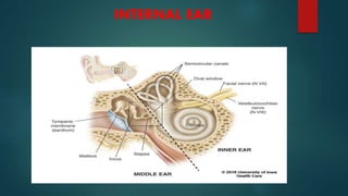
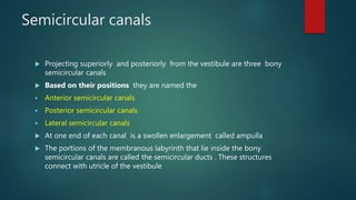
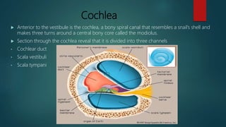
ANATOMY OF THE
INNER EAR
By:
Y.V Vanaja
Lecturer
Vijay Marie College of Nursing
INTERNAL EAR
 The internal ear is also called the labyrinth because of its
complicated series of canals
 Structurally it consists of two main divisions
o Outer Bony labyrinth
o Inner Membranous labyrinth
INTERNAL EAR
Bony Labyrinth
 The bony labyrinth is a series of cavities in the temporal bone divided
into three areas
1) Semicircular Canals both of which contains receptors for
2) Vestibule equilibrium
3) Cochlea - which contains receptors for hearing
 The bony labyrinth is lined with periosteum and contains perilymph
 The fluid which is chemically similar to CSF surrounds the membranous
labyrinth
 The membranous labyrinth is lined by epithelium and contains
endolymph
Vestibule
 It is the oval central portion of the bony labyrinth
 The membranous labyrinth in the vestibule consists of
two sacs called – Utricle
- Saccule
 which are connected by a small duct
Semicircular canals
 Projecting superiorly and posteriorly from the vestibule are three bony
semicircular canals
 Based on their positions they are named the
 Anterior semicircular canals
 Posterior semicircular canals
 Lateral semicircular canals
 At one end of each canal is a swollen enlargement called ampulla
 The portions of the membranous labyrinth that lie inside the bony
semicircular canals are called the semicircular ducts . These structures
connect with utricle of the vestibule
 The vestibular branch of the vestibulocochlear nerve consists of ampullary,
utricular, and saccular nerves.
 These nerves contains both first order neurons and second order neurons
 Cell bodies of the sensory neurons are located in the vestibular ganglia
Cochlea
 Anterior to the vestibule is the cochlea, a bony spiral canal that resembles a snail’s shell and
makes three turns around a central bony core called the modiolus.
 Section through the cochlea reveal that it is divided into three channels
• Cochlear duct
• Scala vestibuli
• Scala tympani
Cochlear Duct or scala media :
 is a continuation of the membranous labyrinth into the cochlea. It is filled with
endolymph.
 The channel above the cochlear duct is the scala vestibule, which ends at the
oval window .
 The channel below is the scala tympani, which ends at the round window
 Both the scala vestibuli and scala tymapani are the part of the bony labyrinth
the cochlea . These chambers are filled with perilymph
 The scala vestibuli and scala tympani are completely separated except for an
opening at the apex of the cochlea, the helicotrema
 The cochlea joins the wall of the vestibule into which the scala vestibule opens
 The vestibular membrane separates the cochlear duct from the scala vestibuli and the basilar
membrane separates the cochlear duct from the scala tympani
 Resting on the basilar membrane is the spiral organ or organ of corti
 The spiral organ is a coiled sheet of epithelial cells, including supporting cells and about
16,000 hair cells, which are receptors for hearing
 There are 2 groups of hair cells:
 The inner hair cells are arranged in single row, where as the outer hair cells are arranged in 3
rows .
 at the apical tip of each hair cell is a hair bundle, consisting of 30-100 stereocilia that extend
into the endolymph of the cochlear duct
 At the their basal ends, inner and outer hair cells synapse both with first –order sensory
neurons and with motor neurons from the cochlear branch of the vestibulocochlear nerve
 The tectorial membrane, a flexible gelatinous membrane, covers the hair cells of the spiral
organ
Internal ear
Internal ear
Internal ear

Internal ear

  • 1.
    ANATOMY OF THE INNEREAR By: Y.V Vanaja Lecturer Vijay Marie College of Nursing
  • 3.
  • 4.
     The internalear is also called the labyrinth because of its complicated series of canals  Structurally it consists of two main divisions o Outer Bony labyrinth o Inner Membranous labyrinth INTERNAL EAR
  • 5.
    Bony Labyrinth  Thebony labyrinth is a series of cavities in the temporal bone divided into three areas 1) Semicircular Canals both of which contains receptors for 2) Vestibule equilibrium 3) Cochlea - which contains receptors for hearing  The bony labyrinth is lined with periosteum and contains perilymph  The fluid which is chemically similar to CSF surrounds the membranous labyrinth  The membranous labyrinth is lined by epithelium and contains endolymph
  • 7.
    Vestibule  It isthe oval central portion of the bony labyrinth  The membranous labyrinth in the vestibule consists of two sacs called – Utricle - Saccule  which are connected by a small duct
  • 8.
    Semicircular canals  Projectingsuperiorly and posteriorly from the vestibule are three bony semicircular canals  Based on their positions they are named the  Anterior semicircular canals  Posterior semicircular canals  Lateral semicircular canals  At one end of each canal is a swollen enlargement called ampulla  The portions of the membranous labyrinth that lie inside the bony semicircular canals are called the semicircular ducts . These structures connect with utricle of the vestibule
  • 9.
     The vestibularbranch of the vestibulocochlear nerve consists of ampullary, utricular, and saccular nerves.  These nerves contains both first order neurons and second order neurons  Cell bodies of the sensory neurons are located in the vestibular ganglia
  • 10.
    Cochlea  Anterior tothe vestibule is the cochlea, a bony spiral canal that resembles a snail’s shell and makes three turns around a central bony core called the modiolus.  Section through the cochlea reveal that it is divided into three channels • Cochlear duct • Scala vestibuli • Scala tympani
  • 12.
    Cochlear Duct orscala media :  is a continuation of the membranous labyrinth into the cochlea. It is filled with endolymph.  The channel above the cochlear duct is the scala vestibule, which ends at the oval window .  The channel below is the scala tympani, which ends at the round window  Both the scala vestibuli and scala tymapani are the part of the bony labyrinth the cochlea . These chambers are filled with perilymph  The scala vestibuli and scala tympani are completely separated except for an opening at the apex of the cochlea, the helicotrema  The cochlea joins the wall of the vestibule into which the scala vestibule opens
  • 14.
     The vestibularmembrane separates the cochlear duct from the scala vestibuli and the basilar membrane separates the cochlear duct from the scala tympani  Resting on the basilar membrane is the spiral organ or organ of corti  The spiral organ is a coiled sheet of epithelial cells, including supporting cells and about 16,000 hair cells, which are receptors for hearing  There are 2 groups of hair cells:  The inner hair cells are arranged in single row, where as the outer hair cells are arranged in 3 rows .  at the apical tip of each hair cell is a hair bundle, consisting of 30-100 stereocilia that extend into the endolymph of the cochlear duct  At the their basal ends, inner and outer hair cells synapse both with first –order sensory neurons and with motor neurons from the cochlear branch of the vestibulocochlear nerve  The tectorial membrane, a flexible gelatinous membrane, covers the hair cells of the spiral organ