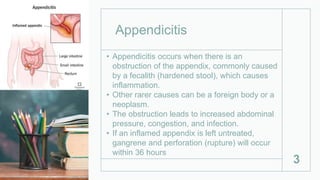
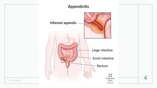
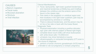
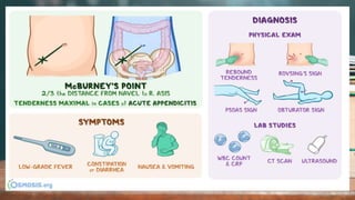
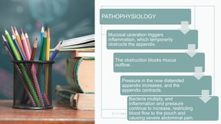
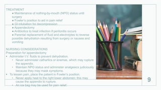
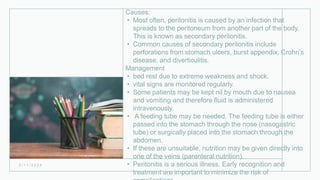
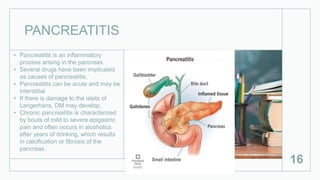
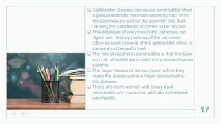
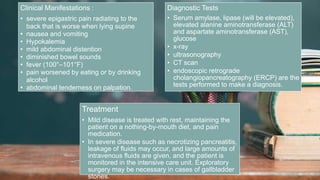
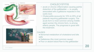
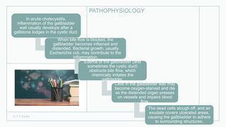
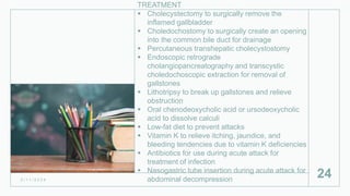
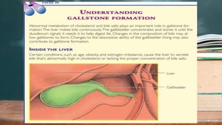
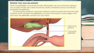
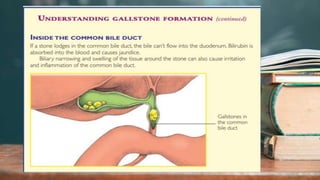
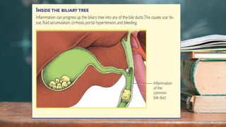
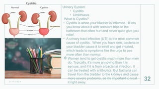
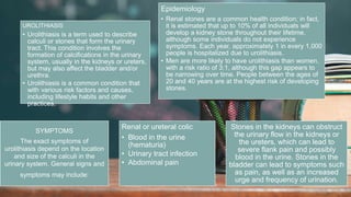
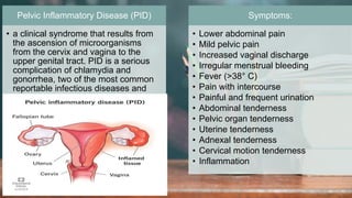
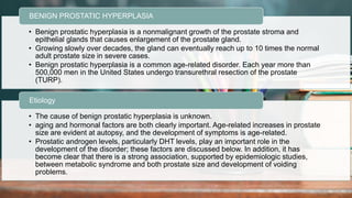
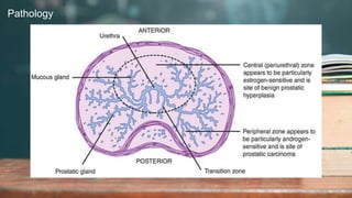
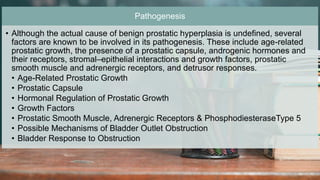
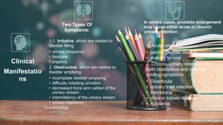
2. 2 / 1 1 / 2 0 2 4
2
a. Appendicitis b. Peritonitis c. Pancreatitis d. Cholecystitis
e. Urinary System
•Cystitis
•Urolithiasis
f. Reproductive
•Pelvic
Inflammatory
•Benign Prostatic
Hypertrophy
3. Appendicitis
• Appendicitis occurs when there is an
obstruction of the appendix, commonly caused
by a fecalith (hardened stool), which causes
inflammation.
• Other rarer causes can be a foreign body or a
neoplasm.
• The obstruction leads to increased abdominal
pressure, congestion, and infection.
• If an inflamed appendix is left untreated,
gangrene and perforation (rupture) will occur
within 36 hours
3
5. Clinical Manifestations
Fever, tachycardia, right lower quadrant tenderness,
and maximal tenderness at McBurney point (halfway
between the umbilicus and the anterior spine of the
ileum) is the classic sign.
Pain starts in the epigastric or umbilical region and
then localizes in the right lower quadrant; pain may be
accompanied by anorexia or vomiting.
There is rebound tenderness, and the person may lie
on his or her side or back with the knees flexed.
Diagnostic Tests
Physical examination including a positive obturator
sign (pain with internal rotation of flexed right thigh)
complete blood count (CBC) will show leukocytosis
with white blood cells >10,000/mm3
urinalysis may show hematuria or pyuria
ultrasonography or computed tomography (CT) scan is
the choice to diagnose.
perform a human chorionic gonadotropin (hCG) test on
a female to rule out pregnancy or ectopic pregnancy
5
CAUSES
● Barium ingestion
● Fecal mass
● Mucosal ulceration
● Stricture
● Viral infection
7. 2 / 1 1 / 2 0 2 4
7
PATHOPHYSIOLOGY
Mucosal ulceration triggers
inflammation, which temporarily
obstructs the appendix.
The obstruction blocks mucus
outflow.
Pressure in the now distended
appendix increases, and the
appendix contracts.
Bacteria multiply, and
inflammation and pressure
continue to increase, restricting
blood flow to the pouch and
causing severe abdominal pain.
8. 2 / 1 1 / 2 0 2 4
8
Pathogenesis And
Complications Of Acute
Appendicitis
9. TREATMENT
● Maintenance of nothing-by-mouth (NPO) status until
surgery
● Fowler’s position to aid in pain relief
● GI intubation for decompression
● Appendectomy
● Antibiotics to treat infection if peritonitis occurs
● Parental replacement of fluid and electrolytes to reverse
possible dehydration resulting from surgery or nausea and
vomiting
NURSING CONSIDERATIONS
Preparation for appendectomy
Administer I.V. fluids to prevent dehydration.
• Never administer cathartics or enemas, which may rupture
the appendix.
• Maintain NPO status and administer analgesics judiciously
because they may mask symptoms.
To lessen pain, place the patient in Fowler’s position.
• Never apply heat to the right lower abdomen; this may
cause the appendix to rupture.
• An ice bag may be used for pain relief.
9
10. Monitor vital signs and intake and output. Give
analgesics as ordered.
Encourage the patient to cough, breathe
deeply, and turn frequently to prevent
pulmonary complications.
Document bowel sounds, passing of flatus,
and bowel movements.
Watch closely for possible surgical
complications. Continuing pain and fever may
signal an abscess.
In appendicitis complicated by peritonitis, a
nasogastric tube may be needed to
decompress the stomach and reduce nausea
and vomiting.
10
After appendectomy
11. PERITONITIS
• Results from contamination of the normally sterile
peritoneal cavity by infection or a chemical irritant.
• refers to the inflammation of the peritoneum, which is the
inner membrane that lines the abdominal cavity and
abdominal organs.
• Peritonitis is typically caused by an infection involving
gastrointestinal or pelvic organs, and it can be life-
threatening if left untreated.
11
14. severe abdominal pain
entire abdomen is tender
board‐like abdomen
Rebound tenderness
inhibited peristalsis, resulting in paralytic
ileus
Bowel sounds are markedly diminished or
absent,
progressive abdominal distention is noted.
Pooling of GI secretions may cause
nausea and vomiting.
Systemic signs of peritonitis
14
Signs and symptoms:
15. Causes:
• Most often, peritonitis is caused by an infection that
spreads to the peritoneum from another part of the body.
This is known as secondary peritonitis.
• Common causes of secondary peritonitis include
perforations from stomach ulcers, burst appendix, Crohn’s
disease, and diverticulitis.
Management
• bed rest due to extreme weakness and shock.
• vital signs are monitored regularly.
• Some patients may be kept nil by mouth due to nausea
and vomiting and therefore fluid is administered
intravenously.
• A feeding tube may be needed. The feeding tube is either
passed into the stomach through the nose (nasogastric
tube) or surgically placed into the stomach through the
abdomen.
• If these are unsuitable, nutrition may be given directly into
one of the veins (parenteral nutrition).
• Peritonitis is a serious illness. Early recognition and
treatment are important to minimize the risk of
2 / 1 1 / 2 0 2 4
16. PANCREATITIS
• Pancreatitis is an inflammatory
process arising in the pancreas.
• Several drugs have been implicated
as causes of pancreatitis,
• Pancreatitis can be acute and may be
interstitial
• If there is damage to the islets of
Langerhans, DM may develop.
• Chronic pancreatitis is characterized
by bouts of mild to severe epigastric
pain and often occurs in alcoholics
after years of drinking, which results
in calcification or fibrosis of the
pancreas.
16
17. Gallbladder disease can cause pancreatitis when
a gallstone blocks the main secretory duct from
the pancreas as well as the common bile duct,
causing the pancreatic enzymes to be blocked.
This blockage of enzymes in the pancreas can
digest and destroy portions of the pancreas.
Often surgical removal of the gallbladder stone or
stones must be performed.
The role of alcohol in pancreatitis is that it is toxic
and can stimulate pancreatic enzymes and cause
spasms.
The large release of the enzymes before they
reach the duodenum is a major component of
this disease.
There are more women with biliary tract
pancreatitis and more men with alcohol-related
pancreatitis.
2 / 1 1 / 2 0 2 4
17
18. 2 / 1 1 / 2 0 2 4
18
Clinical Manifestations :
• severe epigastric pain radiating to the
back that is worse when lying supine
• nausea and vomiting
• Hypokalemia
• mild abdominal distention
• diminished bowel sounds
• fever (100°–101°F)
• pain worsened by eating or by drinking
alcohol
• abdominal tenderness on palpation.
Diagnostic Tests
• Serum amylase, lipase (will be elevated),
elevated alanine aminotransferase (ALT)
and aspartate aminotransferase (AST),
glucose
• x-ray
• ultrasonography
• CT scan
• endoscopic retrograde
cholangiopancreatography (ERCP) are the
tests performed to make a diagnosis.
Treatment
• Mild disease is treated with rest, maintaining the
patient on a nothing-by-mouth diet, and pain
medication.
• In severe disease such as necrotizing pancreatitis,
leakage of fluids may occur, and large amounts of
intravenous fluids are given, and the patient is
monitored in the intensive care unit. Exploratory
surgery may be necessary in cases of gallbladder
stones.
19. 2 / 1 1 / 2 0 2 4
19
Nursing Interventions and Complications
• Controlling pain is a priority for these patients
• Monitoring and maintaining fluid and electrolyte balance.
• nothing by mouth, so maintaining adequate nutrition by nasogastric tube or intravenous fluids is
very important.
• Nurses should educate the patients about pancreatitis and the planned diagnostic tests or surgical
intervention, address the unhealthy lifestyle of the alcoholic, and refer them for treatment or
counseling.
• assess lab values and continually
• assess patients’ vital signs
• perform palpation of the abdomen to check for increased rigidity
• assess for third spacing, falling urine output, or increased abdominal girth and increased pain.
• careful close assessment by nurses is paramount.
• If surgery is indicated, pre- and postoperative teaching and care is important.
20. CHOLECYSTITIS
• acute or chronic inflammation causing painful
distention of the gallbladder — is usually
associated with a gallstone impacted in the
cystic duct.
• Cholecystitis accounts for 10% to 25% of all
patients requiring gallbladder surgery. The
acute form is most common among middle-
aged women the chronic form, is among
elderly people. The prognosis is good with
the treatment.
CAUSES
● Abnormal metabolism of cholesterol and bile
salts
● Gallstones (the most common cause)
● Poor or absent blood flow to the gallbladder
2 / 1 1 / 2 0 2 4
20
21. PATHOPHYSIOLOGY
2 / 1 1 / 2 0 2 4
21
In acute cholecystitis,
inflammation of the gallbladder
wall usually develops after a
gallstone lodges in the cystic duct.
When bile flow is blocked, the
gallbladder becomes inflamed and
distended. Bacterial growth, usually
Escherichia coli, may contribute to the
inflammation.
Edema of the gallbladder (and
sometimes the cystic duct)
obstructs bile flow, which
chemically irritates the
gallbladder.
Cells in the gallbladder wall may
become oxygen-starved and die
as the distended organ presses
on vessels and impairs blood
flow.
The dead cells slough off, and an
exudate covers ulcerated areas,
causing the gallbladder to adhere
to surrounding structures.
22. CLINICAL FINDINGS
• Acute abdominal pain in the right upper quadrant
that may radiate to the back, between the shoulders,
or to the front of the chest secondary to inflammation
and irritation of nerve fibers
• Colic due to the passage of gallstones along the bile
duct
• Nausea and vomiting triggered by the inflammatory
response
• Chills related to fever
• Low-grade fever secondary to inflammation
• Jaundice from obstruction of the common bile duct
by
2 / 1 1 / 2 0 2 4
22
23. TEST RESULTS
X-ray reveals gallstones if they contain enough calcium
to be radiopaque; also helps disclose porcelain
gallbladder (hard, brittle gallbladder due to calcium
deposited in a wall), limy bile, and gallstone ileus.
Ultrasonography detects gallstones as small as 2 mm
and distinguishes between obstructive and
nonobstructive jaundice.
Technetium-labeled scan reveals cystic duct obstruction
and acute or chronic cholecystitis if ultrasound doesn’t
visualize the gallbladder.
Percutaneous transhepatic cholangiography supports
the diagnosis of obstructive jaundice and reveals calculi
in the ducts.
Levels of serum alkaline phosphate, lactate
dehydrogenase, aspartate aminotransferase, and total
bilirubin are high; serum amylase level slightly elevated;
and icteric index elevated.
White blood cell counts are slightly elevated during
cholecystitis attack. 23
24. TREATMENT
Cholecystectomy to surgically remove the
inflamed gallbladder
Choledochostomy to surgically create an opening
into the common bile duct for drainage
Percutaneous transhepatic cholecystostomy
Endoscopic retrograde
cholangiopancreatography and transcystic
choledochoscopic extraction for removal of
gallstones
Lithotripsy to break up gallstones and relieve
obstruction
Oral chenodeoxycholic acid or ursodeoxycholic
acid to dissolve calculi
Low-fat diet to prevent attacks
Vitamin K to relieve itching, jaundice, and
bleeding tendencies due to vitamin K deficiencies
Antibiotics for use during acute attack for
treatment of infection
Nasogastric tube insertion during acute attack for
abdominal decompression
2 / 1 1 / 2 0 2 4
24
25. NURSING CONSIDERATIONS
Care for the patient with cholecystitis focuses on supportive
care and close postoperative observation.
Before surgery, teach the patient to deep breathe, cough,
expectorate, and perform leg exercises that are necessary
after surgery. Also teach splinting, repositioning, and
ambulation techniques. Explain the procedures that will be
performed before, during, and after surgery to help ease the
patient’s anxiety and to help ensure his cooperation.
After surgery, monitor vital signs for signs of bleeding,
infection, or atelectasis.
Evaluate the incision site for bleeding. Serosanguineous
drainage is common during the first 24 to 48 hours if the
patient has a wound drain. If, after a choledochostomy, a T-
tube drain is placed in the duct and attached to a drainage
bag, make sure the drainage tube has no kinks. Also, check
that the connecting tubing from the T tube is well secured to
the patient to prevent dislodgment.
Measure and record T-tube drainage daily (200 to 300 ml is
normal).
Teach patients who will be discharged with a T tube how to
perform dressing changes and routine skin care.
25
26. NURSING CONSIDERATIONS
Monitor intake and output. Allow the patient nothing by
mouth for 24 to 48 hours or until bowel sounds return and
nausea and vomiting cease (postoperative nausea may
indicate a full bladder).
If the patient doesn’t void within 8 hours (or if the amount
voided is inadequate based on I.V. fluid intake), perT-tube
If the patient doesn’t void within 8 hours (or if the amount
voided is inadequate based on I.V. fluid intake), percuss
over the symphysis pubis for bladder distention (especially
in patients receiving anticholinergics). Patients who have
had a laparoscopic cholecystectomy may be discharged the
same day or within 24 hours after surgery. These patients
should have minimal pain, be able to tolerate a regular diet
within 24 hours after surgery and be able to return to normal
activity within 1 week.
Encourage deep breathing and leg exercises every hour.
The patient should ambulate after surgery. Provide elastic
stockings to support leg muscles and promote venous blood
flow, thus preventing stasis and clot formation.
Evaluate the location, duration, and character of pain.
Administer adequate medication to relieve pain, especially
26
27. NURSING CONSIDERATIONS
At discharge, advise the patient against heavy lifting or
straining for 6 weeks. Urge him to walk daily. Tell him that
food restrictions are unnecessary unless he has an
intolerance to a specific food or some underlying
condition (such as diabetes, atherosclerosis, or obesity)
that requires such restriction.
Instruct the patient to notify the surgeon if he has pain for
more than 24 hours, anorexia, nausea or vomiting, fever,
or tenderness in the abdominal area or if he notices
jaundice, as these may indicate a biliary tract injury from
the cholecystectomy, requiring immediate attention.
27
32. Urinary System
• Cystitis
• Urolithiasis
What Is Cystitis?
• Cystitis is when your bladder is inflamed. It lets
you know about it with constant trips to the
bathroom that often hurt and never quite give you
relief.
• A urinary tract infection (UTI) is the most common
cause of cystitis. When you have one, bacteria in
your bladder cause it to swell and get irritated,
which leads to symptoms like the urge to pee
more often than normal.
Women tend to get cystitis much more than men
do. Typically, it’s more annoying than it is
serious, and if it is from a bacterial infection, it
can be treated with antibiotics. But bacteria can
travel from the bladder to the kidneys and cause
more severe problems, so it’s important to treat
it right away.
2 / 1 1 / 2 0 2 4
32
33. Causes:
• bacteria such as E. coli are to blame. They
normally live on your skin and in your intestines,
and they’re not a problem. But if they get into the
urethra, which is the tube that carries pee out of
your body, bacteria can end up in your bladder
and cause issues.
• It’s not as common, but you can also get cystitis
from:
• Chemicals in personal care products, such
as bubble baths, soaps, and spermicides
• Chemotherapy drugs
• Damage from bladder surgery or a catheter --
a tube that helps empty pee from your
bladder
• Radiation to treat cancer around your pelvis
Some people have a condition called interstitial
cystitis, where the bladder is constantly swollen
but there’s no detectable infection. Doctors
aren’t sure what causes it, and it’s much harder
to treat than regular cystitis.
2 / 1 1 / 2 0 2 4
33
34. Cystitis Symptoms
• The symptoms of a bladder infection are similar
to what you will feel when you have a urinary
tract infection (UTI). These symptoms can
include:
• Frequent urination: You may feel the need to
urinate more often.
• Some people also experience urgency
(sudden desire to pee).
• Pain/ burning with urination: During an
infection, you may experience discomfort in
pain in the suprapubic area and burning
during urination.
• Dark or foul-smelling pee.
Other symptoms that can be linked to a more
serious kidney infection include:
• Fever (temperature above 100 degrees
Fahrenheit).
• Chills or rigors (shaking).
• Vomiting.
• Flank pain.
2 / 1 1 / 2 0 2 4
34
35. Diagnosis and Tests:
• Urinalysis: This test involves checking the
appearance, concentration, and content of urine.
• Urine culture: This test determines the type of
bacteria causing the infection and to which
antibiotics the bacteria is sensitive.
Management and Treatment
A bladder infection is typically treated with
antibiotics. Your healthcare provider will
prescribe an antibiotic based on the type of
bacteria found during your urine tests. To
adequately treat the infection, it’s important to
take all of the antibiotics your provider
prescribes.
Some commonly used antibiotics include:
• Nitrofurantoin.
• Sulfonamides (sulfa drugs).
• Amoxicillin.
• Cephalosporins.
• Trimethoprim/sulfamethoxazole (Bactrim®).
• Quinolones, such as ciprofloxacin (Cipro®).
2 / 1 1 / 2 0 2 4
35
36. Prevention:
You can often prevent a bladder infection by
adopting some good lifestyle practices.
These practices include:
Practice good hygiene habits: Make sure you
have good hygiene habits (wiping front to back)
and aggressively treat constipation or diarrhea.
Drink plenty of fluids: Increase your fluid intake
to help flush bacteria out of your bladder.
Estrogen cream for postmenopausal women:
When women go through menopause, their
vaginal tissue changes and the good vaginal
bacteria that normally colonize the vagina cannot
survive. Bacteria can colonize, which causes
bladder infections. To reestablish the good
bacteria, you may need topical estrogen cream.
Talk to your healthcare provider about whether
topical estrogen cream is right for you.
2 / 1 1 / 2 0 2 4
36
37. Prevention
Changing birth control: You may also want to
change your birth control option if you have
frequent bladder infections and UTIs. Women
who use diaphragms and spermicide are at
greater risk of infection. You should also use a
water-based lubricant during sex if needed.
Supplements: There are over-the-counter
supplements (D-mannose and cranberry) that
can help prevent infections in your urinary tract.
If you have frequent UTIs and bladder infections,
talk to your healthcare provider about these
options and make a plan about the best ways to
prevent infections.
2 / 1 1 / 2 0 2 4
37
38. 2 / 1 1 / 2 0 2 4
38
UROLITHIASIS
• Urolithiasis is a term used to describe
calculi or stones that form the urinary
tract. This condition involves the
formation of calcifications in the urinary
system, usually in the kidneys or ureters,
but may also affect the bladder and/or
urethra.
• Urolithiasis is a common condition that
with various risk factors and causes,
including lifestyle habits and other
practices.
Epidemiology
• Renal stones are a common health condition; in fact,
it is estimated that up to 10% of all individuals will
develop a kidney stone throughout their lifetime,
although some individuals do not experience
symptoms. Each year, approximately 1 in every 1,000
people is hospitalized due to urolithiasis.
• Men are more likely to have urolithiasis than women,
with a risk ratio of 3:1, although this gap appears to
be narrowing over time. People between the ages of
20 and 40 years are at the highest risk of developing
stones.
SYMPTOMS
The exact symptoms of
urolithiasis depend on the location
and size of the calculi in the
urinary system. General signs and
symptoms may include:
Renal or ureteral colic
• Blood in the urine
(hematuria)
• Urinary tract infection
• Abdominal pain
Stones in the kidneys can obstruct
the urinary flow in the kidneys or
the ureters, which can lead to
severe flank pain and possibly
blood in the urine. Stones in the
bladder can lead to symptoms such
as pain, as well as an increased
urge and frequency of urination.
40. Diagnosis
The diagnosis of urolithiasis involves a medical history
and a physical examination, in addition to appropriate
testing methods to identify the calculi and make the
appropriate treatment decisions.
Additional diagnostic tests may include:
Urine culture and urinalysis to reveal signs of
hematuria, pyuria, infection, and acidity.
Complete blood count tests to detect elevated white
blood cells indicative of infection.
Intravenous pyelography (IVP) to identify the size and
location of the stone and its effect on the urinary flow.
Computed tomography (CT) scan to visualize the
urinary tract and any obstructions such as in acute
renal colic.
X-ray imaging to identify the size and location of the
stone.
Renal ultrasound to screen for stones and detect
obstruction in the urinary system. 40
41. Diagnosis
The diagnosis of urolithiasis involves a medical history
and a physical examination, in addition to appropriate
testing methods to identify the calculi and make the
appropriate treatment decisions.
Additional diagnostic tests may include:
Urine culture and urinalysis to reveal signs of
hematuria, pyuria, infection, and acidity.
Complete blood count tests to detect elevated white
blood cells indicative of infection.
Intravenous pyelography (IVP) to identify the size and
location of the stone and its effect on the urinary flow.
Computed tomography (CT) scan to visualize the
urinary tract and any obstructions such as in acute
renal colic.
X-ray imaging to identify the size and location of the
stone.
Renal ultrasound to screen for stones and detect
obstruction in the urinary system. 41
42. Diagnosis
The diagnosis of urolithiasis involves a medical history
and a physical examination, in addition to appropriate
testing methods to identify the calculi and make the
appropriate treatment decisions.
Additional diagnostic tests may include:
Urine culture and urinalysis to reveal signs of
hematuria, pyuria, infection, and acidity.
Complete blood count tests to detect elevated white
blood cells indicative of infection.
Intravenous pyelography (IVP) to identify the size and
location of the stone and its effect on the urinary flow.
Computed tomography (CT) scan to visualize the
urinary tract and any obstructions such as in acute
renal colic.
X-ray imaging to identify the size and location of the
stone.
Renal ultrasound to screen for stones and detect
obstruction in the urinary system. 42
43. Diagnosis
The diagnosis of urolithiasis involves a medical history
and a physical examination, in addition to appropriate
testing methods to identify the calculi and make the
appropriate treatment decisions.
Additional diagnostic tests may include:
Urine culture and urinalysis to reveal signs of
hematuria, pyuria, infection, and acidity.
Complete blood count tests to detect elevated white
blood cells indicative of infection.
Intravenous pyelography (IVP) to identify the size and
location of the stone and its effect on the urinary flow.
Computed tomography (CT) scan to visualize the
urinary tract and any obstructions such as in acute
renal colic.
X-ray imaging to identify the size and location of the
stone.
Renal ultrasound to screen for stones and detect
obstruction in the urinary system. 43
44. Diagnosis
The diagnosis of urolithiasis involves a medical history
and a physical examination, in addition to appropriate
testing methods to identify the calculi and make the
appropriate treatment decisions.
Additional diagnostic tests may include:
Urine culture and urinalysis to reveal signs of
hematuria, pyuria, infection, and acidity.
Complete blood count tests to detect elevated white
blood cells indicative of infection.
Intravenous pyelography (IVP) to identify the size and
location of the stone and its effect on the urinary flow.
Computed tomography (CT) scan to visualize the
urinary tract and any obstructions such as in acute
renal colic.
X-ray imaging to identify the size and location of the
stone.
Renal ultrasound to screen for stones and detect
obstruction in the urinary system. 44
45. Management:
• The management of urolithiasis will depend on
the specific patient case, based on factors such
as the presenting symptoms and the size and
location of the stones.
• Most stones are excreted in the urine on their
own and do not require invasive surgical
techniques.
• Keeping hydrated to flush the stones out of the
body is recommended initially if the pain is
manageable for the patient.
• Simple analgesic medication such as
paracetamol can aid in pain relief.
• Antiemetic medications and rehydration therapy
may also be useful.
• Medical expulsive therapy can be used to
facilitate the passing of the stone.
• For larger stones that do not pass
spontaneously, surgery may be needed to
remove the calculi.
2 / 1 1 / 2 0 2 4
45
47. 2 / 1 1 / 2 0 2 4
47
Pelvic Inflammatory Disease (PID)
• a clinical syndrome that results from
the ascension of microorganisms
from the cervix and vagina to the
upper genital tract. PID is a serious
complication of chlamydia and
gonorrhea, two of the most common
reportable infectious diseases and
sexually transmitted diseases (STDs)
in the US.
Symptoms:
• Lower abdominal pain
• Mild pelvic pain
• Increased vaginal discharge
• Irregular menstrual bleeding
• Fever (>38° C)
• Pain with intercourse
• Painful and frequent urination
• Abdominal tenderness
• Pelvic organ tenderness
• Uterine tenderness
• Adnexal tenderness
• Cervical motion tenderness
• Inflammation
48. How do women get pelvic inflammatory disease?
• Women develop PID when certain bacteria, such as Chlamydia
trachomatis (CT) and Neisseria gonorrhoeae (NG), move
upward from a woman’s vagina or cervix into her reproductive
organs. PID can lead to infertility and permanent damage to a
woman’s reproductive organs.
• What causes pelvic inflammatory disease?
• Several different microorganisms can cause or contribute to
PID.
• The sexually transmitted pathogens C. trachomatis and N.
gonorrhoeae have been implicated in a third to half of PID
cases.
• endogenous microorganisms, including gram-positive and
negative anaerobic organisms and aerobic/facultative gram-
positive and negative rods and cocci, found at high levels in
women with bacterial vaginosis, also have been implicated in
the pathogenesis of PID.
• Newer data suggest that Mycoplasma genitalium (Mgen) may
also play a role in PID and may be associated with milder
48
49. 2 / 1 1 / 2 0 2 4
49
Complications of PID include
Tubo-ovarian abscess (TOA) - is a serious short-term complication of PID that is characterized by an inflammatory mass
involving the fallopian tube, ovary, and, occasionally, other adjacent pelvic organs.
Tubal factor infertility
Ectopic pregnancy
Chronic pelvic pain
Diagnoses:
The wide variation in symptoms and signs associated with PID can make diagnosis challenging. No single
historical, physical, or laboratory finding is both sensitive and specific for the diagnosis of PID. Clinicians
should therefore maintain a low threshold for the diagnosis of PID, particularly in young, sexually active
women.
Presumptive treatment for PID should be initiated in sexually active young women and other women at risk for
STDs if they are experiencing pelvic or lower abdominal pain, if no cause for the illness other than PID can be
identified, and if one or more of the following minimum clinical criteria are present on pelvic examination:
• cervical motion tenderness
• uterine tenderness
• adnexal tenderness
The requirement that all three minimum criteria be present before the initiation of empiric treatment could
result in insufficient sensitivity for the diagnosis of PID. After deciding whether to initiate empiric treatment,
clinicians should also consider the risk profile for STDs.
More elaborate diagnostic evaluation frequently is needed because incorrect diagnosis and management of
PID might cause unnecessary morbidity. (ex)
50. • One or more of the following additional criteria
can be used to enhance the specificity of the
minimum clinical criteria and support a diagnosis of
PID:
• oral temperature >101°F (>38.3°C);
• abnormal cervical mucopurulent discharge or
cervical friability;
• presence of abundant numbers of WBC on
saline microscopy of vaginal fluid;
• elevated erythrocyte sedimentation rate;
• elevated C-reactive protein; and
• laboratory documentation of cervical infection
with N. gonorrhoeae or C. trachomatis.
The most specific criteria for diagnosing PID
include:
• endometrial biopsy with histopathologic
evidence of endometritis;
• transvaginal sonography or magnetic
resonance imaging techniques showing
thickened, fluid-filled tubes with or without
free pelvic fluid or tubo-ovarian complex, or
2 / 1 1 / 2 0 2 4
50
51. 2 / 1 1 / 2 0 2 4
51
Treatment
• PID is treated with broad-spectrum antibiotics to
cover likely pathogens.
• Healthcare providers should emphasize to their
patients that although their symptoms may go
away before the infection is cured, they should
finish taking all of the prescribed medicine.
• Additionally, a woman’s sex partner(s) should be
treated to decrease the risk of re-infection, even
if the partner(s) has no symptoms. Although sex
partners may have no symptoms, they may still
be infected with the organisms that can cause
PID.
• In certain cases, clinicians may recommend
hospitalization to treat PID. This decision should
be based on the judgment of the health care
provider and the use of suggested criteria found
in the 2021 STI Treatment Guidelines.7 If a
woman’s symptoms continue, or if an abscess
does not resolve, surgery may be needed.
The decision of whether hospitalization is
necessary should be based on provider judgment
and whether the woman meets any of the following
criteria:
• Surgical emergencies (e.g.,
appendicitis) cannot be excluded
• Tubo-ovarian abscess
• Pregnancy
• Severe illness, nausea and vomiting,
or oral temperature >38.5°C (101°F)
• Unable to follow or tolerate an
outpatient oral regimen
• No clinical response to oral
antimicrobial therapy
52. 2 / 1 1 / 2 0 2 4
52
• Benign prostatic hyperplasia is a nonmalignant growth of the prostate stroma and
epithelial glands that causes enlargement of the prostate gland.
• Growing slowly over decades, the gland can eventually reach up to 10 times the normal
adult prostate size in severe cases.
• Benign prostatic hyperplasia is a common age-related disorder. Each year more than
500,000 men in the United States undergo transurethral resection of the prostate
(TURP).
BENIGN PROSTATIC HYPERPLASIA
• The cause of benign prostatic hyperplasia is unknown.
• aging and hormonal factors are both clearly important. Age-related increases in prostate
size are evident at autopsy, and the development of symptoms is age-related.
• Prostatic androgen levels, particularly DHT levels, play an important role in the
development of the disorder; these factors are discussed below. In addition, it has
become clear that there is a strong association, supported by epidemiologic studies,
between metabolic syndrome and both prostate size and development of voiding
problems.
Etiology
55. 2 / 1 1 / 2 0 2 4
55
Pathogenesis
• Although the actual cause of benign prostatic hyperplasia is undefined, several
factors are known to be involved in its pathogenesis. These include age-related
prostatic growth, the presence of a prostatic capsule, androgenic hormones and
their receptors, stromal–epithelial interactions and growth factors, prostatic
smooth muscle and adrenergic receptors, and detrusor responses.
• Age-Related Prostatic Growth
• Prostatic Capsule
• Hormonal Regulation of Prostatic Growth
• Growth Factors
• Prostatic Smooth Muscle, Adrenergic Receptors & PhosphodiesteraseType 5
• Possible Mechanisms of Bladder Outlet Obstruction
• Bladder Response to Obstruction
56. 2 / 1 1 / 2 0 2 4
56
Clinical
Manifestatio
ns
Two Types Of
Symptoms:
1.1. Irritative, which are related to
bladder filling
• urinary frequency
• Nocturia
• urgency
2. Obstructive, which are related to
bladder emptying
• incomplete bladder emptying
• difficulty initiating urination
• decreased force and caliber of the
urinary stream
• intermittency of the urinary stream
• urinary hesitancy
• dribbling.
In severe cases, prostatic enlargement
may cause either acute or chronic
urinary retention:
1. Acute retention:
• painful bladder dilation
• inability to void
2. Chronic urinary retention has
both obstructive and irritative
voiding symptoms
• Diverticula
• urinary tract infection
• Hematuria
• chronic kidney disease
• Azotemia
57. Laboratory Tests and Evaluation
• blood urea nitrogen (BUN) and serum
creatinine to exclude renal failure,
• urinalysis and urine culture to exclude urinary
tract infection
• CT scan or ultrasound is performed on a man
with benign prostatic hyperplasia, it typically
shows elevation of the bladder base by the
enlarged prostate; trabeculation, thickening,
and diverticula of the bladder wall; elevation
of the ureters; and poor bladder emptying.
• urodynamic evaluation with uroflowmetry and
cystometry
• Pressure–flow studies
• Cystourethroscopy
2 / 1 1 / 2 0 2 4
57
58. TREATMENT
Conservative therapy includes:
● prostate massages
● sitz baths
● fluid restriction to prevent bladder distention
● antimicrobials to treat infection
● regular ejaculation to help relieve prostatic
congestion
● alpha-adrenergic blockers, such as alfuzosin
(Uroxatral), prazosin (Minipress), tamsulosin
(Flomax), and terazosin (Hytrin), to improve urine
flow rates to relieve bladder outlet obstruction by
preventing contractions of the prostatic capsule and
bladder neck
● dutasteride (Avodart) or finasteride (Proscar) to
possibly reduce the size of the prostate in some
patients
● continuous drainage with an indwelling urinary
catheter to alleviate urine retention (high-risk
patients).
2 / 1 1 / 2 0 2 4
58
59. TREATMENT
● transurethral resection (if the prostate weighs
less than 2 oz [56.7 g]); tissue removed with a wire
loop and electric current using a resectoscope
● suprapubic (transvesical) resection (most
common and useful for prostatic enlargement
remaining within the
bladder)
● retropubic (extravesical) resection allowing direct
visualization (potency and continence usually
maintained)
● balloon dilatation of the urethra and prostatic
stents to maintain urethral patency (occasionally)
● laser excision to relieve prostatic enlargement
● nerve-sparing surgical techniques to reduce
common complications such as erectile
dysfunction.
2 / 1 1 / 2 0 2 4
59
60. 2 / 1 1 / 2 0 2 4
60
NURSING CONSIDERATIONS
• Monitor and record the patient’s vital signs,
intake and output, and daily weight,
watching closely for signs of post-
obstructive diuresis (such as increased
urine output and hypotension) that may lead
to serious dehydration, reduced blood
volume, shock, electrolyte loss, and anuria.
• Insert an indwelling urinary catheter for
urine retention (usually difficult in a patient
with BPH). Using a catheter coudé may
make insertion easier.
• If the catheter can’t be passed
transurethrally, assist with suprapubic
cystostomy under local anesthetic (watching
for rapid bladder decompression).
After Prostatic Surgery, Interventions
May Include The Following:
• Maintain patient comfort, watch for and
prevent postoperative complications,
observe for immediate dangers of prostatic
bleeding (shock, hemorrhage), check the
catheter often (every 15 minutes for the first
2 to 3 hours) for patency and urine color,
and check dressings for bleeding.
• Postoperatively, many urologists insert a
three-way catheter and establish continuous
bladder irrigation.
• Watch for septic shock (the most serious
complication of prostatic surgery).
• Administer belladonna and opium
suppositories or other anticholinergics, as
ordered, to relieve painful bladder spasms
that commonly occur after transurethral
resection.
61. NURSING CONSIDERATIONS
• After an open procedure, take patient comfort
measures
• Continue infusing I.V. fluids until the patient can drink
sufficient fluids (2 to 3 qt [2 to 3 L] per day) to maintain
adequate hydration.
• Administer stool softeners and laxatives, as ordered, to
prevent straining.
• Reassure the patient that temporary frequency,
dribbling, and occasional hematuria will likely occur
after the catheter is removed.
• Reinforce prescribed limits on activity.
• Instruct the patient about the prescribed oral antibiotic
drug regimen and the indications for using gentle
laxatives.
• Urge the patient to seek medical care immediately if he
can’t void or if he passes bloody urine or develops a
fever.
• Encourage annual digital rectal examinations and
screening for PSA to identify a possible malignancy.
2 / 1 1 / 2 0 2 4
61
The vermiform appendix is a small fingerlike projection attached to the cecum (a blind pouch that forms the first portion of the large intestine).
It is the most common abdominal surgical emergency and affects about 10% of the population aged 10–30 years.
4. Children who perforate may develop generalized peritonitis, which may result in mortality.
3 - In a patient whose nausea and abdominal rigidity have subsided, these signs indicate readiness to resume oral fluids.
4 - The complaint that “something gave way” may mean wound dehiscence. If an abscess or peritonitis develops, incision and drainage may be necessary. Frequently assess the dressing for wound drainage.
4 - If so, record drainage and give good mouth and nose care.
Peritonitis results from contamination of the normally sterile peritoneal cavity by infection or a chemical irritant. Chemical peritonitis often precedes bacterial peritonitis.
Perforation of a peptic ulcer or rupture of the gall bladder releases gastric juices (hydrochloric acid and pepsin) or bile into the peritoneal cavity, causing an acute inflammatory response.
Bacterial peritonitis is usually caused by infection by Escherichia coli, Klebsiella, Proteus or Pseudomonas bacteria, which normally inhabit the bowel.
Inflammatory and immune defense mechanisms are activated when bacteria enter the peritoneal space. These defences can effectively eliminate small numbers of bacteria, but may be overwhelmed by massive or continued contamination. When this occurs, mast cells release histamine and other vasoactive substances, causing local vasodilation and increased capillary permeability. Polymorphonuclear leukocytes (a type of WBC) infiltrate the peritoneum to phagocytize bacteria and foreign matter.
Fibrinogen‐rich plasma exudate promotes bacterial destruction and forms fibrin clots to seal off and segregate the bacteria. This process helps limit and localize the infection, allowing host defences to eradicate it. Continued contamination, however, leads to generalized inflammation of the peritoneal cavity.
The inflammatory process causes fluid to shift into the peritoneal space (third spacing). Circulating blood volume is depleted, leading to hypovolaemia.
Septicaemia, a systemic disease caused by pathogens or their toxins in the blood, may follow.
Peritonitis that develops without an abdominal rupture (spontaneous peritonitis) is usually a complication of liver disease, such as cirrhosis. Advanced cirrhosis causes a large amount of fluid build‐up in the abdominal cavity (ascites). That fluid build‐up is
susceptible to bacterial infection
acute, abrupt onset of diffuse
with guarding or rigidity of abdominal muscles.
Tenderness may be present over the area of inflammation
Systemic signs of peritonitis include fever, malaise, tachycardia and tachypnoea, restlessness, and possible disorientation. oliguric (scanty urine
Surgery
The cause of peritonitis may also need to be surgically treated. For example, if a burst appendix caused peritonitis, the appendix will need to be removed. Some people develop abscesses (pus‐filled swellings) in their peritoneum that need to be drained with a needle. This is carried out using an ultrasound scanner to guide the needle to the abscess.
Gallstone disease and excessive alcohol use account for 70%–80% of cases of pancreatitis.
People with genetic hyperlipidemia with very high triglyceride levels can also develop pancreatitis.
Several drugs have been implicated as causes of pancreatitis, such as sulfonamides, methyldopa, metronidazole, erythromycin, and acetaminophen
Pancreatitis can be acute and may be interstitial, causing fluid accumulation and swelling due to the release of digestive enzymes, or necrotizing, causing death and tissue damage
In an acute situation, analgesic medications such as nonsteroidal anti-inflammatory drugs (NSAIDs) or codeine can be administered to relieve pain
For example, the presence of signs of lower-genital–tract inflammation (predominance of leukocytes in vaginal secretions, cervical exudates, or cervical friability), in addition to one of the three minimum criteria, increases the specificity of the diagnosis.
Most women with PID have either mucopurulent cervical discharge or evidence of WBCs on a microscopic evaluation of a saline preparation of vaginal fluid (i.e., wet prep). If the cervical discharge appears normal and no WBCs are observed on the wet prep of vaginal fluid, the diagnosis of PID is unlikely, and alternative causes of pain should be considered. A wet prep of vaginal fluid also can detect the presence of concomitant infections (e.g., BV and trichomoniasis).
Several types of antibiotics can cure PID. Antibiotic treatment does not, however, reverse any scarring that has already been caused by the infection. For this reason, it is critical that a woman receive care immediately if she has pelvic pain or other symptoms of PID. Prompt antibiotic treatment could prevent severe damage to the reproductive organs.
The normal prostate is composed of both stromal (smooth muscle) and epithelial (glandular) elements. Growth of each of these elements—alone or in combination—can result in hyperplastic nodules and ultimately the symptoms of benign prostatic hyperplasia. Pathologically, the hyperplastic gland is enlarged, with a firm, rubbery consistency. Although small nodules are often present throughout the gland, benign prostatic hyperplasia arises most commonly in the periurethral and transition zones of the gland (Figure 23–10). With advancing age, there is an increase in the overall size of the transition zone, as well as an increase in the number—and later the size—of nodules. The urethra is compressed and has a slit-like appearance.
Histologically, benign prostatic hyperplasia is a true hyperplastic process with an increase in prostatic cell number. The prostatic nodules are composed of both hyperplastic glands and hyperplastic stromal muscle. Most periurethral nodules are stromal in character, but transition zone nodules most often consist of glandular tissue. The glands become larger than normal, with stromal muscle between the proliferative glands. Perhaps as much as 40% of the hyperplastic prostate is smooth muscle. The cellular proliferation leads to a tight packing of glands within a given area. There is an increase in the height of the lining epithelium and the epithelium often shows papillary projections (Figure 23–11). Individual epithelial cells also show some hypertrophy.
In men with benign prostatic hyperplasia, the bladder can show both detrusor (bladder wall) smooth muscle hypertrophy and trabeculation associated with an increase in collagen deposition. This is due to the increased bladder pressure created by urinary system obstruction.
The size of the prostate does not always correlate with the degree of obstruction. The amount of periurethral and transition zone tissue may relate more to the degree of obstruction than the overall prostate size. However, the idea that the clinical symptoms of benign prostatic hyperplasia are due simply to a mass-related increase in urethral resistance is probably too simplistic. Instead, some of its symptoms may be due to obstruction-induced detrusor dysfunction and neural alterations in the bladder and prostate. This has been demonstrated in men with lower urinary tract symptoms undergoing urodynamic testing, which measures the perfusion pressure of the urethra.
The presence of a capsule around the prostate is thought to play a role in development of obstructive symptoms. Besides human males, dogs are the only animals known to develop benign prostatic hyperplasia. However, the canine prostate lacks a capsule, and dogs do not develop obstructive symptoms. In men, the capsule presumably causes the “pressure” created by the expanded periurethral–-transition zone tissue that is transmitted to the urethra, leading to an increase in urethral resistance. Surgical incision of the prostatic capsule or removal of the obstructing portion of the prostate, whether by transurethral resection or by open prostatectomy, is effective in relieving symptoms. The presence of a capsule around the prostate is thought to play a role in development of obstructive symptoms. Besides human males, dogs are the only animals known to develop benign prostatic hyperplasia. However, the canine prostate lacks a capsule, and dogs do not develop obstructive symptoms. In men, the capsule presumably causes the “pressure” created by the expanded periurethral–-transition zone tissue that is transmitted to the urethra, leading to an increase in urethral resistance. Surgical incision of the prostatic capsule or removal of the obstructing portion of the prostate, whether by transurethral resection or by open prostatectomy, is effective in relieving symptoms.
The development of benign prostatic hyperplasia requires testicular androgens as well as aging. There are several lines of evidence for this relationship. First, men who are castrated before puberty or who have disorders of impaired androgen production or action do not develop benign prostatic hyperplasia. Second, the prostate, unlike other androgen-dependent organs, maintains its ability to respond to androgens throughout life. Androgens are required for normal cell proliferation and differentiation in the prostate. They may also actively inhibit cell turnover and death. Finally, androgen deprivation at various levels of the hypothalamic–pituitary–testicular axis can reduce prostate size and improve obstructive symptoms
Evidence suggests that prostatic growth is under the direct control of specific growth factors and only indirectly modulated by androgens. According to this evidence, growth factors from both the fibroblast growth factor (FGF) family and the transforming growth factor (TGF) “superfamily” act together to regulate growth.
Prostatic smooth muscle represents a significant proportion of the gland. Urethral elasticity and the degree of bladder outlet obstruction are undoubtedly influenced by the relative content of smooth muscle within the prostate in patients with benign prostatic hyperplasia.
There are several ways in which benign prostatic hyperplasia might cause obstruction of the bladder neck. The prominent median lobe may simply act as a ball valve; restriction may occur from the nondistensible capsule; static obstruction may result from the enlarged prostate surrounding the prostatic urethra; and dynamic obstruction may occur from an inability to relax prostatic smooth muscle.
Many of the clinical symptoms of benign prostatic hyperplasia are related to obstruction-induced changes in bladder function rather than to outflow obstruction per se. Thus, one-third of men continue to have significant voiding problems even after surgical relief of obstruction
Irritative symptoms occur as a consequence of bladder hypertrophy and dysfunction and include urinary frequency, nocturia, and urgency. These observations may be more related to the bladder’s response to the obstruction, rather than to the direct effects of the obstruction itself.
Obstructive symptoms result from the distortion and narrowing of the bladder neck and prostatic urethra
Acute urinary retention is often precipitated by prostate swelling caused by the infarction of a nodule or by certain medications.
Chronic urinary retention has both obstructive and irritative voiding symptoms. Occasionally, a patient presents with marked urinary retention, yet few if any symptoms. Complications of chronic bladder dilation include hypertrophy of the bladder wall musculature and the development of diverticula; urinary tract infection as a result of the stagnant bladder urine; hematuria, particularly with infarction of a prostatic nodule; and chronic kidney disease and azotemia from bilateral hydroureter and hydronephrosis. Bladder dilation can be treated with a permanent catheter or by teaching the patient the technique of intermittent self catheterization to empty the bladder about every 4 hours.
Elevations of BUN or serum creatinine from benign prostatic hyperplasia occur only rarely.
CT scans and ultrasounds are usually not performed in patients with normal findings on these simple laboratory tests. reserved for patients with hematuria or suspected hydronephrosis
Uncommonly, in a neglected patient, the CT or ultrasound shows hydronephrosis, putting the patient at risk for acute kidney failure
The most useful technique for assessing the significance of benign prostatic hyperplasia is urodynamic evaluation with uroflowmetry and cystometry. In these tests, the patient voids and various measurements are made. In uroflowmetry, the maximal urinary flow rate is recorded. If the peak flow rate is less than 10 mL/s, the patient is considered to have significant bladder outlet obstruction. However, the patient must void at least 150 mL for the measurement to be considered reliable.
Pressure–flow studies are simultaneous recordings of urinary bladder pressure and urinary flow rates, which provide information about urethral resistance. These pressure–flow studies can help determine which patients are less likely to benefit from prostatic surgery by providing information on detrusor function.
Cystourethroscopy is usually reserved for patients who have hematuria that remains unexplained despite a CT or ultrasound or preoperatively for patients who require TURP. Validated symptom scores, an estimation of prostate volume, and a determination of serum prostate-specific antigen can help predict the progression of benign prostatic hyperplasia. New ultrasound techniques also hold promise.
Surgery is the only effective therapy to relieve acute urine retention, hydronephrosis, severe hematuria, recurrent UTIs, and other intolerable symptoms. The following procedures involve open surgical removal:
POST OP
..
Keep the catheter open at a rate sufficient to maintain clear, light-pink returns; watch for fluid overload from absorption of the irrigating fluid into systemic circulation; observe an indwelling regular catheter closely (if used); and irrigate a catheter with stopped drainage caused by clots with 80 to 100 ml of normal saline solution, as ordered, maintaining strict sterile technique.
Immediately report severe chills, sudden fever, tachycardia, hypotension, or other signs of shock; start rapid infusion of I.V. antibiotics as ordered; watch for signs and symptoms of a pulmonary embolus, heart failure, and renal failure; and monitor vital signs, central venous pressure, and arterial pressure continuously. (Supportive care in the intensive care unit may be needed.)
..
POST OP
such as providing suppositories (except after perineal prostatectomy), analgesic medication to control incisional pain, and frequent dressing changes. Suppositories and rectal temperatures are sometimes contraindicated following open prostatic procedures—confirm orders with the physician.
..
Don’t check for fecal impaction because a rectal examination may precipitate bleeding.)
..
such as lifting, strenuous exercise, and long automobile rides that increase bleeding tendency; caution the patient to restrict sexual activity for several weeks after discharge.