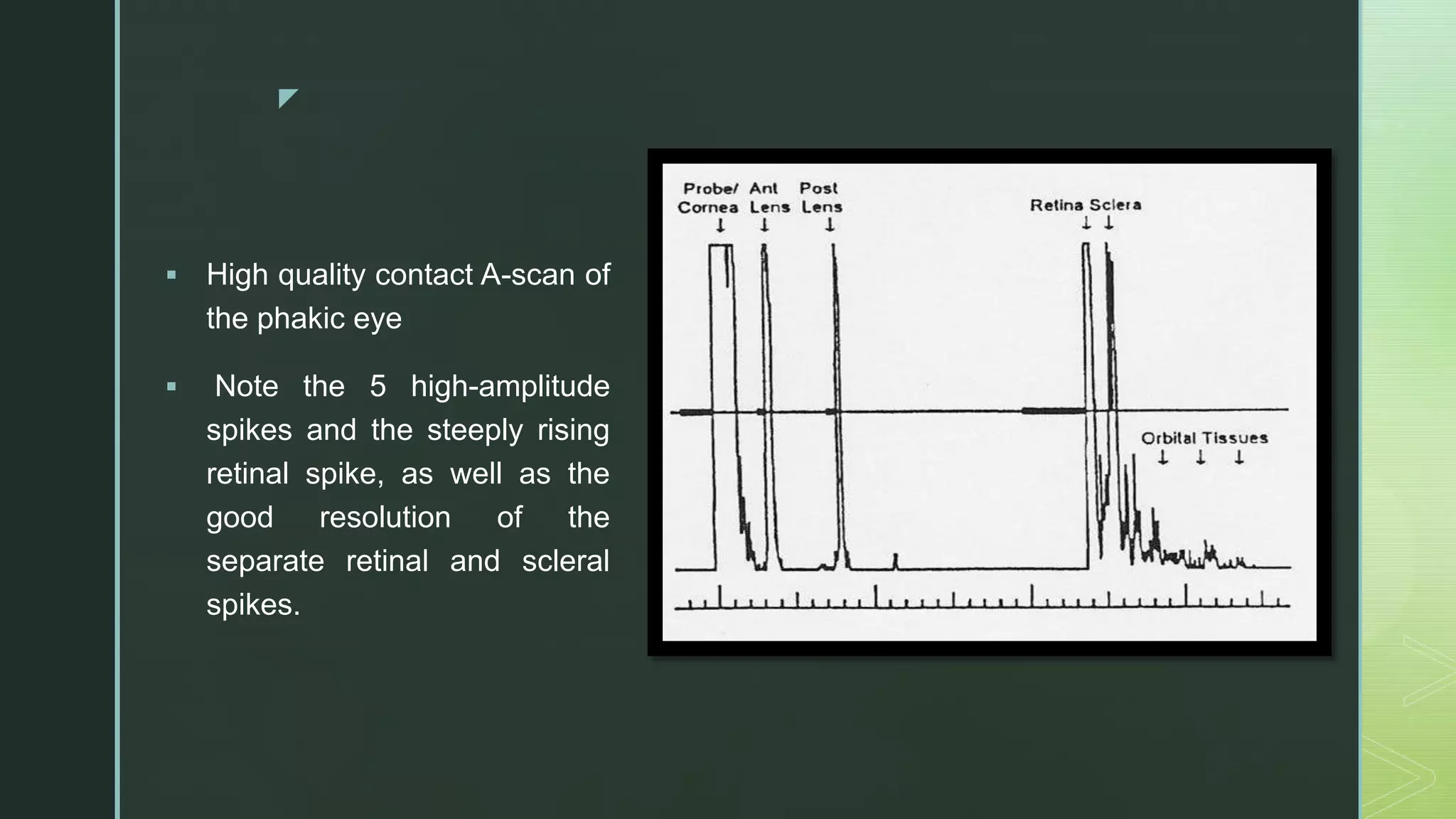
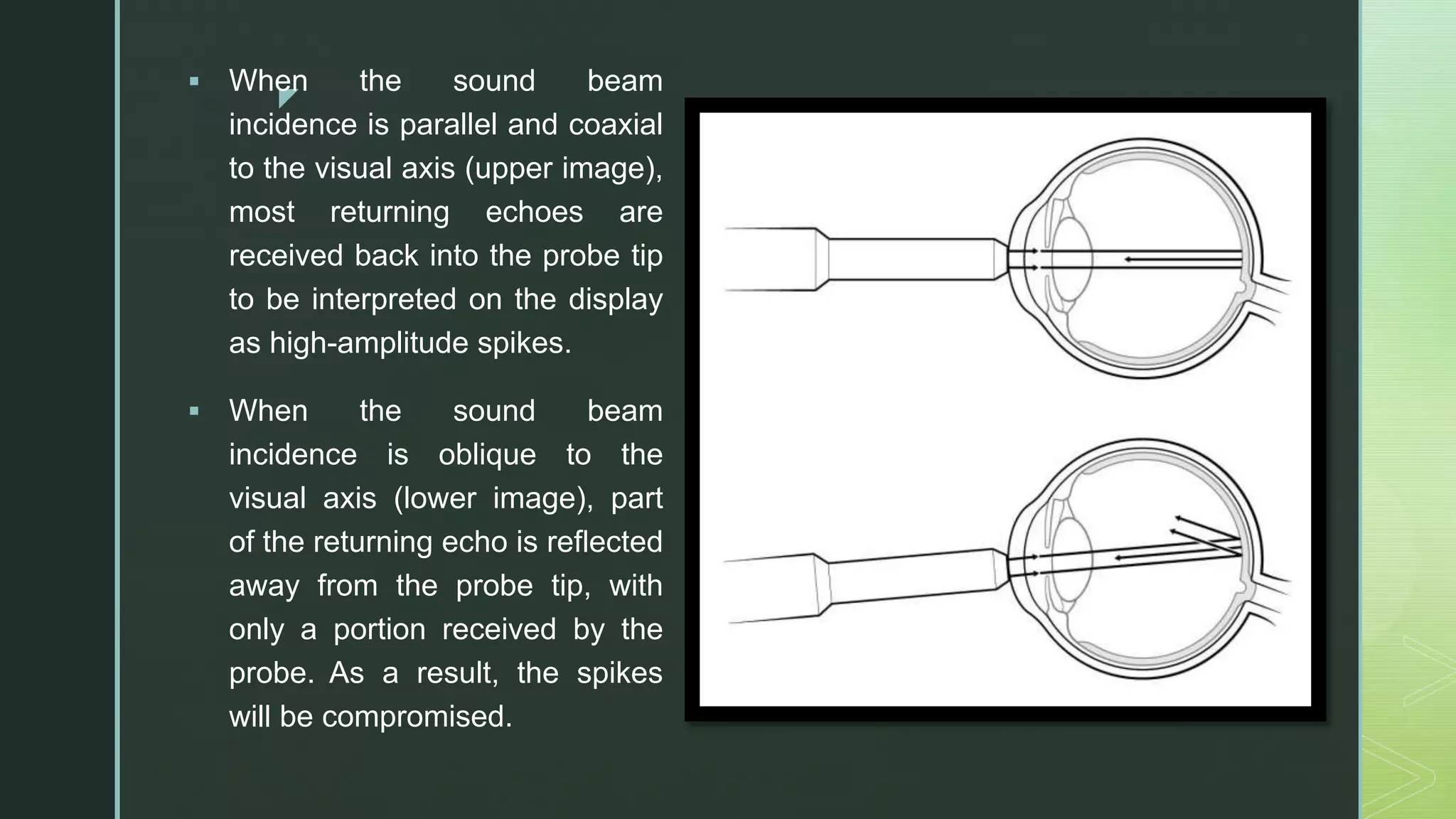
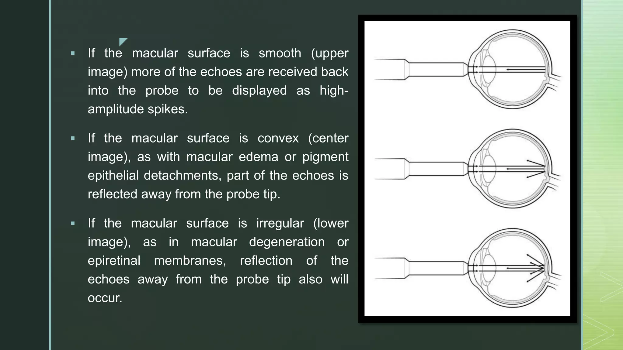
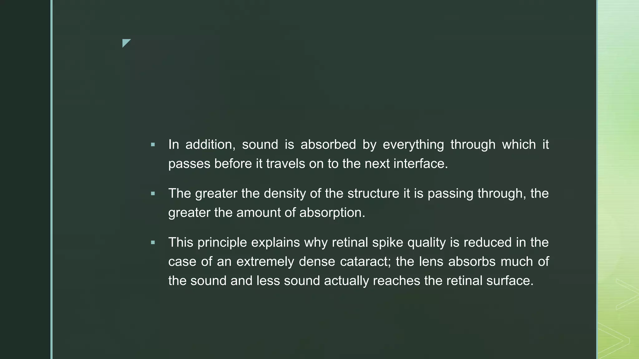
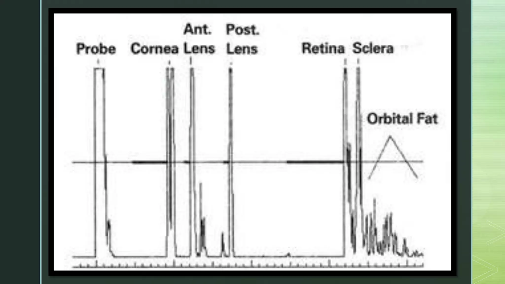
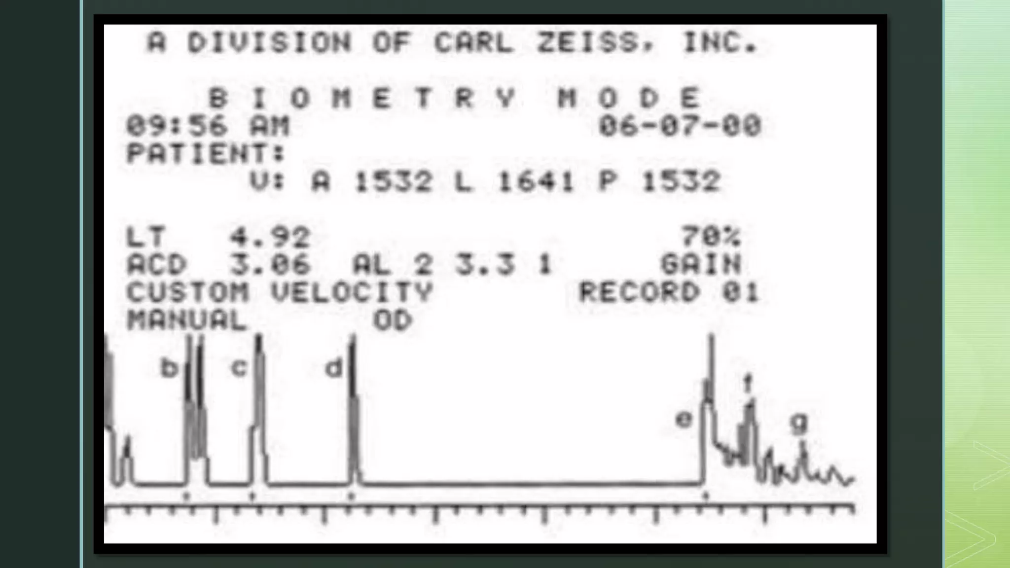
z
A- scan
By
Dr. Alshymaa Moustafa
Ophthalmology Specialist
z
Principles Of A- mode ultrasonography
 A-scan is also known as the amplitude scan.
 In the A-scan, a single sound beam is sent from the transducer, and the
returned echoes are converted into a series of two- dimensional wave-like
spikes with the height proportional to the strength of the echo.
 The strength of the echo depends on various tissue and tissue interface
factors
1. Relative difference between the tissue interfaces
2. Angle of the incident sound beam
3. Size and shape of the interfaces
4. Density of the media
z
 High quality contact A-scan of
the phakic eye
 Note the 5 high-amplitude
spikes and the steeply rising
retinal spike, as well as the
good resolution of the
separate retinal and scleral
spikes.
z
 If the ocular tissue interfaces are very different, the reflected
echoes will be of higher amplitude, resulting in a taller spike on the
A-wave scan, but if the interfaces are similar, the spike will be
shorter in height.
 In addition, the spike height is also impacted by the angle of the
sound wave hitting the interface, and the height of the spike will be
affected in presence of denser media, such as dense cataracts or
dense vitreous hemorrhages.
 A-scan is employed for measuring the axial length of the eye.
 In addition, corneal pachymetry can also be performed using A-
scan.
z
 When the sound beam
incidence is parallel and coaxial
to the visual axis (upper image),
most returning echoes are
received back into the probe tip
to be interpreted on the display
as high-amplitude spikes.
 When the sound beam
incidence is oblique to the
visual axis (lower image), part
of the returning echo is reflected
away from the probe tip, with
only a portion received by the
probe. As a result, the spikes
will be compromised.
z
 If the macular surface is smooth (upper
image) more of the echoes are received back
into the probe to be displayed as high-
amplitude spikes.
 If the macular surface is convex (center
image), as with macular edema or pigment
epithelial detachments, part of the echoes is
reflected away from the probe tip.
 If the macular surface is irregular (lower
image), as in macular degeneration or
epiretinal membranes, reflection of the
echoes away from the probe tip also will
occur.
z
 In addition, sound is absorbed by everything through which it
passes before it travels on to the next interface.
 The greater the density of the structure it is passing through, the
greater the amount of absorption.
 This principle explains why retinal spike quality is reduced in the
case of an extremely dense cataract; the lens absorbs much of
the sound and less sound actually reaches the retinal surface.
z
z
z
Thanks

A scan

  • 1.
    z A- scan By Dr. AlshymaaMoustafa Ophthalmology Specialist
  • 2.
    z Principles Of A-mode ultrasonography  A-scan is also known as the amplitude scan.  In the A-scan, a single sound beam is sent from the transducer, and the returned echoes are converted into a series of two- dimensional wave-like spikes with the height proportional to the strength of the echo.  The strength of the echo depends on various tissue and tissue interface factors 1. Relative difference between the tissue interfaces 2. Angle of the incident sound beam 3. Size and shape of the interfaces 4. Density of the media
  • 3.
    z  High qualitycontact A-scan of the phakic eye  Note the 5 high-amplitude spikes and the steeply rising retinal spike, as well as the good resolution of the separate retinal and scleral spikes.
  • 4.
    z  If theocular tissue interfaces are very different, the reflected echoes will be of higher amplitude, resulting in a taller spike on the A-wave scan, but if the interfaces are similar, the spike will be shorter in height.  In addition, the spike height is also impacted by the angle of the sound wave hitting the interface, and the height of the spike will be affected in presence of denser media, such as dense cataracts or dense vitreous hemorrhages.  A-scan is employed for measuring the axial length of the eye.  In addition, corneal pachymetry can also be performed using A- scan.
  • 5.
    z  When thesound beam incidence is parallel and coaxial to the visual axis (upper image), most returning echoes are received back into the probe tip to be interpreted on the display as high-amplitude spikes.  When the sound beam incidence is oblique to the visual axis (lower image), part of the returning echo is reflected away from the probe tip, with only a portion received by the probe. As a result, the spikes will be compromised.
  • 6.
    z  If themacular surface is smooth (upper image) more of the echoes are received back into the probe to be displayed as high- amplitude spikes.  If the macular surface is convex (center image), as with macular edema or pigment epithelial detachments, part of the echoes is reflected away from the probe tip.  If the macular surface is irregular (lower image), as in macular degeneration or epiretinal membranes, reflection of the echoes away from the probe tip also will occur.
  • 7.
    z  In addition,sound is absorbed by everything through which it passes before it travels on to the next interface.  The greater the density of the structure it is passing through, the greater the amount of absorption.  This principle explains why retinal spike quality is reduced in the case of an extremely dense cataract; the lens absorbs much of the sound and less sound actually reaches the retinal surface.
  • 8.
  • 9.
  • 11.