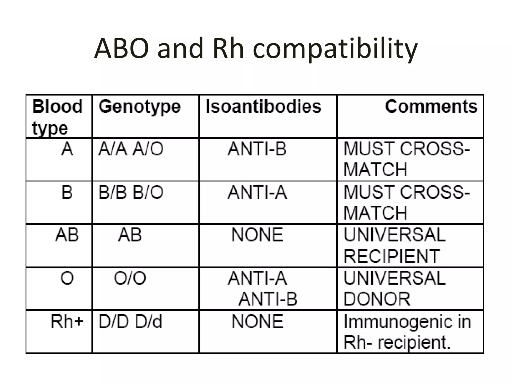
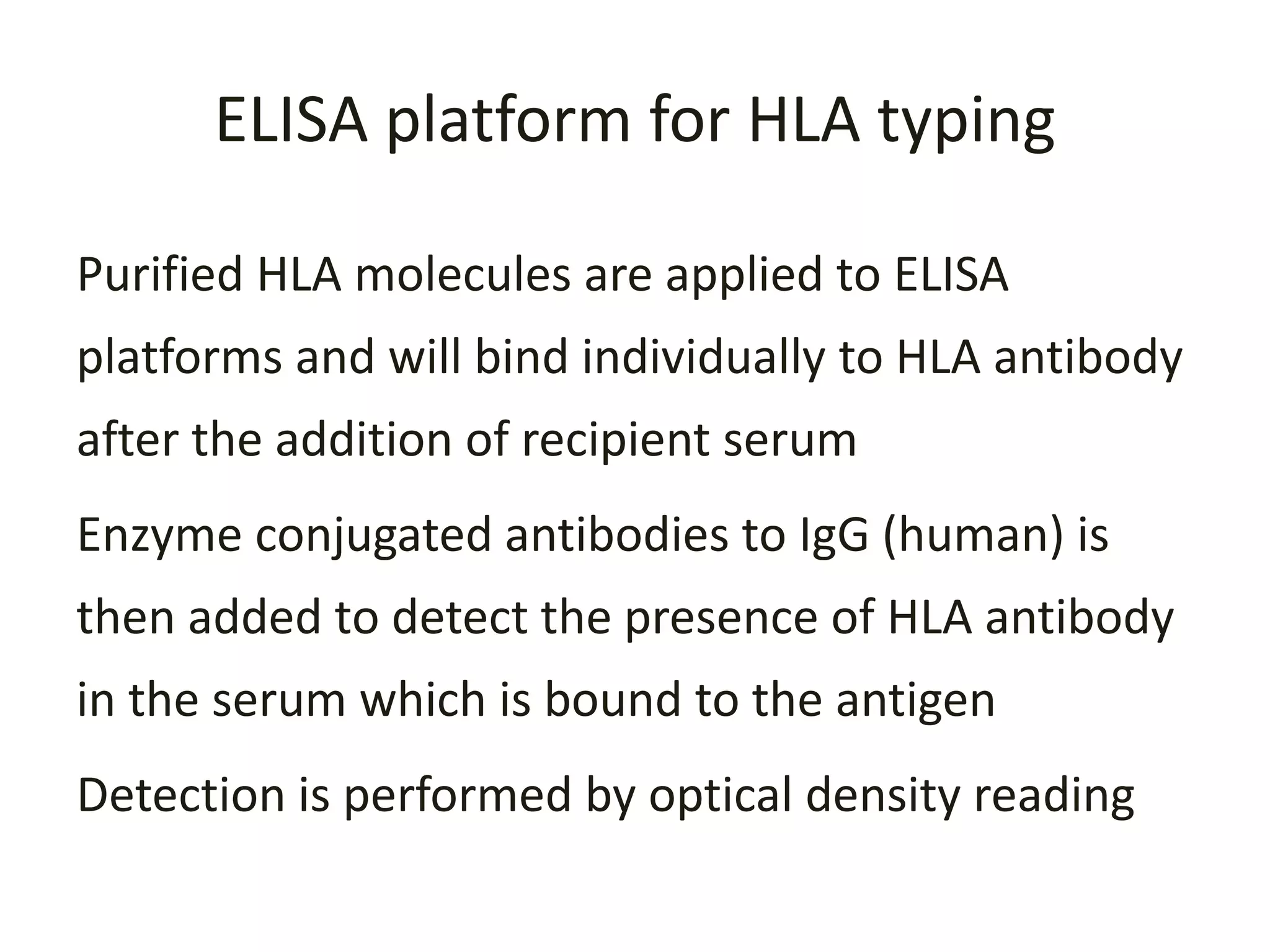
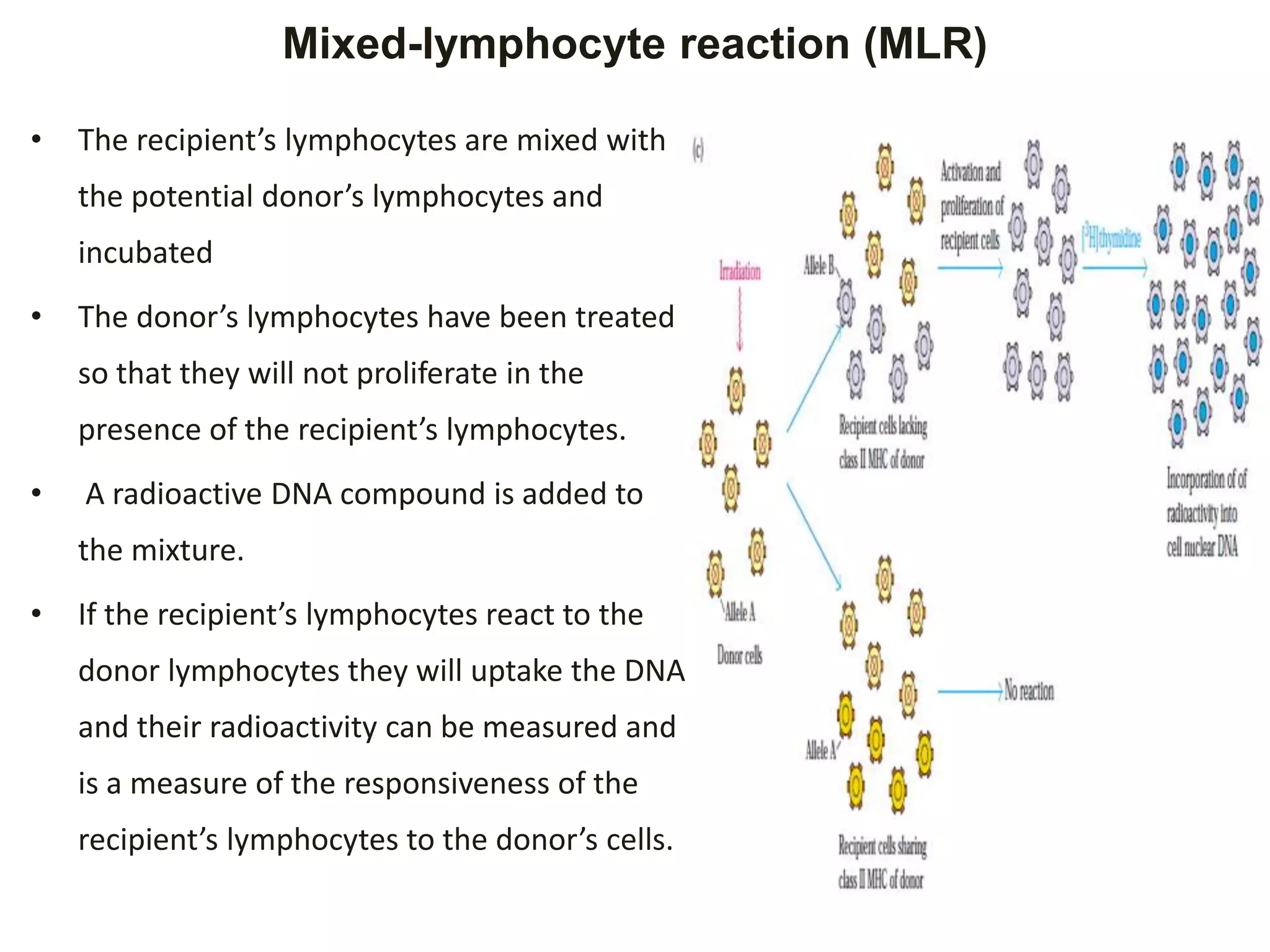
1. Introduction
• The adaptive immunity is the main response exerted to
the transplanted tissue
• Both cell mediated( CD4+ and CD8) as well as humoral
immunoresponse recognize the MHC molecules/HLA
expressed on the surface of donor cells
• These surface molecules induce an antigenic stimulus
that cause the rejection immune response to grafted
tissue or organ
• HLA are expressed on almost all nucleated cells, and are
the major molecules that initiate graft rejection
• Therefore, both ABO blood grouping and HLA class I and
II matching is important in organ transplantation
2. Introduction cont..
• The following are varieties of transplantation
antigens that induce immunoresponse against
grafted tissue/organ including the
The major histocompatibility molecules
The minor histocompatibility antigens,
The ABO blood group antigens and Rh
Endothelial cell antigens
3. Methods for HLA-typing
• Several methods including :-
1. Serological methods
– Complement mediated cytotoxicity
– ELIZA
2. Molecular techniques such as
– sequence-specific priming (SSP),
– sequence-specific oligonucleotide probing (SSOP),
– Sequence based typing (SBT) and
– reference strand-based conformation analysis (RSCA)
method
5. HLA class I typing
• There are three classical loci/allelic diversity at
HLA class I
– HLA-A,
– HLA -B, and
– HLA-Cw.
6. HLA class II typing
• There are five loci/allelic diversity at class II
– HLA-DR,
– HLA-DQ,
– HLA-DP,
– HLA-DM, and
– HLA-DO
• The HLA-DM and HLA-DO, are used in the internal
processing of antigens, loading the antigenic peptides
generated from pathogens onto the HLA molecules of
APCs
• The contribution of the allelic diversity of class I and II
genes to immune recognition and alloreactivity can be
analyzed by serological methods and molecular methods
as preciously mentioned
7. HLA typing and its influence on organ
transplantation
• The selection of the optimal donor is based on
high resolution HLA typing
• Because HLA mismatches result graft rejection
• HLA mismatches may occur at antigenic or allelic
level
– Antigenic mismatch are characterized by amino acid
substitutions in both peptide binding and T-cell
recognition regions
– Mismatch at allelic level are characterized by amino-
acid substitution in the peptide binding regions only
8. Types tissue typing
ABO and Rh compatibility (cross-matching)
testing
HLA I and HLA II typing
10. ELISA platform for HLA typing
Purified HLA molecules are applied to ELISA
platforms and will bind individually to HLA antibody
after the addition of recipient serum
Enzyme conjugated antibodies to IgG (human) is
then added to detect the presence of HLA antibody
in the serum which is bound to the antigen
Detection is performed by optical density reading
11. Mixed-lymphocyte reaction (MLR)
• The recipient’s lymphocytes are mixed with
the potential donor’s lymphocytes and
incubated
• The donor’s lymphocytes have been treated
so that they will not proliferate in the
presence of the recipient’s lymphocytes.
• A radioactive DNA compound is added to
the mixture.
• If the recipient’s lymphocytes react to the
donor lymphocytes they will uptake the DNA
and their radioactivity can be measured and
is a measure of the responsiveness of the
recipient’s lymphocytes to the donor’s cells.
12. Lymphocyte microcytotoxicity testing (LMT)
• If the lymphocyte cells express the
MHC allele for which a particular
monoclonal antibody is specific, then
the cells will be lysed upon addition of
complement and these dead cells will
take up a dye such as trypan blue.
• HLA typing based on antibody-
mediated microcytotoxicity can thus
indicate the presence or absence of
various MHC alleles.
Editor's Notes
MLR determine and quantify identity of class II HLA between a potential donor and recipient when a fully HLA-compatible donor is not available.
Lymphocytes from the donor are irradiated or treated with mitomycin C to prevent cell division and then added to recipient cells
If the class II HLA on the two cell populations are different, the recipient cells rapidly divide and take up large quantities of radioactive nucleotides into the newly synthesized nuclear DNA.
The amount of radioactive nucleotide uptake is roughly proportionate to the MHC class II differences between the donor and recipient lymphocytes