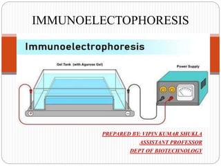
PREPARED BY: VIPIN KUMAR SHUKLA
ASSISTANT PROFESSOR
DEPT OF BIOTECHNOLOGY
IMMUNOELECTOPHORESIS
Introduction:
 Immunoelectrophoresis refers to precipitation in agar under an
electric field.
 It is a process of combination of Immuno-diffusion and
electrophoresis.
 An antigen mixture is first separated into its component parts by
electrophoresis and then tested by double Immuno-diffusion.
 Antigens are placed into wells cut in a gel (without antibody)
and electrophoresed. A trough is then cut in the gel into which
antibodies are placed.
 The antibodies diffuse laterally to meet diffusing antigen, and
lattice formation and precipitation occur permitting
determination of the nature of the antigens.
 The term “Immunoelectrophoresis” was first coined by Grabar
and Williams in 1953.
Principle:
 When electric current is applied to a slide layered
with gel, antigen mixture placed in wells is
separated into individual antigen components
according to their charge and size.
 Following electrophoresis, the separated antigens
are reacted with specific antisera placed in troughs
parallel to the electrophoretic migration and
diffusion is allowed to occur.
 Antiserum present in the trough moves toward the
antigen components resulting in formation of
separate precipitin lines in 18-24 hrs, each
indicating reaction between individual proteins
with its antibody.
Procedure:
 Agarose gel is prepared on a glass slide put in a
horizontal position.
 Using sample template, wells are borne on the
application zone carefully.
 The sample is diluted 2:3 with protein diluents
solution (20μl antigen solution +10 μl diluents).
 Using a 5 μl pipette, 5 μl of control and sample is
applied across each corresponding slit (Control slit
and Sample slit).
 The gel is placed into the electrophoresis chamber
with the samples on the cathode side, and
electrophoresis run for 20 mins/ 100 volts.
Continued……
 After electrophoresis completes, 20 μl of the
corresponding antiserum is added to troughs in
moist chamber and incubated for 18- 20 hours at
room temperature on a horizontal position.
 The agarose gel is placed on a horizontal position,
and dried with blotter sheets.
 The gel in saline solution is soaked for 10 minutes
and the drying and washing repeated twice again.
 The gel is dried at a temperature less than 70°C
and may be stained with protein staining solution
for about 3 minutes.
Results:
 Presence of elliptical precipitin arcs represents
antigen antibody interaction.
 Absence of formation of precipitate suggests no
reaction.
 Different antigens (proteins) can be identified
based on the intensity, shape, and position of the
precipitation lines.
Applications:
 The test helps in identification and approximate
quantization of various proteins present in the
serum, in protein identification and in
immunology.
 Immunoelectrophoresis is used in patients with
suspected monoclonal and polyclonal
gammopathies.
Continued……
 The medical diagnostic use is of value where
certain proteins are suspected of being absent (e.g.,
hypogammaglobulinemia) or overproduced (e.g.,
multiple myeloma).
 This method is useful to monitor antigen and
antigen-antibody purity and to identify a single
antigen in a mixture of antigens.
Continued…..
 The method is used to detect normal as well as abnormal
proteins, such as myeloma proteins in human serum.
 Used to analyze complex protein mixtures containing
different antigens.
Immunoelectophoresis

Immunoelectophoresis

  • 1.
    PREPARED BY: VIPINKUMAR SHUKLA ASSISTANT PROFESSOR DEPT OF BIOTECHNOLOGY IMMUNOELECTOPHORESIS
  • 2.
    Introduction:  Immunoelectrophoresis refersto precipitation in agar under an electric field.  It is a process of combination of Immuno-diffusion and electrophoresis.  An antigen mixture is first separated into its component parts by electrophoresis and then tested by double Immuno-diffusion.  Antigens are placed into wells cut in a gel (without antibody) and electrophoresed. A trough is then cut in the gel into which antibodies are placed.  The antibodies diffuse laterally to meet diffusing antigen, and lattice formation and precipitation occur permitting determination of the nature of the antigens.  The term “Immunoelectrophoresis” was first coined by Grabar and Williams in 1953.
  • 3.
    Principle:  When electriccurrent is applied to a slide layered with gel, antigen mixture placed in wells is separated into individual antigen components according to their charge and size.  Following electrophoresis, the separated antigens are reacted with specific antisera placed in troughs parallel to the electrophoretic migration and diffusion is allowed to occur.  Antiserum present in the trough moves toward the antigen components resulting in formation of separate precipitin lines in 18-24 hrs, each indicating reaction between individual proteins with its antibody.
  • 5.
    Procedure:  Agarose gelis prepared on a glass slide put in a horizontal position.  Using sample template, wells are borne on the application zone carefully.  The sample is diluted 2:3 with protein diluents solution (20μl antigen solution +10 μl diluents).  Using a 5 μl pipette, 5 μl of control and sample is applied across each corresponding slit (Control slit and Sample slit).  The gel is placed into the electrophoresis chamber with the samples on the cathode side, and electrophoresis run for 20 mins/ 100 volts.
  • 6.
    Continued……  After electrophoresiscompletes, 20 μl of the corresponding antiserum is added to troughs in moist chamber and incubated for 18- 20 hours at room temperature on a horizontal position.  The agarose gel is placed on a horizontal position, and dried with blotter sheets.  The gel in saline solution is soaked for 10 minutes and the drying and washing repeated twice again.  The gel is dried at a temperature less than 70°C and may be stained with protein staining solution for about 3 minutes.
  • 7.
    Results:  Presence ofelliptical precipitin arcs represents antigen antibody interaction.  Absence of formation of precipitate suggests no reaction.  Different antigens (proteins) can be identified based on the intensity, shape, and position of the precipitation lines.
  • 8.
    Applications:  The testhelps in identification and approximate quantization of various proteins present in the serum, in protein identification and in immunology.  Immunoelectrophoresis is used in patients with suspected monoclonal and polyclonal gammopathies.
  • 9.
    Continued……  The medicaldiagnostic use is of value where certain proteins are suspected of being absent (e.g., hypogammaglobulinemia) or overproduced (e.g., multiple myeloma).  This method is useful to monitor antigen and antigen-antibody purity and to identify a single antigen in a mixture of antigens.
  • 10.
    Continued…..  The methodis used to detect normal as well as abnormal proteins, such as myeloma proteins in human serum.  Used to analyze complex protein mixtures containing different antigens.