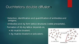
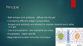
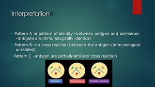
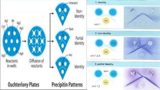
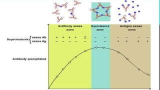
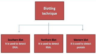
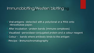
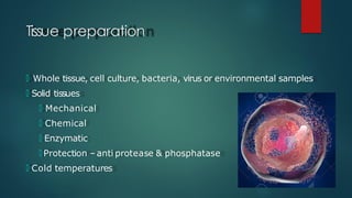
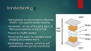
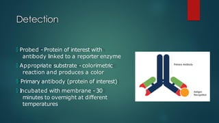
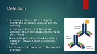
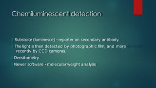
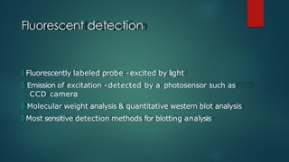
Diagnosis of disease using
immunological methods- Ouchterlony
double diffusion, Immunoblotting
UNIT-5
R.T.VINODHINI
Immunodiffusion
🞅 Study reaction between antigens and
antibodies
🞅 Observing precipitates formed
🞅 Combination of specific antigens and
antibodies
🞅 Diffused in a gel
🞅 Separately placed
Ouchterlony double diffusion
🞅 Detection, identification and quantification of antibodies and
antigens.
🞅 Antibodies and Ag form lattice structures (visible precipitate).
🞅 Formation of Ab-Ag lattice depends on,
• Ab must be bivalent.
• Ag must be bivalent or polyvalent.
Principle
🞅 Both antigen and antibody - diffuse into the gel
🞅 Comparing different antigen preparations
🞅 Antigen and antibody are allowed to migrate towards each other
in a gel
🞅 Line of precipitation - two reactants are meet
🞅 Precipitation - highly specific
🞅 Diagnosisand protein detection technique
Required materials
🞅 Chemicals:
🞅 Agarose
🞅 Assay Buffer(PBS)
🞅 Anti-serum
🞅 T
est antigens
🞅 pH - 7.2
Required materials
🞅 Equipments:
🞅 I
ncubator (37°C)
🞅 Micro pipette
🞅 Tips
🞅 Weighing balance
🞅 Gel punch with syringe
Required materials
🞅 Glassware's:
🞅 Glass plate
🞅 Conical flask
🞅 Measuring cylinder
🞅 Reagents:
🞅 Alcohol
🞅 Distilled water
Procedure
🞅 25 ml of 1.2 %
agarose (0.3g/25ml) in assay buffer
🞅 Boiling to dissolve the agarose completely
🞅 Cool - 55 to 60°C
🞅 Pour 4ml/plate
🞅 Gel to set - 30 minutes
🞅 Punch wells
Procedure
🞅 Fill the centre well 10 micro liter of anti-serum
🞅 Upper T
wo wells with 10 ml of each antigen
🞅 Glass plate - moist chamber over night at 37°C
🞅 Opaque precipitin lines - Antigen and anti-sera wells
Specific Objective
 🞅 To interpret the results obtained in Ouchterlony double diffusion
method (U/C)
Interpretation
🞅 Pattern A or pattern of identity -between antigen and anti-serum
-antigens are immunologically identical
🞅 Pattern B–no cross reaction between the antigen (immunological
unrelated)
🞅 Pattern C - antigen are partially similar or cross reaction
Applications
🞅 Identification of Fungal antigen. Eg - Coccidiomycosis, Histoplasma.
🞅 Measuring hormones serum proteins, drugs, etc.
🞅 Clinical laboratory - determination of Immunoglobulin levels in
patient sample.
Immunoblotting/Western blotting
🞅 Viral antigens - detected with a polyclonal or a MAb onto
nitrocellulose paper.
🞅 After incubation - protein bands (immune complexes)
🞅 Visualized - peroxidase-conjugated protein and a colour reagent
🞅 Colour - bands where antibody binds to the antigen
🞅 Principe - I
mmunochromatography
Procedure
🞅 T
issue preparation
🞅 Gel electrophoresis
🞅 T
ransfer/blotting
🞅 Blocking
🞅 Hybridization
🞅 Detection
Tissue preparation
🞅 Whole tissue, cell culture, bacteria, virus or environmental samples
🞅 Solid tissues
🞅 Mechanical
🞅 Chemical
🞅 Enzymatic
🞅 Protection –anti protease & phosphatase
🞅 Cold temperatures
Gel electrophoresis
🞅 SDSPAGE
🞅 Separation - molecularweight, electric
charge or a combination
Transfer/blotting
🞅 Nitrocellulose or polyvinylidene difluoride
(PVDF) –non specific protein binding
🞅 Membrane - on top of the gel & stack of
filter papers placed on top of that
🞅 Placed in a buffer solution
🞅 Moves up the paper by capillary action
bringing the proteins with it
🞅 Electrobloting - electric current to pull
proteins from the gel into membrane
Blocking
🞅 Prevent interactions between the membrane and the antibody
used
🞅 Bovin Serum Albumin (BSA) or non-fat dry milk with Tween20
(detergent)
🞅 The protein in the dilute solution attaches to the membrane in
all places where the target proteins have not attached
🞅 Reduces "noise" in the final product
🞅 Leading to clearer results & eliminates false positives
Detection
🞅 Probed - Protein of interest with
antibody linked to a reporter enzyme
🞅 Appropriate substrate - colorimetric
reaction and produces a color
🞅 Primary antibody (protein of interest)
🞅 I
ncubated with membrane - 30
minutes to overnight at different
temperatures
Detection
🞅 Secondary antibody {
After rinsing the
membrane to remove unbound primary
antibody)
🞅 Secondary antibody +biotin/reporter
enzymes (alkalin phosphatase/horseradish
peroxidase)
🞅 Horseradish peroxidase-linked secondary
antibody - cleave a chemiluminescent
agent
🞅 Luminescence in proportion to the amount
of protein
Analysis
🞅 Colorimetric detection
🞅 Chemiluminescent detection
🞅 Radioactive detection
🞅 Fluorescent detection
Colorimetric detection
🞅 Incubation with substrate -reporter enzyme (such as peroxidase)
that is bound to the secondary antibody
🞅 Converts soluble dye - insoluble form of a different color
🞅 Precipitates next to the enzyme & stains the membrane
🞅 Densitometry or spectrophotometry
Chemiluminescent detection
🞅 Substrate (luminesce) - reporter on secondary antibody.
🞅 The light is then detected by photographic film, and more
recently by CCD cameras.
🞅 Densitometry.
🞅 Newer software - molecularweight analysis
Radioactive detection
🞅 Radioactive labels - no enzyme substrates
🞅 Placement of medical X-ray film directly against the western blot
🞅 Develops as it isexposed to the label and creates dark regions
🞅 Corresponds - protein bands of interest
🞅 Very expensive, health and safety risks are high
Fluorescent detection
🞅 Fluorescently labeled probe - excited by light
🞅 Emission of excitation -detected by a photosensor such as
CCD camera
🞅 Molecular weight analysis & quantitative western blot analysis
🞅 Most sensitive detection methods for blotting analysis
Thank you

ouchterlony double diffusion method and immunoblotting

  • 1.
    Diagnosis of diseaseusing immunological methods- Ouchterlony double diffusion, Immunoblotting UNIT-5 R.T.VINODHINI
  • 2.
    Immunodiffusion 🞅 Study reactionbetween antigens and antibodies 🞅 Observing precipitates formed 🞅 Combination of specific antigens and antibodies 🞅 Diffused in a gel 🞅 Separately placed
  • 3.
    Ouchterlony double diffusion 🞅Detection, identification and quantification of antibodies and antigens. 🞅 Antibodies and Ag form lattice structures (visible precipitate). 🞅 Formation of Ab-Ag lattice depends on, • Ab must be bivalent. • Ag must be bivalent or polyvalent.
  • 4.
    Principle 🞅 Both antigenand antibody - diffuse into the gel 🞅 Comparing different antigen preparations 🞅 Antigen and antibody are allowed to migrate towards each other in a gel 🞅 Line of precipitation - two reactants are meet 🞅 Precipitation - highly specific 🞅 Diagnosisand protein detection technique
  • 5.
    Required materials 🞅 Chemicals: 🞅Agarose 🞅 Assay Buffer(PBS) 🞅 Anti-serum 🞅 T est antigens 🞅 pH - 7.2
  • 6.
    Required materials 🞅 Equipments: 🞅I ncubator (37°C) 🞅 Micro pipette 🞅 Tips 🞅 Weighing balance 🞅 Gel punch with syringe
  • 7.
    Required materials 🞅 Glassware's: 🞅Glass plate 🞅 Conical flask 🞅 Measuring cylinder 🞅 Reagents: 🞅 Alcohol 🞅 Distilled water
  • 8.
    Procedure 🞅 25 mlof 1.2 % agarose (0.3g/25ml) in assay buffer 🞅 Boiling to dissolve the agarose completely 🞅 Cool - 55 to 60°C 🞅 Pour 4ml/plate 🞅 Gel to set - 30 minutes 🞅 Punch wells
  • 9.
    Procedure 🞅 Fill thecentre well 10 micro liter of anti-serum 🞅 Upper T wo wells with 10 ml of each antigen 🞅 Glass plate - moist chamber over night at 37°C 🞅 Opaque precipitin lines - Antigen and anti-sera wells
  • 10.
    Specific Objective  🞅To interpret the results obtained in Ouchterlony double diffusion method (U/C)
  • 11.
    Interpretation 🞅 Pattern Aor pattern of identity -between antigen and anti-serum -antigens are immunologically identical 🞅 Pattern B–no cross reaction between the antigen (immunological unrelated) 🞅 Pattern C - antigen are partially similar or cross reaction
  • 14.
    Applications 🞅 Identification ofFungal antigen. Eg - Coccidiomycosis, Histoplasma. 🞅 Measuring hormones serum proteins, drugs, etc. 🞅 Clinical laboratory - determination of Immunoglobulin levels in patient sample.
  • 16.
    Immunoblotting/Western blotting 🞅 Viralantigens - detected with a polyclonal or a MAb onto nitrocellulose paper. 🞅 After incubation - protein bands (immune complexes) 🞅 Visualized - peroxidase-conjugated protein and a colour reagent 🞅 Colour - bands where antibody binds to the antigen 🞅 Principe - I mmunochromatography
  • 17.
    Procedure 🞅 T issue preparation 🞅Gel electrophoresis 🞅 T ransfer/blotting 🞅 Blocking 🞅 Hybridization 🞅 Detection
  • 18.
    Tissue preparation 🞅 Wholetissue, cell culture, bacteria, virus or environmental samples 🞅 Solid tissues 🞅 Mechanical 🞅 Chemical 🞅 Enzymatic 🞅 Protection –anti protease & phosphatase 🞅 Cold temperatures
  • 19.
    Gel electrophoresis 🞅 SDSPAGE 🞅Separation - molecularweight, electric charge or a combination
  • 20.
    Transfer/blotting 🞅 Nitrocellulose orpolyvinylidene difluoride (PVDF) –non specific protein binding 🞅 Membrane - on top of the gel & stack of filter papers placed on top of that 🞅 Placed in a buffer solution 🞅 Moves up the paper by capillary action bringing the proteins with it 🞅 Electrobloting - electric current to pull proteins from the gel into membrane
  • 21.
    Blocking 🞅 Prevent interactionsbetween the membrane and the antibody used 🞅 Bovin Serum Albumin (BSA) or non-fat dry milk with Tween20 (detergent) 🞅 The protein in the dilute solution attaches to the membrane in all places where the target proteins have not attached 🞅 Reduces "noise" in the final product 🞅 Leading to clearer results & eliminates false positives
  • 22.
    Detection 🞅 Probed -Protein of interest with antibody linked to a reporter enzyme 🞅 Appropriate substrate - colorimetric reaction and produces a color 🞅 Primary antibody (protein of interest) 🞅 I ncubated with membrane - 30 minutes to overnight at different temperatures
  • 23.
    Detection 🞅 Secondary antibody{ After rinsing the membrane to remove unbound primary antibody) 🞅 Secondary antibody +biotin/reporter enzymes (alkalin phosphatase/horseradish peroxidase) 🞅 Horseradish peroxidase-linked secondary antibody - cleave a chemiluminescent agent 🞅 Luminescence in proportion to the amount of protein
  • 24.
    Analysis 🞅 Colorimetric detection 🞅Chemiluminescent detection 🞅 Radioactive detection 🞅 Fluorescent detection
  • 25.
    Colorimetric detection 🞅 Incubationwith substrate -reporter enzyme (such as peroxidase) that is bound to the secondary antibody 🞅 Converts soluble dye - insoluble form of a different color 🞅 Precipitates next to the enzyme & stains the membrane 🞅 Densitometry or spectrophotometry
  • 26.
    Chemiluminescent detection 🞅 Substrate(luminesce) - reporter on secondary antibody. 🞅 The light is then detected by photographic film, and more recently by CCD cameras. 🞅 Densitometry. 🞅 Newer software - molecularweight analysis
  • 27.
    Radioactive detection 🞅 Radioactivelabels - no enzyme substrates 🞅 Placement of medical X-ray film directly against the western blot 🞅 Develops as it isexposed to the label and creates dark regions 🞅 Corresponds - protein bands of interest 🞅 Very expensive, health and safety risks are high
  • 28.
    Fluorescent detection 🞅 Fluorescentlylabeled probe - excited by light 🞅 Emission of excitation -detected by a photosensor such as CCD camera 🞅 Molecular weight analysis & quantitative western blot analysis 🞅 Most sensitive detection methods for blotting analysis
  • 29.