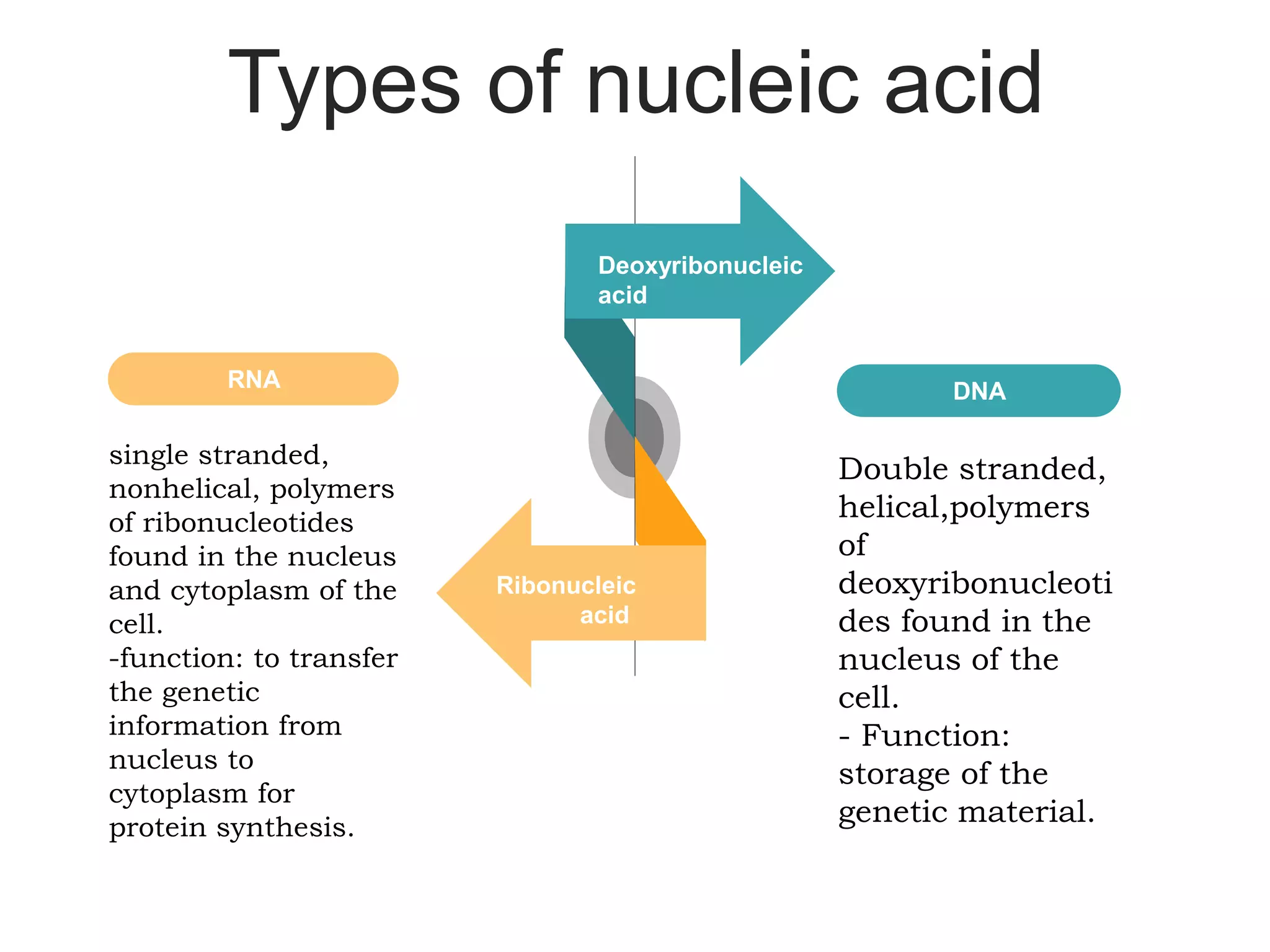
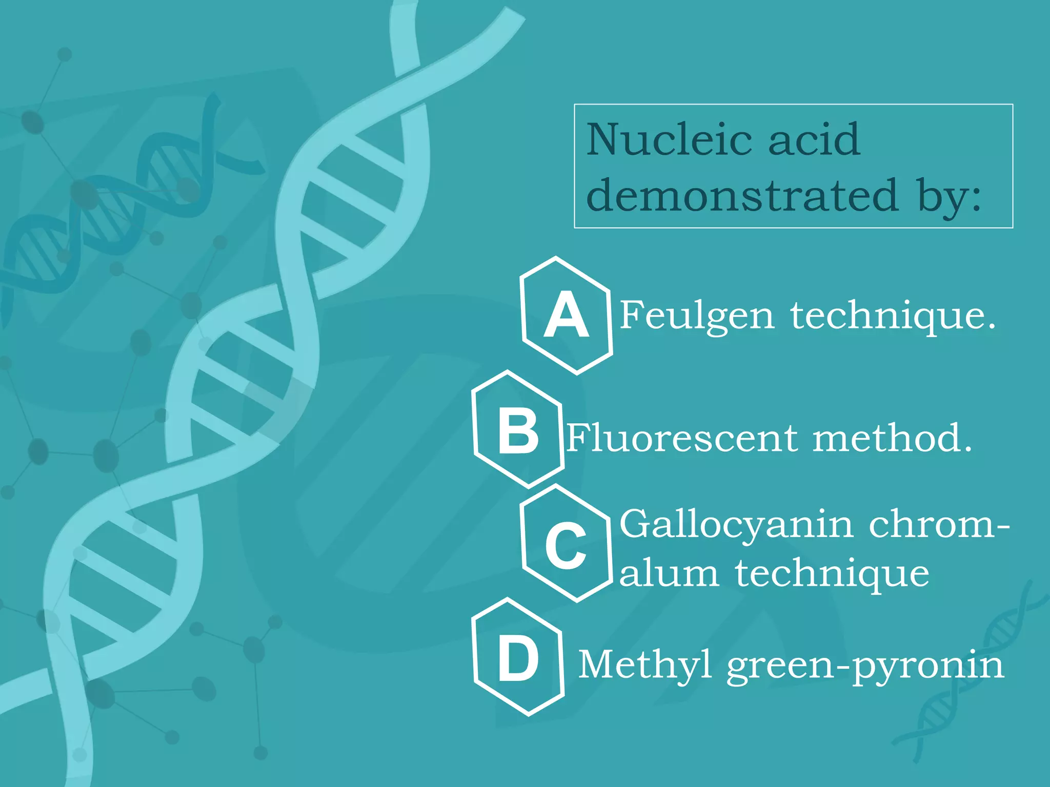
Demonstration of
nucleic acids
By: Omnia Elamin
Objectives :
Define the nucleic acid and
describe its composition.
0
1
Describe the diagnostic
application for the demonstration
of the nucleic acid.
0
2
List the methods for the
demonstration and describe their
principles.
0
3
Define the enzymatic extraction
and describe its function
0
4
Composition
of nucleic
acids:
You can simply impress your
audience and add a unique
zing and appeal to your
Presentations. Easy to change
colors, photos and Text. Get a
modern PowerPoint
Presentation that is beautifully
designed. You can simply
impress your audience and add
a unique zing and appeal to
your Presentations. Easy to
change colors, photos and Text.
Get a modern PowerPoint
Presentation that is beautifully
designed.
-Linear polymers of nucleotides.
-Each nucleotide consists of 3
components:
1- nitrogenous base.
2- pentose sugar.
3- phosphate group.
Types of nucleic acid
Double stranded,
helical,polymers
of
deoxyribonucleoti
des found in the
nucleus of the
cell.
- Function:
storage of the
genetic material.
DNA
single stranded,
nonhelical, polymers
of ribonucleotides
found in the nucleus
and cytoplasm of the
cell.
-function: to transfer
the genetic
information from
nucleus to
cytoplasm for
protein synthesis.
RNA
Deoxyribonucleic
acid
Ribonucleic
acid
Demonstration
of the nucliec
acid:
commonly used medical process in
the
medical diagnosis of tumors
in which a dye color is
applied on the sample tissues to
locate the diseased or tumorous cells
or other pathological cell
Why do we demonstrate the
nucleic acids?
Fixation
- Pathologic diagnosis requires tissue fixation for histologic
and immunohistologic analysis, and formalin is routinely
used for
this.
- In general terms, the nucleic acids are best preserved
in alcoholic and acidic fixatives, a good example
being Carnoy’s fluid which contains both alcohol
and glacial acetic acid
Decalcification
- strong acids tend to degrade DNA, making the decalcified
bone sections suboptimal for testing.
- Weak acids, although slower than the strong acid agents
in the decalcifying process, are much gentler in action and
less likely to interfere with nuclear staining
The monument to the
laboratory mouse
A little break!
Modern
Portfolio
Presenta
tion
You can simply impress your
audience and add a unique
zing and appeal to your
Presentations. Easy to change
colors, photos and Text. Get a
modern PowerPoint
Presentation that is beautifully
designed. You can simply
impress your audience and add
a unique zing and appeal to
your Presentations. Easy to
change colors, photos and Text.
Get a modern PowerPoint
Presentation that is beautifully
designed.
Nucleic acid
demonstrated by:
Feulgen technique.
Fluorescent method.
Gallocyanin chrom-
alum technique
Methyl green-pyronin
A
B
C
D
Feulgen reaction:
Mild acid hydrolysis, employing 1 M
hydrochloric acid at 60°C, is used to break
the purine-deoxyribose bond
the resulting ‘exposed’ aldehydes are then
demonstrated by the use of Schiff’s reagen
(plasmal reaction).
The method of Feulgen and Rossenbeck
(1924) is the
standard technique for demonstrating
deoxyribose.
Feulgen reaction:
Phospholipid may give feulgen reaction in
cryostat technique but not in paraffin technique.
Feulgen reaction used in conjunction with
micro-dinstomitry to study cancer (DNA
content in certain lymphoma is inverse to the
tumor prognosis).
Feulgen-naphthoic acid-hydrazide method
This technique can be used as control method for standard feulgen reaction
2-hydroxy-3-naphthoic acid
is coupled with fast blue B
0
4
The reaction gives blue to
bluish purple colour.
0
5
0
6
The sections are hydrolyzed in
1M HCl as in felgen reaction
0
1
This hydrolysis produces
aldehyde group
0
2
The aldehyde couples with
2-hydroxy-3-naphthoic acid
0
3
Gallocyanine-chrome
alum method:
It gives blue colour with both DNA and RNA.
The phosphoric acid of the nucleic acid of
both DNA and RNA combined with
gallocynin-chrome alum at acid PH.
Fluorescent method:
Acidine orange is an organic compound. It
interacts with DNA and RNA by interaction or
electrostatic attraction respectively.
DNA stained yellow-green and RNA stained red.
The most used method is acidine orange
technique.
Methyl green-pyronin:
Methyl green is specific for phosphate
radicals in the DNA double helix staining it
green-blue.
Pyronin does not posses this affinity and
binds to the remaining negatively charged
RNA staining it red.
Classical histological staining technique using
two basic (cationic) dyes for the demonstration
and defferentiation of DNA and RNA.
Methyl green is an impure dye containing
methyl violet which removed by washing by
chloroform, at PH 4,8.
Methods used for digestion of one of the nucleic
acid to make the applied technique specific for
the other one.
Digestion methods
for nucleic acid:
0
1
0
2
Enzymatic
method:
Chemical extraction
method:
Specific enzymes can be
used to digest DNA and
RNA in tissue section
-Pure deoxyribonuclease :
removes DNA.
-- ribonuclease : removes
RNA
1- percloric acid.
2- tri-chlora acetic acid.
3- hydrochloric acid
Thank You
For your attention

Nucleic acids (DNA+RNA) demonstration in histopathology

  • 1.
  • 2.
    Objectives : Define thenucleic acid and describe its composition. 0 1 Describe the diagnostic application for the demonstration of the nucleic acid. 0 2 List the methods for the demonstration and describe their principles. 0 3 Define the enzymatic extraction and describe its function 0 4
  • 3.
    Composition of nucleic acids: You cansimply impress your audience and add a unique zing and appeal to your Presentations. Easy to change colors, photos and Text. Get a modern PowerPoint Presentation that is beautifully designed. You can simply impress your audience and add a unique zing and appeal to your Presentations. Easy to change colors, photos and Text. Get a modern PowerPoint Presentation that is beautifully designed. -Linear polymers of nucleotides. -Each nucleotide consists of 3 components: 1- nitrogenous base. 2- pentose sugar. 3- phosphate group.
  • 4.
    Types of nucleicacid Double stranded, helical,polymers of deoxyribonucleoti des found in the nucleus of the cell. - Function: storage of the genetic material. DNA single stranded, nonhelical, polymers of ribonucleotides found in the nucleus and cytoplasm of the cell. -function: to transfer the genetic information from nucleus to cytoplasm for protein synthesis. RNA Deoxyribonucleic acid Ribonucleic acid
  • 5.
  • 6.
    commonly used medicalprocess in the medical diagnosis of tumors in which a dye color is applied on the sample tissues to locate the diseased or tumorous cells or other pathological cell Why do we demonstrate the nucleic acids?
  • 7.
    Fixation - Pathologic diagnosisrequires tissue fixation for histologic and immunohistologic analysis, and formalin is routinely used for this. - In general terms, the nucleic acids are best preserved in alcoholic and acidic fixatives, a good example being Carnoy’s fluid which contains both alcohol and glacial acetic acid
  • 8.
    Decalcification - strong acidstend to degrade DNA, making the decalcified bone sections suboptimal for testing. - Weak acids, although slower than the strong acid agents in the decalcifying process, are much gentler in action and less likely to interfere with nuclear staining
  • 9.
    The monument tothe laboratory mouse A little break!
  • 10.
    Modern Portfolio Presenta tion You can simplyimpress your audience and add a unique zing and appeal to your Presentations. Easy to change colors, photos and Text. Get a modern PowerPoint Presentation that is beautifully designed. You can simply impress your audience and add a unique zing and appeal to your Presentations. Easy to change colors, photos and Text. Get a modern PowerPoint Presentation that is beautifully designed.
  • 11.
    Nucleic acid demonstrated by: Feulgentechnique. Fluorescent method. Gallocyanin chrom- alum technique Methyl green-pyronin A B C D
  • 12.
    Feulgen reaction: Mild acidhydrolysis, employing 1 M hydrochloric acid at 60°C, is used to break the purine-deoxyribose bond the resulting ‘exposed’ aldehydes are then demonstrated by the use of Schiff’s reagen (plasmal reaction). The method of Feulgen and Rossenbeck (1924) is the standard technique for demonstrating deoxyribose.
  • 13.
    Feulgen reaction: Phospholipid maygive feulgen reaction in cryostat technique but not in paraffin technique. Feulgen reaction used in conjunction with micro-dinstomitry to study cancer (DNA content in certain lymphoma is inverse to the tumor prognosis).
  • 14.
    Feulgen-naphthoic acid-hydrazide method Thistechnique can be used as control method for standard feulgen reaction 2-hydroxy-3-naphthoic acid is coupled with fast blue B 0 4 The reaction gives blue to bluish purple colour. 0 5 0 6 The sections are hydrolyzed in 1M HCl as in felgen reaction 0 1 This hydrolysis produces aldehyde group 0 2 The aldehyde couples with 2-hydroxy-3-naphthoic acid 0 3
  • 15.
    Gallocyanine-chrome alum method: It givesblue colour with both DNA and RNA. The phosphoric acid of the nucleic acid of both DNA and RNA combined with gallocynin-chrome alum at acid PH.
  • 16.
    Fluorescent method: Acidine orangeis an organic compound. It interacts with DNA and RNA by interaction or electrostatic attraction respectively. DNA stained yellow-green and RNA stained red. The most used method is acidine orange technique.
  • 17.
    Methyl green-pyronin: Methyl greenis specific for phosphate radicals in the DNA double helix staining it green-blue. Pyronin does not posses this affinity and binds to the remaining negatively charged RNA staining it red. Classical histological staining technique using two basic (cationic) dyes for the demonstration and defferentiation of DNA and RNA. Methyl green is an impure dye containing methyl violet which removed by washing by chloroform, at PH 4,8.
  • 18.
    Methods used fordigestion of one of the nucleic acid to make the applied technique specific for the other one. Digestion methods for nucleic acid:
  • 19.
    0 1 0 2 Enzymatic method: Chemical extraction method: Specific enzymescan be used to digest DNA and RNA in tissue section -Pure deoxyribonuclease : removes DNA. -- ribonuclease : removes RNA 1- percloric acid. 2- tri-chlora acetic acid. 3- hydrochloric acid
  • 20.