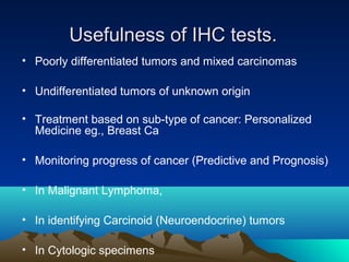
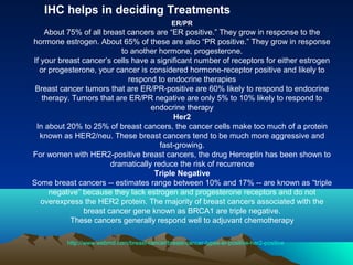
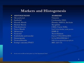
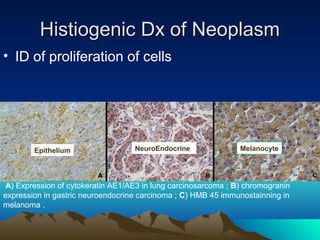
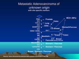
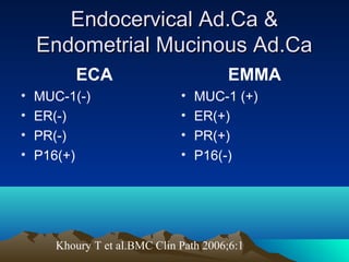
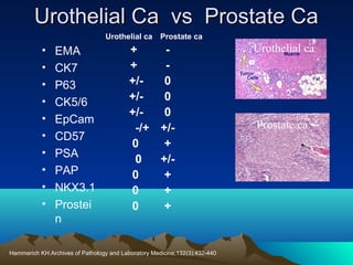
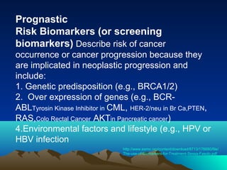
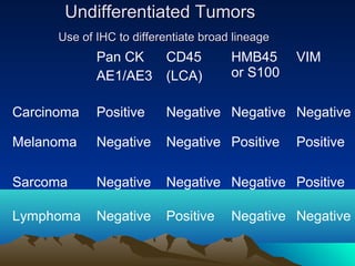
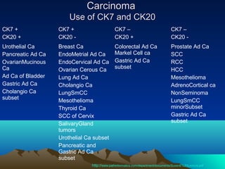
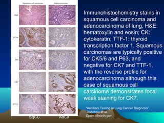
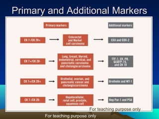
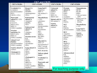
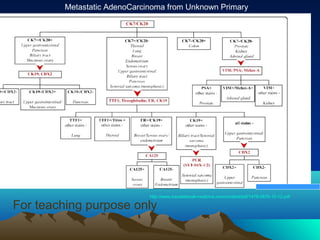
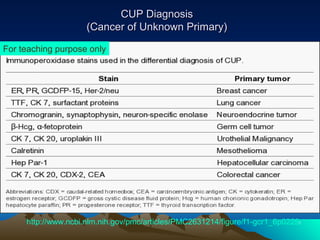
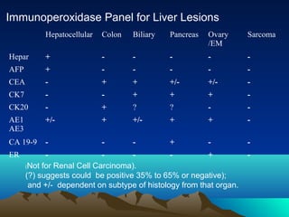
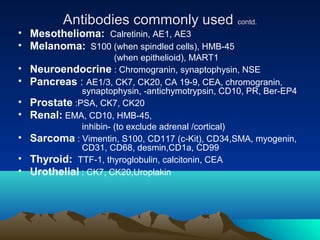
IHC is an important diagnostic tool that can provide valuable information to pathologists. When used along with H&E staining and other special stains, IHC helps identify cell types and origins that can aid in cancer diagnosis, classification, and determining appropriate treatment. Different tumor types express specific protein markers detectable by IHC. While IHC results must be interpreted carefully, they can help diagnose poorly differentiated tumors, identify primary sites of metastatic cancers, and guide personalized cancer treatment decisions. Large antibody panels are used for different cancer types to provide diagnostic, prognostic and predictive information through protein expression profiling.
Biomarkers have a diversified role in diagnosis, prognostication and risk stratification. This presentation aims to compile the basic information and new literature on various biomarkers pertaining to cancer care.
Therapeutic Cancer Vaccines: A Future of Possibilities Haunted By A History o...Michael Sheckler
These slides provide an overview of 100 therapeutic cancer vaccines in development, a look at some of the failures, what's been and is being done to address the clinical development of these vaccines and a snapshot of some deals, terms and the number of companies seeking commercializations partners.
Robert Anders, MD, PhD, Julie R. Brahmer, MD, MSc, and Christopher D. Gocke, MD, prepared useful Practice Aids pertaining to immunotherapy and biomarker testing for this CME/MOC/CC activity titled "Keeping Up With Advances in Cancer Immunotherapy and Biomarker Testing: Implications for Pathologists at the Forefront of the Emerging Precision Immuno-Oncology Era." For the full presentation, monograph, complete CME/MOC/CC information, and to apply for credit, please visit us at http://bit.ly/2L7zlSy. CME/MOC/CC credit will be available until May 2, 2020.
This presentation is part of MIU CE Pharmacy Program and is designed primarily for pharmacists with the following learning objectives:
1- Explain the mechanisms of action behind immune response to cancer and the application of immunotherapy in cancer treatment
2- Distinguish new and emerging immunotherapy classes and individual agents efficacy, safety to therapy in cancer treatment
3-Strategies to counsel and assist patients to overcome barriers to therapy, including Treatment side effects to improve adherence to therapy
This is needs to be updated as new Biomarkers and Dx antibodies come into existence in remarkable numbers every day! You have to construct an algorithm based on your need and availability of antibodies. Selection of Antibodies for the algorithm also depends on your ability to recognize the characterization of the specific antibody.
Target Audience: Oncology fellows and Oncologists
Carcinoma of unknown primary is a challenging scenario often encountered in Oncology practice. This slide presentation discusses favorable and unfavorable presentations of CUP and it's management
Report Back from SGO 2024: What’s the Latest in Cervical Cancer?bkling
Are you curious about what’s new in cervical cancer research or unsure what the findings mean? Join Dr. Emily Ko, a gynecologic oncologist at Penn Medicine, to learn about the latest updates from the Society of Gynecologic Oncology (SGO) 2024 Annual Meeting on Women’s Cancer. Dr. Ko will discuss what the research presented at the conference means for you and answer your questions about the new developments.
Acute scrotum is a general term referring to an emergency condition affecting the contents or the wall of the scrotum.
There are a number of conditions that present acutely, predominantly with pain and/or swelling
A careful and detailed history and examination, and in some cases, investigations allow differentiation between these diagnoses. A prompt diagnosis is essential as the patient may require urgent surgical intervention
Testicular torsion refers to twisting of the spermatic cord, causing ischaemia of the testicle.
Testicular torsion results from inadequate fixation of the testis to the tunica vaginalis producing ischemia from reduced arterial inflow and venous outflow obstruction.
The prevalence of testicular torsion in adult patients hospitalized with acute scrotal pain is approximately 25 to 50 percent
Ethanol (CH3CH2OH), or beverage alcohol, is a two-carbon alcohol
that is rapidly distributed in the body and brain. Ethanol alters many
neurochemical systems and has rewarding and addictive properties. It
is the oldest recreational drug and likely contributes to more morbidity,
mortality, and public health costs than all illicit drugs combined. The
5th edition of the Diagnostic and Statistical Manual of Mental Disorders
(DSM-5) integrates alcohol abuse and alcohol dependence into a single
disorder called alcohol use disorder (AUD), with mild, moderate,
and severe subclassifications (American Psychiatric Association, 2013).
In the DSM-5, all types of substance abuse and dependence have been
combined into a single substance use disorder (SUD) on a continuum
from mild to severe. A diagnosis of AUD requires that at least two of
the 11 DSM-5 behaviors be present within a 12-month period (mild
AUD: 2–3 criteria; moderate AUD: 4–5 criteria; severe AUD: 6–11 criteria).
The four main behavioral effects of AUD are impaired control over
drinking, negative social consequences, risky use, and altered physiological
effects (tolerance, withdrawal). This chapter presents an overview
of the prevalence and harmful consequences of AUD in the U.S.,
the systemic nature of the disease, neurocircuitry and stages of AUD,
comorbidities, fetal alcohol spectrum disorders, genetic risk factors, and
pharmacotherapies for AUD.
Prix Galien International 2024 Forum ProgramLevi Shapiro
June 20, 2024, Prix Galien International and Jerusalem Ethics Forum in ROME. Detailed agenda including panels:
- ADVANCES IN CARDIOLOGY: A NEW PARADIGM IS COMING
- WOMEN’S HEALTH: FERTILITY PRESERVATION
- WHAT’S NEW IN THE TREATMENT OF INFECTIOUS,
ONCOLOGICAL AND INFLAMMATORY SKIN DISEASES?
- ARTIFICIAL INTELLIGENCE AND ETHICS
- GENE THERAPY
- BEYOND BORDERS: GLOBAL INITIATIVES FOR DEMOCRATIZING LIFE SCIENCE TECHNOLOGIES AND PROMOTING ACCESS TO HEALTHCARE
- ETHICAL CHALLENGES IN LIFE SCIENCES
- Prix Galien International Awards Ceremony
New Drug Discovery and Development .....NEHA GUPTA
The "New Drug Discovery and Development" process involves the identification, design, testing, and manufacturing of novel pharmaceutical compounds with the aim of introducing new and improved treatments for various medical conditions. This comprehensive endeavor encompasses various stages, including target identification, preclinical studies, clinical trials, regulatory approval, and post-market surveillance. It involves multidisciplinary collaboration among scientists, researchers, clinicians, regulatory experts, and pharmaceutical companies to bring innovative therapies to market and address unmet medical needs.
ARTIFICIAL INTELLIGENCE IN HEALTHCARE.pdfAnujkumaranit
Artificial intelligence (AI) refers to the simulation of human intelligence processes by machines, especially computer systems. It encompasses tasks such as learning, reasoning, problem-solving, perception, and language understanding. AI technologies are revolutionizing various fields, from healthcare to finance, by enabling machines to perform tasks that typically require human intelligence.
The prostate is an exocrine gland of the male mammalian reproductive system
It is a walnut-sized gland that forms part of the male reproductive system and is located in front of the rectum and just below the urinary bladder
Function is to store and secrete a clear, slightly alkaline fluid that constitutes 10-30% of the volume of the seminal fluid that along with the spermatozoa, constitutes semen
A healthy human prostate measures (4cm-vertical, by 3cm-horizontal, 2cm ant-post ).
It surrounds the urethra just below the urinary bladder. It has anterior, median, posterior and two lateral lobes
It’s work is regulated by androgens which are responsible for male sex characteristics
Generalised disease of the prostate due to hormonal derangement which leads to non malignant enlargement of the gland (increase in the number of epithelial cells and stromal tissue)to cause compression of the urethra leading to symptoms (LUTS
NVBDCP.pptx Nation vector borne disease control programSapna Thakur
NVBDCP was launched in 2003-2004 . Vector-Borne Disease: Disease that results from an infection transmitted to humans and other animals by blood-feeding arthropods, such as mosquitoes, ticks, and fleas. Examples of vector-borne diseases include Dengue fever, West Nile Virus, Lyme disease, and malaria.
Title: Sense of Smell
Presenter: Dr. Faiza, Assistant Professor of Physiology
Qualifications:
MBBS (Best Graduate, AIMC Lahore)
FCPS Physiology
ICMT, CHPE, DHPE (STMU)
MPH (GC University, Faisalabad)
MBA (Virtual University of Pakistan)
Learning Objectives:
Describe the primary categories of smells and the concept of odor blindness.
Explain the structure and location of the olfactory membrane and mucosa, including the types and roles of cells involved in olfaction.
Describe the pathway and mechanisms of olfactory signal transmission from the olfactory receptors to the brain.
Illustrate the biochemical cascade triggered by odorant binding to olfactory receptors, including the role of G-proteins and second messengers in generating an action potential.
Identify different types of olfactory disorders such as anosmia, hyposmia, hyperosmia, and dysosmia, including their potential causes.
Key Topics:
Olfactory Genes:
3% of the human genome accounts for olfactory genes.
400 genes for odorant receptors.
Olfactory Membrane:
Located in the superior part of the nasal cavity.
Medially: Folds downward along the superior septum.
Laterally: Folds over the superior turbinate and upper surface of the middle turbinate.
Total surface area: 5-10 square centimeters.
Olfactory Mucosa:
Olfactory Cells: Bipolar nerve cells derived from the CNS (100 million), with 4-25 olfactory cilia per cell.
Sustentacular Cells: Produce mucus and maintain ionic and molecular environment.
Basal Cells: Replace worn-out olfactory cells with an average lifespan of 1-2 months.
Bowman’s Gland: Secretes mucus.
Stimulation of Olfactory Cells:
Odorant dissolves in mucus and attaches to receptors on olfactory cilia.
Involves a cascade effect through G-proteins and second messengers, leading to depolarization and action potential generation in the olfactory nerve.
Quality of a Good Odorant:
Small (3-20 Carbon atoms), volatile, water-soluble, and lipid-soluble.
Facilitated by odorant-binding proteins in mucus.
Membrane Potential and Action Potential:
Resting membrane potential: -55mV.
Action potential frequency in the olfactory nerve increases with odorant strength.
Adaptation Towards the Sense of Smell:
Rapid adaptation within the first second, with further slow adaptation.
Psychological adaptation greater than receptor adaptation, involving feedback inhibition from the central nervous system.
Primary Sensations of Smell:
Camphoraceous, Musky, Floral, Pepperminty, Ethereal, Pungent, Putrid.
Odor Detection Threshold:
Examples: Hydrogen sulfide (0.0005 ppm), Methyl-mercaptan (0.002 ppm).
Some toxic substances are odorless at lethal concentrations.
Characteristics of Smell:
Odor blindness for single substances due to lack of appropriate receptor protein.
Behavioral and emotional influences of smell.
Transmission of Olfactory Signals:
From olfactory cells to glomeruli in the olfactory bulb, involving lateral inhibition.
Primitive, less old, and new olfactory systems with different path
These simplified slides by Dr. Sidra Arshad present an overview of the non-respiratory functions of the respiratory tract.
Learning objectives:
1. Enlist the non-respiratory functions of the respiratory tract
2. Briefly explain how these functions are carried out
3. Discuss the significance of dead space
4. Differentiate between minute ventilation and alveolar ventilation
5. Describe the cough and sneeze reflexes
Study Resources:
1. Chapter 39, Guyton and Hall Textbook of Medical Physiology, 14th edition
2. Chapter 34, Ganong’s Review of Medical Physiology, 26th edition
3. Chapter 17, Human Physiology by Lauralee Sherwood, 9th edition
4. Non-respiratory functions of the lungs https://academic.oup.com/bjaed/article/13/3/98/278874
Lung Cancer: Artificial Intelligence, Synergetics, Complex System Analysis, S...Oleg Kshivets
RESULTS: Overall life span (LS) was 2252.1±1742.5 days and cumulative 5-year survival (5YS) reached 73.2%, 10 years – 64.8%, 20 years – 42.5%. 513 LCP lived more than 5 years (LS=3124.6±1525.6 days), 148 LCP – more than 10 years (LS=5054.4±1504.1 days).199 LCP died because of LC (LS=562.7±374.5 days). 5YS of LCP after bi/lobectomies was significantly superior in comparison with LCP after pneumonectomies (78.1% vs.63.7%, P=0.00001 by log-rank test). AT significantly improved 5YS (66.3% vs. 34.8%) (P=0.00000 by log-rank test) only for LCP with N1-2. Cox modeling displayed that 5YS of LCP significantly depended on: phase transition (PT) early-invasive LC in terms of synergetics, PT N0—N12, cell ratio factors (ratio between cancer cells- CC and blood cells subpopulations), G1-3, histology, glucose, AT, blood cell circuit, prothrombin index, heparin tolerance, recalcification time (P=0.000-0.038). Neural networks, genetic algorithm selection and bootstrap simulation revealed relationships between 5YS and PT early-invasive LC (rank=1), PT N0—N12 (rank=2), thrombocytes/CC (3), erythrocytes/CC (4), eosinophils/CC (5), healthy cells/CC (6), lymphocytes/CC (7), segmented neutrophils/CC (8), stick neutrophils/CC (9), monocytes/CC (10); leucocytes/CC (11). Correct prediction of 5YS was 100% by neural networks computing (area under ROC curve=1.0; error=0.0).
CONCLUSIONS: 5YS of LCP after radical procedures significantly depended on: 1) PT early-invasive cancer; 2) PT N0--N12; 3) cell ratio factors; 4) blood cell circuit; 5) biochemical factors; 6) hemostasis system; 7) AT; 8) LC characteristics; 9) LC cell dynamics; 10) surgery type: lobectomy/pneumonectomy; 11) anthropometric data. Optimal diagnosis and treatment strategies for LC are: 1) screening and early detection of LC; 2) availability of experienced thoracic surgeons because of complexity of radical procedures; 3) aggressive en block surgery and adequate lymph node dissection for completeness; 4) precise prediction; 5) adjuvant chemoimmunoradiotherapy for LCP with unfavorable prognosis.
Couples presenting to the infertility clinic- Do they really have infertility...Sujoy Dasgupta
Dr Sujoy Dasgupta presented the study on "Couples presenting to the infertility clinic- Do they really have infertility? – The unexplored stories of non-consummation" in the 13th Congress of the Asia Pacific Initiative on Reproduction (ASPIRE 2024) at Manila on 24 May, 2024.
263778731218 Abortion Clinic /Pills In Harare ,sisternakatoto
263778731218 Abortion Clinic /Pills In Harare ,ABORTION WOMEN’S CLINIC +27730423979 IN women clinic we believe that every woman should be able to make choices in her pregnancy. Our job is to provide compassionate care, safety,affordable and confidential services. That’s why we have won the trust from all generations of women all over the world. we use non surgical method(Abortion pills) to terminate…Dr.LISA +27730423979women Clinic is committed to providing the highest quality of obstetrical and gynecological care to women of all ages. Our dedicated staff aim to treat each patient and her health concerns with compassion and respect.Our dedicated group ABORTION WOMEN’S CLINIC +27730423979 IN women clinic we believe that every woman should be able to make choices in her pregnancy. Our job is to provide compassionate care, safety,affordable and confidential services. That’s why we have won the trust from all generations of women all over the world. we use non surgical method(Abortion pills) to terminate…Dr.LISA +27730423979women Clinic is committed to providing the highest quality of obstetrical and gynecological care to women of all ages. Our dedicated staff aim to treat each patient and her health concerns with compassion and respect.Our dedicated group of receptionists, nurses, and physicians have worked together as a teamof receptionists, nurses, and physicians have worked together as a team wwww.lisywomensclinic.co.za/
Immunohistochemistry is an important complimentary tool for diagnosis of cancer"
1. “ IHC is an Important Complimentary Tool
for Diagnosis of Cancer”
Lawrence T. Richards
M.S.,H.T(ASCP), QIHC(ASCP)
Consultant,
Bio Marker Assay Development,
Santa Barbara, California.
For teaching purpose only
2. “ The Diagnostic Power of any
Immunohistochemical Procedure is
no Greater than the wisdom of the
Pathologist interpreting it.”
Dr.Allen M.Gown
3. IHC as a compliment to H&E
• Is H&E alone not enough?
• Is Special Stain not enough?
• Is IHC alone enough?
4. Special Stain
Antibody for Yeast
Budding yeast GMS Stain and H&E (Blastomycosis).
AFB
HP Alcian YellowToluidine Blue
5. Special stain vs IHC
H.pylori
Alcian Yellow-Toluidine Blue
H.Pylori antibody
6.
7. Usefulness of IHC tests.
• Poorly differentiated tumors and mixed carcinomas
• Undifferentiated tumors of unknown origin
• Treatment based on sub-type of cancer: Personalized
Medicine eg., Breast Ca
• Monitoring progress of cancer (Predictive and Prognosis)
• In Malignant Lymphoma,
• In identifying Carcinoid (Neuroendocrine) tumors
• In Cytologic specimens
8. IHC helps in deciding Treatments
ER/PR
About 75% of all breast cancers are “ER positive.” They grow in response to the
hormone estrogen. About 65% of these are also “PR positive.” They grow in response
to another hormone, progesterone.
If your breast cancer’s cells have a significant number of receptors for either estrogen
or progesterone, your cancer is considered hormone-receptor positive and likely to
respond to endocrine therapies
Breast cancer tumors that are ER/PR-positive are 60% likely to respond to endocrine
therapy. Tumors that are ER/PR negative are only 5% to 10% likely to respond to
endocrine therapy
Her2
In about 20% to 25% of breast cancers, the cancer cells make too much of a protein
known as HER2/neu. These breast cancers tend to be much more aggressive and
fast-growing.
For women with HER2-positive breast cancers, the drug Herceptin has been shown to
dramatically reduce the risk of recurrence
Triple Negative
Some breast cancers -- estimates range between 10% and 17% -- are known as “triple
negative” because they lack estrogen and progesterone receptors and do not
overexpress the HER2 protein. The majority of breast cancers associated with the
breast cancer gene known as BRCA1 are triple negative.
These cancers generally respond well to adjuvant chemotherapy
http://www.webmd.com/breast-cancer/breast-cancer-types-er-positive-her2-positive
9.
10. Histiogenic Dx of Neoplasm
• ID of proliferation of cells
Epithelium
NeuroEndocrine
Melanocyte
A) Expression of cytokeratin AE1/AE3 in lung carcinosarcoma ; B) chromogranin
expression in gastric neuroendocrine carcinoma ; C) HMB 45 immunostainning in
melanoma .
11.
12. Metastatic Adenocarcinoma of
unknown origin
with site specific markers
PSA
(-) +
TTF-1
(-) +
GCDFP15 +
(-)
CDX2 / CK20
Colon
Lung
CDX2
CK 7
Mesothelin
(-) +
(-) +
Mesothelin
(-) +
Lysozyme
+
(-)
CA125
+
(-)
(-) +
ER
MC5+ (98%)
Prostate
+
Breast
Stomach/Pancreas
+
(-)
Ovary
(-)
Breast
Pancreas,(Ovary serous)
Stomach / Pancreas
Stomach / Pancreas
Breast / Stomach / Pancreas
Source: www.clincancerres.aacrjournals.org/egi/content/full/11/10/3766
Colon
14. Prostate Ca or Benign ?
• Prostate
Cancer
•
•
•
•
•
•
•
•
EpCam +
ATM +
AMACR +
PSA + / (-)
34ßE12 (-) almost all
p63 (-) almost all
Prostein +
NKX3.1 +
• Benign Prostate
•
•
•
•
•
•
•
•
EpCam (-)
ATM (-) / +
AMACR (-)
PSA + / (-)
34ßE12 +
p63 +
Prostein +
NKX3.1 +
ATM=ataxia-telangiectasia mutated;AMACR=alpha-methylacyl-CoA racemase;Ep-Cam=epithelial transmembrane glycoprotein
Hammerich KH et al.Application of Immunohistochemistry to the Genitourinary System(Prostate, Urinary
Bladder, Testis, and Kidney)Archives of Pathology and Laboratory Medicine;132:432-440,2007
15. Urothelial Ca vs Prostate Ca
Urothelial ca
•
•
•
•
•
•
•
•
•
•
EMA
CK7
P63
CK5/6
EpCam
CD57
PSA
PAP
NKX3.1
Prostei
n
Prostate ca
+
+
+/0
+/0
+/0
-/+ +/0
+
0
+/0
+
0
+
0
+
Hammerich KH.Archives of Pathology and Laboratory Medicine;132(3):432-440
Urothelial ca
Prostate ca
16. Prognastic
Risk Biomarkers (or screening
biomarkers) Describe risk of cancer
occurrence or cancer progression because they
are implicated in neoplastic progression and
include:
1. Genetic predisposition (e.g., BRCA1/2)
2. Over expression of genes (e.g., BCRABLTyrosin Kinase Inhibitor in CML, HER-2/neu in Br Ca,PTEN,
RAS,Colo Rectal Cancer AKTin Pancreatic cancer)
4.Environmental factors and lifestyle (e.g., HPV or
HBV infection
http://www.esmo.org/content/download/8713/176680/file/
The-use-of-Biomarkers-for-Treatment-Sessa-Fasolo.pdf
17. Undifferentiated Tumors
Use of IHC to differentiate broad lineage
Pan CK
AE1/AE3
CD45
(LCA)
HMB45
or S100
VIM
Carcinoma
Positive
Negative Negative Negative
Melanoma
Negative
Negative Positive
Sarcoma
Negative
Negative Negative Positive
Lymphoma
Negative
Positive
Positive
Negative Negative
18. Carcinoma
Use of CK7 and CK20
CK7 +
CK20 +
CK7 +
CK20 -
CK7 –
CK20 +
CK7 –
CK20 -
Urothelial Ca
Pancreatic Ad Ca
OvarianMucinous
Ca
Ad Ca of Bladder
Gastric Ad Ca
Cholangio Ca
subset
Breast Ca
EndoMetrial Ad Ca
EndoCervical Ad Ca
Ovarian Cerous Ca
Lung Ad Ca
Cholangio Ca
LungSmCC
Mesothelioma
Thyroid Ca
SCC of Cervix
SalivaryGland
tumors
Urothelial Ca subset
Pancreatic and
Gastric Ad Ca
subset
Colorectal Ad Ca
Markel Cell ca
Gastric Ad Ca
subset
Prostate Ad Ca
SCC
RCC
HCC
Mesothelioma
AdrenoCortical ca
NonSeminoma
LungSmCC
minorSubset
Gastric Ad Ca
subset
http://www.pathinformatics.com/department/documents/SusanE%20Lecture.pdf
19. Immunohistochemistry stains in
squamous cell carcinoma and
adenocarcinoma of lung. H&E:
hematoxylin and eosin; CK:
cytokeratin; TTF-1: thyroid
transcription factor 1. Squamous
carcinomas are typically positive
for CK5/6 and P63, and
negative for CK7 and TTF-1,
with the reverse profile for
adenocarcinoma although this
case of squamous cell
carcinoma demonstrates focal
weak staining for CK7.
SqCC
AdCa
“Ancillary Testing in Lung Cancer Diagnosis”
Dublinski et al.
Openi.nlm.nih.gov
21. CK7 and CK20
For teaching purpose only
http://www.pathinformatics.com/department/documents/SusanE%20Lecture.pdf
22. Metastatic AdenoCarcinoma from Unknown Primary
Tumors from Unknown Primary Site
http://www.translational-medicine.com/content/pdf/1479-5876-10-12.pdf
For teaching purpose only
23. CUP Diagnosis
(Cancer of Unknown Primary)
For teaching purpose only
http://www.ncbi.nlm.nih.gov/pmc/articles/PMC2631214/figure/f1-gcr1_6p0229/
24. Prostate Ca vs Urothelial Ca
Cancer PSA/ CKPSAP hmw
CK7 CK20
Prostate
+
Adenoca few -
few +
foc +
foc +
Urothelial Ca
+
few -
+
rare -
+/-
CK17 T -Mod CEAm CD57
Leu 7
6%+
-
foc +
+
few -
+/-
+
60-90% foc +
http://www.ihcworld.com/_newsletter/2001/focus_mar_2001.pdf
+
few-
25. Immunoperoxidase Panel for Liver Lesions
Hepatocellular
Colon
Biliary
Pancreas
Ovary
/EM
Sarcoma
Hepar
+
-
-
-
-
-
AFP
+
-
-
-
-
-
CEA
-
+
+
+/-
+/-
-
CK7
-
-
+
+
+
-
CK20
-
+
?
?
-
-
AE1
AE3
+/-
+
+/-
+
+
-
CA 19-9
-
-
-
+
-
-
ER
+
(Not for Renal Cell Carcinoma).
(?) suggests could be positive 35% to 65% or negative);
and +/- dependent on subtype of histology from that organ.
-
26. Napsin A + TTF-1
Napsin A, an aspartic acid protease whose
expression in the lung is regulated by
TTF-1, has also shown promise in helping
to differentiate primary lung from
metastatic adenocarcinomas. While
Napsin A expression may also be seen in
normal kidney and in a proportion of renal
tumors, positivity for both TTF-1 and
napsin A is a strong indication that an
adenocarcinoma originated from lung .
27. Examples of common panels of Antibodies Used
Generic T-cell Vs B-Cell : CD3, CD20, CD45
Folicular Lymphoma Vs Hyperplasia: Bcl2, Bcl6, CD3, CD10,CD20
Low Grade B Lymphoma: CD3, CD5, CD10, CD20, CD23,
CD43, Bcl2, Bcl6,
MALT Lymphoma: CD3, Cd5, CD20, Bcl2, ISH Kappa and Lambda
Hodgkin’s Lymphoma: CD3, CD15, CD20, CD30, CD45
Myeloma: CD138, ISH Kappa and Lambda
Carcinoma Vs Lymphoma: CD3, CD20, CD45, PanCK
Metastatic Carcinoma: CK7, CK20, TTF-1
GIST: CD117, CD34, S100, Desmin, SMA
Mesothelioma: PanCK, CK5/6, Calret, TTF-1, CEA, CD15
If male add PSA/ if female add BRST2
28. Antibodies commonly used
•
Breast
ER, PR, gross cystic fluid protein, CK7,CK20, E-cadherin
•
Colon and other GI tract
CEA (monoclonal), CK7, CK20
•
Germ cell
PLAP, -fetoprotein, -HCG, AE1/3
• Hepatocellular
Hepar, -fetoprotein, CK7, CK20, AE1 and AE3 (separately)
• Lung
TTF-1, CK7, CK20, CEA, Ber-EP4, chromogranin, synaptophysin, S100
• Lymphoma
LCA (CD45RB monoclonal), CD3, CD20, CD30, ALK-1,
myeloperoxidase, and light chains, Bcl-2
30. Conclusion
Thank you for giving me an opportunity to
share my knowledge with you all.
It is my hope that that this presentation will
fulfill the goal of providing the attendees a
little insight into the powerful tool of IHC in
diagnosis of difficult cases and help in
monitoring disease progression in patients
and course correction in treatment
procedures.
When morphology and routine staining cannot provide all the diagnostic answers, pathologists turn to advanced staining techniques like Special Stains, Immunohistochemistry (IHC) and In Situ Hybridization (ISH).
Immunohistochemistry has become the “Gold Standard” for many diagnostic situations. However, the vast majority of cases do not require any additional stains other than the H & E. or special Stains.
In the words of Dr.Rodney Miller : “….a Pathologist must be committed to remaining current with ever-expanding IHC literature, since awareness of the specificities of the antibodies and expected patterns of reactivity in tissue is critical to employ this technique effectively and avoid diagnostic or interpretive errors.”.
In most cases YES. But we need all three. Special stains and IHC both compliment H&E in making the correct diagnosis. In the following few slides I will show cause why and how special stain and IHC compliment H&E and then I will move on to Immunohistochemistry and panels in diagnosis of cancer.
Special stains remain an important tool for many Pathologists providing a powerful complement to H&E.
Micro-organisms are easily and clearly identified using Special Stains than with IHC.
Special stain gives a better contrast and morphology of the spirochetes than IHC.
Adenocarcinoma stained with special Stain and MUC-1
When Pathologists come across a case of poorly differentiated carcinoma or undifferentiated carcinoma of unknown origin, an additional test like special stains or IHC or both is necessary for a correct diagnosis of the disease.
IHC is also very useful when it is important to know the sub-type of the cancer for selecting the drug of choice or to monitor treatment based on the progress of the disease.
1) Sometimes it is difficult to identify if the tumor is from epithelial or from connective tissues or from plasma cells or lymphatic or from mixed cell types. IHC is very useful in these cases to identify if it is a carcinoma or sarcoma or lymphoma or mixed like carcinosarcoma or adeno-squamous carcinoma etc
2) IHC is a reliable tool in deciding if the tumor originated as a primary or it is metastatic from another organ.
3)In Treatments: BRCA1,BRCA2 for Hereditary BrCa, Her2 for Herceptin treatment, BCR for CML(Gleevac)
4) IHC play an increasingly important role in the clinical management of breast cancer(ER,Ki67),VEGF in Melanoma, P63 in Head and neck cancer.
5) without IHC it is difficult to diagnose correctly any Hematolymphoid proliferations. http://www.pathologyportal.org/96th/pdf/companion02h03.pdf
6)However PSAP will stain carcinoid tumors of unknown origin so should not conclude that it is from prostate. If you include an nuero endocrine group antibody you will get the correct Dx
7) Intraoperative SLN evaluation by imprint cytology with immunohistochemistry achieves a more accurate diagnosis of metastasis than imprint cytology alone
These classifications provide doctors with valuable information about how the tumor acts and what kind of treatments may work best.
In general, surgical and radiation treatments are similar for these different types of breast cancer. But drug treatments -- such as chemotherapy, endocrine therapies, and other medications -- are usually different. These treatments are targeted to the specific type of cancer.
Markers for Tissue of origin
The immunohistochemical reactions can be used in different situations within research or pathological anatomy laboratories. The most important are: 1) histogenetic diagnosis of morphologically non-differentiated neoplasias ; 2) subtyping of neoplasias (such as lymphomas, for example); 3) characterization of primary site of malignant neoplasias; 4) research for prognostic factors and therapeutic indications of some diseases; 5) discrimination of benign versus the malignant nature of certain cell proliferations ; identification of structures, organisms and materials secreted by cells.
An Algorithm can be designed broadly or based on information available to the Pathologist about the patient he or she can design a simple algorithm to R/O some doubts and confirm some Dx.
<number>
Example of most common
-Applying this panel and the algorithm correctly classified 88% of the original known tumors.(352 known Ad ca were used)However,HCC expressed none of these markers. If you do TTF-1 first then remember that TTF-1 will show cytoplasmic reactivity in a granualr pattern in most of Prostate Ad Ca. True staining should be Nuclear. Some tumors had similar profiles that were difficult to separate: pancreas and stomach (but we can say with some degree of confident that if CK7 is negative ca of pancreas unlikely, CA 125 pos and mesothelin pos then more likely pancreas. ER,mesothelin and CA125 together indicate ovary.Cocktails: Mammaglobin + ER is good cocktail for br ca.
MC5 + will increase the specificity upto 98%
Explanation: GCDFP 15 +, CDX2 (-), at this stage if we do MC5 and if that is positive then we can say with 98% certainity that the primary tumor is breast.
Here is another scenario:
GCDFP15(-), CDX2(-),ER + BUT Mesothelin (-) = Breast.
<number>
Dx difficult when tumor involves the lower uterine segmnt or upper endocx.It becomes more difficult in the mucinous subtype in which stroma is absent, (one of the feature that suggests an EM origin).
This differentiation is very important due to its clinical significance,that is their differences in management of the disease and prognosis. While the treatment of EM ca starts with surgical staging and intraoperative assessment of the grade and extent of tumor in the uterus, primary ECA is treated by an initial radical hysterectomy and pelvic lymph adenectomy with or without adjuvant radiation.
All four ab as a panel gives 100% differential Dx. However any two markers MUC1 and p16, MUC1 with ER/PR also gives best results.
You can include Vim also EMMA(70%) and ECA(7%)
<number>
Panel of Abs is very useful when you have a dilemma to treat or not decision to make in a not so sure diagnosis.
Is it BPH (Benign Prostatic Hyperplasia? Or PIN Prostate Intra epithelial Neoplsm - a precursor to Prostate cancer?
<number>
In most cases of a poorly differentiated carcinoma within the area of prostate/urinary bladder, morphological features on the H & E alone is sufficient. However if overlapping morphologic features of both neoplsms are present, making determination of site of origin is very difficult and may necessitate IHC confirmation.
For example: BRCA1/2 predisposes to Breast cancer.Over expression of HER2/neu in 30% of Breast cancer patients, amplification of RAS in colorectal cases and over expression and suppression of PTEN vs AKT in cancer modulations. Presence of HPV in Head and neck Sq.Cell Carcinoma and Genital warts and cervical cancer.
CML-Chronic Myelogenous Leukemia results in Philadelphia Chromosome(http://www.nejm.org/doi/full/10.1056/NEJMct071828 Drug: Imatinib
CRC:ColoRectal Cancer
PanCK(AE1/AE3) can detect most of carcinoma. CD45 will be positive for 95% of Lymphoma, mMelanoma will be 95 to 100% positive with HMB45. There are a few exemptions to the above. However, PanCK is positive for Gilial Tumors and Schwanoma and mesenchymal tumors. And..Negative for Hepato Cellular Carcinoma. PanCK is also positive for mixed tumors like Sarcomatoidcarcinoma. In Renal Cell carcinoma, Endometrial adenoCarcinoma, Ovarian carcinoma and Thyroid carcinoma, Vimentin is co-expressed with Cytokeratins.
Expression of CK7 and CK20 can differentiate most of Carcinoma from each other.
Sq CC Vs AdCa
Primary and Addl Markers
However each group can be further differentiated by using specific Antibodies for each disease. (Please refer to hand out for details).
Algorithmic immunohistochemical analysis of undifferentiated carcinomas. CA indicates carcinoma; adenoCA, adenocarcinoma; SmCC,
small cell carcinoma; SCC, squamous cell carcinoma; RCC, renal cell carcinoma; HCC, hepatocellular carcinoma; ¶, seminoma is keratin negative,
OCT3/4 positive; *NE markers, neuroendocrine markers, including synaptophysin, chromogranin, and CD56; , undifferentiated anaplastic thyroid
carcinoma is often negative for thyroid transcription factor 1 (TTF-1) and thyroglobulin; and , characteristic canalicular pattern.
Here is another algorithm. As you can see most of the antibodies used are common ones routinely used in cancer diagnosis. Diagnostic algorithm to characterize metastatic adenocarcinoma from unknown primary. (CK: cytokeratin; TTF1: thyroid
transcription factor 1; ER: estrogen receptor; CA125: cancer antigen 125; tireo: tireoglobulin; VIM: vimentin; PSA: prostate specific antigen)
With the help of IHC as a tool in diagnosis 90% of cancer can be correctly guessed.
KEY: + (few -) = Large majority of cases positive, a few negative cases may be seen
- (foc +) = Largely negative, but scattered positive tumor cells may be seen
+/- = Tumor may be either positive or negative
+ (rare-) = Tumor positive, but rare negative cases may be encountered
Liver Lesion panel
J. Ye, J. J. Findeis-Hosey, Q. Yang et al., “Combination of napsin A and TTF-1 immunohistochemistry helps in differentiating primary lung adenocarcinoma from metastatic carcinoma in the lung,” Applied Immunohistochemistry and Molecular Morphology, vol. 19, no. 4, pp. 313–317, 2011.
Hodgekin’s:NLPHL with LCA+, CD20+, EMA+/-,
EBER –
z
CHL with a LCA -, CD30+, CD15+,
EBER+/-
Breast
ER, + or -
PR, + or -
GCDFP ;gross cystic fluid protein, 75 to 95% +
CK7, CK20 + or – from Colon or Lung
E-cadherin + DCIS and - ILC
http://www.webmd.com/breast-cancer/breast-cancer-types-er-positive-her2-positive
Lung: Ber-EP4 + reactive mesothelial cells from cancer cells.
I thank NYU for giving me an opportunity to work on Immunohistochemistry from its infant stages (1984 …)
I thank DAKO for hiring me to develop new antibodies which were flooding the market after completion of human genome mapping.
I also thank Quest Dx for asking me to join their Clinical Trial team for Bio-Marker assay development for leading Bio Pharmas like Pfizer, Scherring-Plough, Merck, Astra-zenka, Biogen-Idec, Roche etc etc