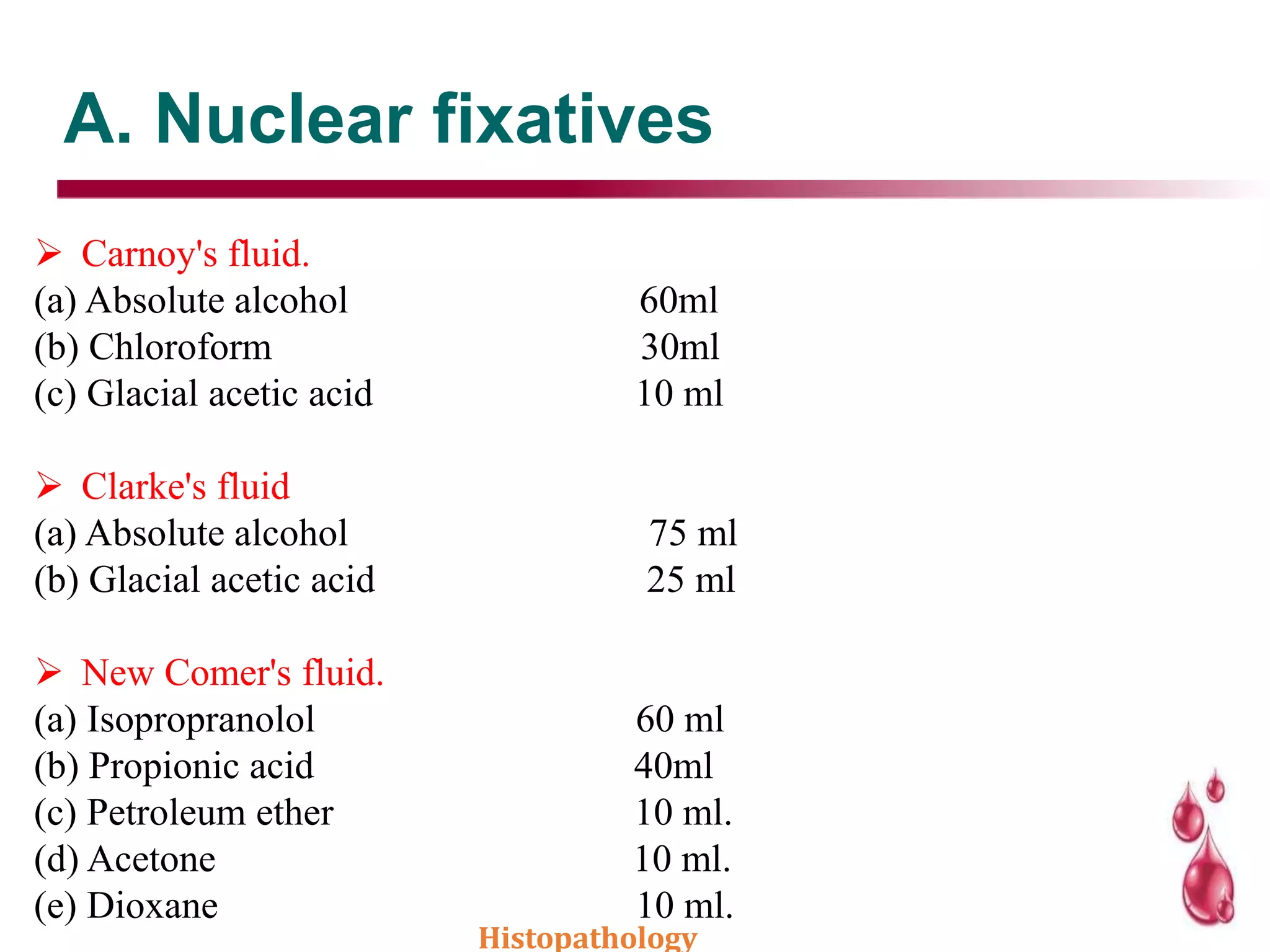
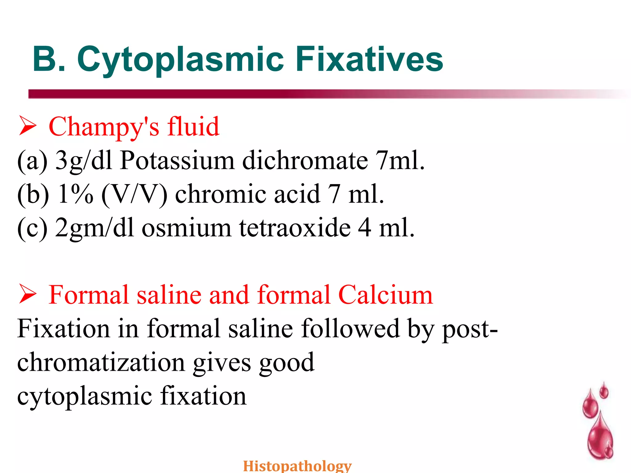
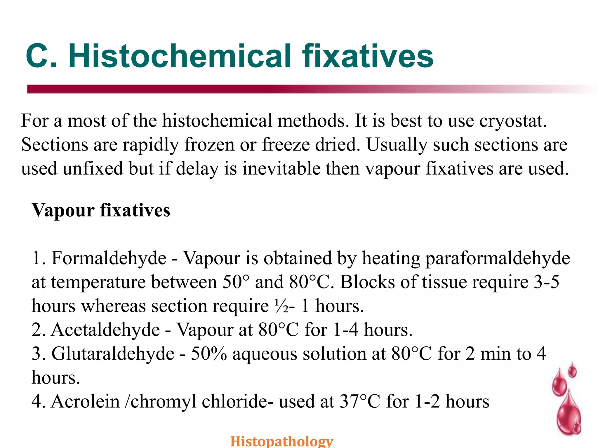
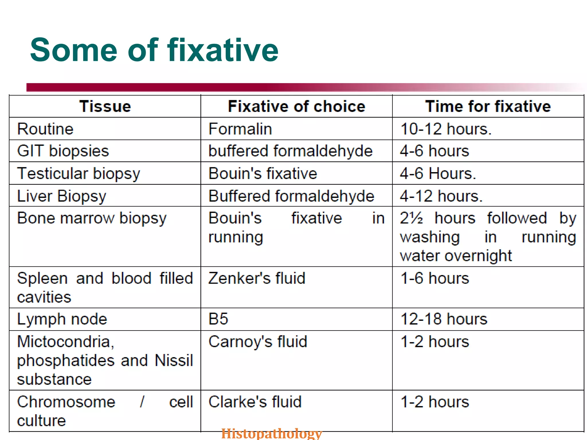
Objectives
Histopathology
2
The objective of the subsequent discussions will be to
acquaint the staff with their responsibility; the basic details
of tissue handling, processing and staining
Introduction .’
• Histopathology- Definition it is a branch of pathology
which deals with the study of disease in a tissue section.
• The tissue undergoes a series of steps before it reaches
the examiners desk to be thoroughly examined
microscopically to arrive at a particular diagnosis.
• To achieve this it is important that the tissue must be
prepared in such a manner that it is sufficiently thick or
thin to be examined microscopically and all the
structures in a tissue may be differentiated.
• . Histopathology
Definition
 Histopathology is examination of diseased tissues for
presence or absence of changes in their structure due to
disease processes.
 The term histochemistry means study of chemical
nature of the tissue
components by histological methods.
 The cell is the single structural unit of all tissues. The
study of cell is called cytology.
 A tissue is a group of cells specialized and
differentiated to perform a specialized function.
Collection of different type of cells forms an organ.
Histopathology
Types of Histological preparation
1. Whole mounts- These are preparation entire animal
eg. fungus, parasite. These preparations should be no
more than 0.2-0.5 mm in thickness.
2. Sections- The majority of the preparations in histology
are sections. The tissue is cut in about 3-5 mm thick
pieces processed and 5 microns thick sections are cut on
a microtome.
3. Smears- Smears are made from blood, bone marrow
or any fluid such as pleural or ascitic fluid..
Tissue Sampling
1. As biopsy- A small piece of lesions or tumor
which in sent for diagnosis before final removal
of the lesion or the tumor (Incisional biopsy).
2. If the whole of the tumor or lesion is sent for
examination and diagnosis by the pathologist, it is
called excisional biopsy.
3. Tissues from the autopsy are sent for the study of
disease and its course, for the advancement of
medicine. Histopathology
Tissue Fixation
 Any tissue removed from the body start decomposing
immediately because of loss of blood supply and
oxygen, accumulation of products of metabolism,
action of autolytic enzymes and putrefaction by
bacteria.
 This process of decomposition is prevented by fixation
 Definition It is a complex series of chemical events
which brings about changes in the various chemical
constituents of cell like hardening, however the cell
morphology and structural detail is preserved.
Histopathology
Principle of fixation
 The fixative brings about crosslinking of proteins which produces
denaturation or coagulation of proteins so that the semifluid state is
converted into semisolid state; so that it maintains everything in
vivo in relation to each other. Thus semisolid state facilitate easy
manipulation of tissue.
 During fixation ,tissues are fixed in complete physical and partly
chemical state to prevent autolysis or decomposition
 By keeping I fixatives soft tissue like brain and breast ,etc…will
become firm in consistency ,so it can be grossed easily
 Fixative available as solution or vapors
Histopathology
Effects of fixation
1. To preserve the tissue in as lf like manner as possible.
2. To prevent postmortem changes like autolysis and putrefaction. Autolysis is the
lysis or dissolution of cells by enzymatic action probably as a result of rupture of
lysosomes. Putrefaction The breakdown of tissue by bacterial action often with
formation of gas.
3. Preservation of chemical compounds and micro-anatomic constituents so that
further histochemistry is possible.
4. Hardening : the hardening effect of fixatives allows easy manipulation of soft
tissue like brain, intestines etc.
5. Solidification: Converts the normal semifluid consistency of cells (gel) to an
irreversible semisolid consistency (solid).
6. Optical differentiation - it alters to varying degrees the refractive indices of the
various components of cells and tissues so that unstained components are more
easily visualized than when unfixed.
7. Effects of staining - certain fixatives like formaldehyde intensifies the staining
character of tissue especially with haematoxylin.
Histopathology
Properties of fixatives
1. Coagulation and precipitation as described above.
2. Faster a fixative can penetrate the tissue better it is penetration
power depends upon the molecular weight e.g. formalin fixes
faster than osimic acid.
3. Solubility of fixatives - All fixatives should be soluble in a
suitable solvent, preferably in water so that adequate
concentrations can be prepared.
4. Concentration - It is important that the concentration of
fixative is isotonic or hypotonic
5. Reaction - Most fixatives are acidic. It may help in fixation but
can affect staining so has to be neutralized e.g. formalin is
neutralized by adding of calcium carbonate.
Histopathology
Classification of fixatives
• Fixatives may be simple or compound
 simple fixatives consists of one substance (e.g.formalin)
 Compound fixative has two or more substances
(e.g.Bouin’s/zenker’s)
• Fixatives can also be divided into the following categories
 Micro-anatomical fixative which preserve the anatomy of the
tissue
 Cytological fixatives which may be cytoplasmic or nuclear and
preserve respective intracellular constituents
 Histochemical fixatives employed for demonstration of
histochemical constituent and enzymes
Histopathology
Aldehydes fixative
 Formaldehyde - Formaldehyde is a gas but is soluble
in water to the extent of 37-40% w/v. This solution of
formaldehyde in water is called formalin or full
strength formalin.
 Formalin is one of the commonly used fixative in all
laboratories since it is cheap penetrates rapidly and
does not over harden the tissues.
 For routine fixation ,10% formalin is used which is
prepared by disolving 10ml of commercially formalin
in 90ml of water
FORMALIN
Histopathology
Cont.
• It preserves the proteins by forming cross-linkage with them and the
tissue component.
• It denatures the proteins.
• Glycogen is partially preserved hence formalin is not a fixative
choice for carbohydrates.
• Some enzymes can be demonstrated in formalin fixed tissues.
• It neither preserves nor destroys fat. Complex lipids are fixed but has
no effect on neutral fat. After formalin fixation fat may be
demonstrated in frozen section.
Demerits or disadvantage of formalin
• Causes excessive hardening of tissues
• Causes irritation of skin ,mucous membranes and conjunctiva
• If blood is present it turns tissue black by formalin pigment
Histopathology
Aldehydes fixative
GLUTARALDEHYDE
 Is available as 25% solution for fixation it
is used in 4% concentration.
 It is less irritating and less allergic
 It doesn’t cause much shrinkage of tissue
 Once the tissue is fixed we can keep it for
months before processing
 it is best for electron microscopy and nerve
biopsy Histopathology
Alcohol (Ethyl Alcohol)
Methyl and ethyl alcohols are used in PCR, In situ
hybridization, immunofluorescence and in enzyme studies
It acts as a reducing agents, become oxidized to
acetaldehyde and then to acetic acid.
• It is slow to penetrate, hardens and shrinks the tissue.
• Alcohol penetrates rapidly in presence of other fixative
hence in combination e.g. Carnoy's fixative is used to
increase the speed of tissue processing.
• It is a fat solvent hence it dissolve fats and lipids
• Methyl alcohol is used for fixing blood and bone marrow
smears.
Histopathology
Acetone / Mercuric Chloride (HgCl2)
Acetone : Cold acetone is sometimes used as a fixative for the histochemical
demonstration of some tissue enzymes like phosphatases and lipases.
Its mode of action as fixative is similar to that of alcohol
Mercuric Chloride (HgCl2)
Mercuric chloride is a very good salt employed in fixing but is rarely used
alone because it causes shrinkage of the tissue.
• It brings about precipitation of the proteins which are required to be
removed before staining by using potassium iodide in which they are soluble.
• The size (thickness) of the tissue to be fixed in mercuric chloride is
important, since if the tissue is more than 4 mm, then it hardens the tissue at
the periphery whereas the centre remains soft & under fixed.
Histopathology
Picric acid /Potassium dichromate
Picric acid - It produces marked cells shrinkage hence it is not used alone.
It has to be stored in a damp place because of its explosive nature it is
preferably stored under a layer of water.
Note : All the tissues fixed in picric acid containing fixatives should be
thoroughly washed to remove the yellow discolouration to ensure proper
staining of tissue sections. If the fixative is not removed by washing
thoroughly with time even the embedded tissue looses its staining quality.
Potassium dichromate
It fixes the cytoplasm without precipitation. Valuable in mixtures for the
fixation of lipids especially phospholipids. Used for fixing phosphatides and
mitochondria.
Note - Thorough washing of the tissue fixed in dichromate is required to
avoid forming an oxide in alcohol which cannot be removed later.
Histopathology
Osimium tetraoxide / Acetic acid
Osimium tetraoxide - It is a strong oxidizing agent and brings about
fixation by forming cross links with proteins.
• It gives excellent preservation of details of a cell, therefore
exclusively used for electron microscopy.
• It fixes fat e.g. myelin.
• It also demonstrates fat when 0.5-2% aqueous solution is used it
gives a black colour to fat.
Acetic acid - It causes the cells to swell hence can never be used
alone but should be used with fixatives causing cell shrinkage
Histopathology
Compound fixatives
1.MICROANATOMICAL FIXATIVES
 10% (v/v) formalin in 0.9% sodium chloride (normal saline).
 Buffered formation
 Formal calcium (Lillie : 1965)
 Buffered formal sucrose (Holt and Hicks, 1961)
 Alcoholic formalin
 Acetic alcoholic formalin
 Formalin ammonium bromide
 Heidenhain Susa (Mercuric chloride 4.5gm,Sodium chloride 0.5
gm,Trichloroacetic acid 2.0 gm, Acetic acid 4.0)
 Zenker's fluid (Mercuric chloride 5gm/Potassium dichromate 2.5
gm/Sodium sulphate 1.0 gm
 Distilled water to 100 ml/Add immediately before use : Glacial
acetic acid : 5 ml) Histopathology
cont.’
Zenker formal (Helly's fluid)- (Mercuric chloride - 5
gm /Potassium dichromate 2.5 gm/Sodium sulphate 1.0
gm/Distilled water to 100 ml/Add formalin immediately
before use 5 ml)
B5 stock solution (Mercuric chloride 12 gm/Sodium
acetate 2.5gm/Distilled water 200ml/B5 Working
solution/B5 stock solution 20ml/Formalin (40% w/v
formaldehyde) 2 ml)
Bouin's fluid (Saturated aqueous picric acid
75ml/Formalin 25ml/Glacial acetic acid 5 ml)
Gender's fluid (Saturated picric acid in 95% v/v/ alcohol
80ml/ Formalin 15ml/Glacial acetic acid 5ml)
Histopathology
Cont.’
2. CYTOLOGICAL FIXATIVES
Subdivided into
(A)Nuclear fixatives
(B) Cytoplasmic fixatives
Histopathology
A. Nuclear fixatives
 Carnoy's fluid.
(a) Absolute alcohol 60ml
(b) Chloroform 30ml
(c) Glacial acetic acid 10 ml
 Clarke's fluid
(a) Absolute alcohol 75 ml
(b) Glacial acetic acid 25 ml
 New Comer's fluid.
(a) Isopropranolol 60 ml
(b) Propionic acid 40ml
(c) Petroleum ether 10 ml.
(d) Acetone 10 ml.
(e) Dioxane 10 ml.
Histopathology
B. Cytoplasmic Fixatives
 Champy's fluid
(a) 3g/dl Potassium dichromate 7ml.
(b) 1% (V/V) chromic acid 7 ml.
(c) 2gm/dl osmium tetraoxide 4 ml.
 Formal saline and formal Calcium
Fixation in formal saline followed by post-
chromatization gives good
cytoplasmic fixation
Histopathology
C. Histochemical fixatives
For a most of the histochemical methods. It is best to use cryostat.
Sections are rapidly frozen or freeze dried. Usually such sections are
used unfixed but if delay is inevitable then vapour fixatives are used.
Vapour fixatives
1. Formaldehyde - Vapour is obtained by heating paraformaldehyde
at temperature between 50° and 80°C. Blocks of tissue require 3-5
hours whereas section require ½- 1 hours.
2. Acetaldehyde - Vapour at 80°C for 1-4 hours.
3. Glutaraldehyde - 50% aqueous solution at 80°C for 2 min to 4
hours.
4. Acrolein /chromyl chloride- used at 37°C for 1-2 hours
Histopathology
Some of fixative
Histopathology
Knowledge checking
1) What is a fixative? How will you classify them?
2) What is an ideal fixative?
3) What are the advantages and demerits of formalin
?
4) Where glutaraldehyde and alcohols are used as
fixatives ?
5) What are metallic fixatives and where they are
used?
6) What are metallic fixatives and where they are
used
Histopathology
Question ?
Histopathology
2
8
Histopathology
Decalcification
Definition Decalcification is a process of complete removal
of calcium salt from the tissues like bone and teeth and
other calcified tissues following fixation.
• Decalcification is done to assure that the specimen is
soft enough to allow cutting with the microtome knife.
Unless the tissues in completely decalcified the sections
will be torn and ragged and may damage the cutting
edge of microtome knife.
• Since calcium-rich bones are exceedingly difficult to
study, scientists use bone decalcification to make
specimens available for their research.
Histopathology
Compact bone (Decalcification)
Histopathology
Different methods of decalcification
◦1. Acid decalcification
◦2. Ion exchange resin
◦3. Electrical ionization
◦4. Chelating methods
◦5. Surface decalcification
Histopathology
Steps of decalcification
1. To ensure adequate fixation and complete removal of the
calcium it is important that the slices are 4-5 mm thick.
Calcified tissue needs 2-3 hours only, for complete
decalcification to be achieved so it in necessary to check
the decalcification after 2-3 hours.
2. Fixative of choice for bone or bone marrow is Zenker
formal or Bouin's fluid. Unfixed tissue tends be damaged 4
times greater during decalcification than a properly fixed
tissue.
Histopathology
Histopathology
Acid decalcification
1. Aqueous Nitric Acid-
◦ Nitric acid - 5 ml
◦Distilled water - 100 ml
◦If tissue is left for long time in the solution, the tissue may be damaged.
◦Yellow colour of nitric acid should be removed with urea. But this solution
◦gives good nuclear staining and also rapid action.
2.Hydrochloric acid –
5-10% aqueous solution decalcification slower than nitric acid but still rapid. Fairly
good nuclear staining.
Weak acid e.g. formic, acetic and picric acid of these formic acids is
extensively used as acid decalcifier.
3.Formic acid
1. Brings out fairly rapid decalcification.
2. Nuclear staining in better.
3. But requires neutralization and thorough washing prior to dehydration.
Fluids
1. Gooding and Stelwart's fluid.
90% formic acid 5-10ml.
Formalin 5ml
Distilled water to 100 ml.
2. Evans and Krajian fluid
20% aqueous trisodium citrate 65 ml
90% formic acid 35 ml
This solution has a pH of - 2-3
Histopathology
Surface decalcification
 The surface of the block to be decalcified is
trimmed with scalpel.
 The block is then placed in acid solution at 1%
hydrochloric acid face downwards so that acid
bathes the cut surface for 15- 60 min.
 As penetration and decalcification is only
sufficient for a few sections be cut the block
shall be carefully oriented in microtome to avoid
wastage of decalcified tissue.Histopathology
Ion exchange resins
 Ion exchange resins in decalcifying fluids are used to remove
calcium
 ion from the fluid. Therefore ensuring a rapid rate of solubility of
calcium from tissue and reduction in time of decalcification.
 The resins an ammoniated salt of sulfonated resin along with various
concentrations of formic acid are used.
 The resin in layered on the bottom of a container to a depth of = ½
inch, the specimen is allowed to rest in it.
 After use, the resin may be regenerated by washing twice with dilute
N/10 HCL followed by three washes in distilled water. Use of Ion
exchange resin has advantage of (ii) faster decalcification (ii) tissue
preservation and (iii) cellular details better preserved.
Histopathology
Ion exchange resins
Histopathology
Chelating agents
 Chelating agents are organic compounds which
have the power of binding certain metals.
 Ethylene-diamene-tetra-aceticacid, disodium salt
called Versenate has the power of capturing
metallic ions.
 This is a slow process but has little or no effect on
other tissue elements.
 Some enzymes are still active after EDTA
decalcification.
Histopathology
Determination of end point of
decalcification
1. Flexibility method
Bending, needling or by use of scalpel if it bends easily that
means decalcification is complete.
Unreliable, causes damage and distortion of tissue
2. X-ray method
Best method for determining complete decalcification but
very costly. Tissue fixed in mercuric chloride containing
fixatives cannot be tested as they will be radio opaque.
3. Chemical Method
It is done to detect calcium in the decalcifying fluid when no
further calcium is detected, decalcification in considered
complete.
Histopathology
Factors affecting rate of
Decalcification
◦1. Concentration of decalcifying solution-Increased
concentration of the decalcifying agent fastens the
reaction.
◦2. Temperature-The rate of decalcification increases
with rise of temperature.
◦3. Density of bone-Harder bone takes longer time to
decalcify.
◦4. Thickness of the tissue-Small tissue pieces
decalcify earlier.
◦5. Agitation-Agitation increases the rate of
decalcification.
Histopathology
Knowledge checking
1) Define decalcification? What are the aim of it
2) What is an ideal decalcification methods?
3) What are the advantages and demerits of ion
exchanges resins ?
4) How can you determines the good
decalcification?
5) Which cheating agent used to decalcification ?
Histopathology
Question?
Histopathology
Books

Lecture 1.introduction to histopathology ii

  • 2.
    Objectives Histopathology 2 The objective ofthe subsequent discussions will be to acquaint the staff with their responsibility; the basic details of tissue handling, processing and staining
  • 3.
    Introduction .’ • Histopathology-Definition it is a branch of pathology which deals with the study of disease in a tissue section. • The tissue undergoes a series of steps before it reaches the examiners desk to be thoroughly examined microscopically to arrive at a particular diagnosis. • To achieve this it is important that the tissue must be prepared in such a manner that it is sufficiently thick or thin to be examined microscopically and all the structures in a tissue may be differentiated. • . Histopathology
  • 4.
    Definition  Histopathology isexamination of diseased tissues for presence or absence of changes in their structure due to disease processes.  The term histochemistry means study of chemical nature of the tissue components by histological methods.  The cell is the single structural unit of all tissues. The study of cell is called cytology.  A tissue is a group of cells specialized and differentiated to perform a specialized function. Collection of different type of cells forms an organ. Histopathology
  • 5.
    Types of Histologicalpreparation 1. Whole mounts- These are preparation entire animal eg. fungus, parasite. These preparations should be no more than 0.2-0.5 mm in thickness. 2. Sections- The majority of the preparations in histology are sections. The tissue is cut in about 3-5 mm thick pieces processed and 5 microns thick sections are cut on a microtome. 3. Smears- Smears are made from blood, bone marrow or any fluid such as pleural or ascitic fluid..
  • 6.
    Tissue Sampling 1. Asbiopsy- A small piece of lesions or tumor which in sent for diagnosis before final removal of the lesion or the tumor (Incisional biopsy). 2. If the whole of the tumor or lesion is sent for examination and diagnosis by the pathologist, it is called excisional biopsy. 3. Tissues from the autopsy are sent for the study of disease and its course, for the advancement of medicine. Histopathology
  • 7.
    Tissue Fixation  Anytissue removed from the body start decomposing immediately because of loss of blood supply and oxygen, accumulation of products of metabolism, action of autolytic enzymes and putrefaction by bacteria.  This process of decomposition is prevented by fixation  Definition It is a complex series of chemical events which brings about changes in the various chemical constituents of cell like hardening, however the cell morphology and structural detail is preserved. Histopathology
  • 8.
    Principle of fixation The fixative brings about crosslinking of proteins which produces denaturation or coagulation of proteins so that the semifluid state is converted into semisolid state; so that it maintains everything in vivo in relation to each other. Thus semisolid state facilitate easy manipulation of tissue.  During fixation ,tissues are fixed in complete physical and partly chemical state to prevent autolysis or decomposition  By keeping I fixatives soft tissue like brain and breast ,etc…will become firm in consistency ,so it can be grossed easily  Fixative available as solution or vapors Histopathology
  • 9.
    Effects of fixation 1.To preserve the tissue in as lf like manner as possible. 2. To prevent postmortem changes like autolysis and putrefaction. Autolysis is the lysis or dissolution of cells by enzymatic action probably as a result of rupture of lysosomes. Putrefaction The breakdown of tissue by bacterial action often with formation of gas. 3. Preservation of chemical compounds and micro-anatomic constituents so that further histochemistry is possible. 4. Hardening : the hardening effect of fixatives allows easy manipulation of soft tissue like brain, intestines etc. 5. Solidification: Converts the normal semifluid consistency of cells (gel) to an irreversible semisolid consistency (solid). 6. Optical differentiation - it alters to varying degrees the refractive indices of the various components of cells and tissues so that unstained components are more easily visualized than when unfixed. 7. Effects of staining - certain fixatives like formaldehyde intensifies the staining character of tissue especially with haematoxylin. Histopathology
  • 10.
    Properties of fixatives 1.Coagulation and precipitation as described above. 2. Faster a fixative can penetrate the tissue better it is penetration power depends upon the molecular weight e.g. formalin fixes faster than osimic acid. 3. Solubility of fixatives - All fixatives should be soluble in a suitable solvent, preferably in water so that adequate concentrations can be prepared. 4. Concentration - It is important that the concentration of fixative is isotonic or hypotonic 5. Reaction - Most fixatives are acidic. It may help in fixation but can affect staining so has to be neutralized e.g. formalin is neutralized by adding of calcium carbonate. Histopathology
  • 11.
    Classification of fixatives •Fixatives may be simple or compound  simple fixatives consists of one substance (e.g.formalin)  Compound fixative has two or more substances (e.g.Bouin’s/zenker’s) • Fixatives can also be divided into the following categories  Micro-anatomical fixative which preserve the anatomy of the tissue  Cytological fixatives which may be cytoplasmic or nuclear and preserve respective intracellular constituents  Histochemical fixatives employed for demonstration of histochemical constituent and enzymes Histopathology
  • 12.
    Aldehydes fixative  Formaldehyde- Formaldehyde is a gas but is soluble in water to the extent of 37-40% w/v. This solution of formaldehyde in water is called formalin or full strength formalin.  Formalin is one of the commonly used fixative in all laboratories since it is cheap penetrates rapidly and does not over harden the tissues.  For routine fixation ,10% formalin is used which is prepared by disolving 10ml of commercially formalin in 90ml of water FORMALIN Histopathology
  • 13.
    Cont. • It preservesthe proteins by forming cross-linkage with them and the tissue component. • It denatures the proteins. • Glycogen is partially preserved hence formalin is not a fixative choice for carbohydrates. • Some enzymes can be demonstrated in formalin fixed tissues. • It neither preserves nor destroys fat. Complex lipids are fixed but has no effect on neutral fat. After formalin fixation fat may be demonstrated in frozen section. Demerits or disadvantage of formalin • Causes excessive hardening of tissues • Causes irritation of skin ,mucous membranes and conjunctiva • If blood is present it turns tissue black by formalin pigment Histopathology
  • 14.
    Aldehydes fixative GLUTARALDEHYDE  Isavailable as 25% solution for fixation it is used in 4% concentration.  It is less irritating and less allergic  It doesn’t cause much shrinkage of tissue  Once the tissue is fixed we can keep it for months before processing  it is best for electron microscopy and nerve biopsy Histopathology
  • 15.
    Alcohol (Ethyl Alcohol) Methyland ethyl alcohols are used in PCR, In situ hybridization, immunofluorescence and in enzyme studies It acts as a reducing agents, become oxidized to acetaldehyde and then to acetic acid. • It is slow to penetrate, hardens and shrinks the tissue. • Alcohol penetrates rapidly in presence of other fixative hence in combination e.g. Carnoy's fixative is used to increase the speed of tissue processing. • It is a fat solvent hence it dissolve fats and lipids • Methyl alcohol is used for fixing blood and bone marrow smears. Histopathology
  • 16.
    Acetone / MercuricChloride (HgCl2) Acetone : Cold acetone is sometimes used as a fixative for the histochemical demonstration of some tissue enzymes like phosphatases and lipases. Its mode of action as fixative is similar to that of alcohol Mercuric Chloride (HgCl2) Mercuric chloride is a very good salt employed in fixing but is rarely used alone because it causes shrinkage of the tissue. • It brings about precipitation of the proteins which are required to be removed before staining by using potassium iodide in which they are soluble. • The size (thickness) of the tissue to be fixed in mercuric chloride is important, since if the tissue is more than 4 mm, then it hardens the tissue at the periphery whereas the centre remains soft & under fixed. Histopathology
  • 17.
    Picric acid /Potassiumdichromate Picric acid - It produces marked cells shrinkage hence it is not used alone. It has to be stored in a damp place because of its explosive nature it is preferably stored under a layer of water. Note : All the tissues fixed in picric acid containing fixatives should be thoroughly washed to remove the yellow discolouration to ensure proper staining of tissue sections. If the fixative is not removed by washing thoroughly with time even the embedded tissue looses its staining quality. Potassium dichromate It fixes the cytoplasm without precipitation. Valuable in mixtures for the fixation of lipids especially phospholipids. Used for fixing phosphatides and mitochondria. Note - Thorough washing of the tissue fixed in dichromate is required to avoid forming an oxide in alcohol which cannot be removed later. Histopathology
  • 18.
    Osimium tetraoxide /Acetic acid Osimium tetraoxide - It is a strong oxidizing agent and brings about fixation by forming cross links with proteins. • It gives excellent preservation of details of a cell, therefore exclusively used for electron microscopy. • It fixes fat e.g. myelin. • It also demonstrates fat when 0.5-2% aqueous solution is used it gives a black colour to fat. Acetic acid - It causes the cells to swell hence can never be used alone but should be used with fixatives causing cell shrinkage Histopathology
  • 19.
    Compound fixatives 1.MICROANATOMICAL FIXATIVES 10% (v/v) formalin in 0.9% sodium chloride (normal saline).  Buffered formation  Formal calcium (Lillie : 1965)  Buffered formal sucrose (Holt and Hicks, 1961)  Alcoholic formalin  Acetic alcoholic formalin  Formalin ammonium bromide  Heidenhain Susa (Mercuric chloride 4.5gm,Sodium chloride 0.5 gm,Trichloroacetic acid 2.0 gm, Acetic acid 4.0)  Zenker's fluid (Mercuric chloride 5gm/Potassium dichromate 2.5 gm/Sodium sulphate 1.0 gm  Distilled water to 100 ml/Add immediately before use : Glacial acetic acid : 5 ml) Histopathology
  • 20.
    cont.’ Zenker formal (Helly'sfluid)- (Mercuric chloride - 5 gm /Potassium dichromate 2.5 gm/Sodium sulphate 1.0 gm/Distilled water to 100 ml/Add formalin immediately before use 5 ml) B5 stock solution (Mercuric chloride 12 gm/Sodium acetate 2.5gm/Distilled water 200ml/B5 Working solution/B5 stock solution 20ml/Formalin (40% w/v formaldehyde) 2 ml) Bouin's fluid (Saturated aqueous picric acid 75ml/Formalin 25ml/Glacial acetic acid 5 ml) Gender's fluid (Saturated picric acid in 95% v/v/ alcohol 80ml/ Formalin 15ml/Glacial acetic acid 5ml) Histopathology
  • 21.
    Cont.’ 2. CYTOLOGICAL FIXATIVES Subdividedinto (A)Nuclear fixatives (B) Cytoplasmic fixatives Histopathology
  • 22.
    A. Nuclear fixatives Carnoy's fluid. (a) Absolute alcohol 60ml (b) Chloroform 30ml (c) Glacial acetic acid 10 ml  Clarke's fluid (a) Absolute alcohol 75 ml (b) Glacial acetic acid 25 ml  New Comer's fluid. (a) Isopropranolol 60 ml (b) Propionic acid 40ml (c) Petroleum ether 10 ml. (d) Acetone 10 ml. (e) Dioxane 10 ml. Histopathology
  • 23.
    B. Cytoplasmic Fixatives Champy's fluid (a) 3g/dl Potassium dichromate 7ml. (b) 1% (V/V) chromic acid 7 ml. (c) 2gm/dl osmium tetraoxide 4 ml.  Formal saline and formal Calcium Fixation in formal saline followed by post- chromatization gives good cytoplasmic fixation Histopathology
  • 24.
    C. Histochemical fixatives Fora most of the histochemical methods. It is best to use cryostat. Sections are rapidly frozen or freeze dried. Usually such sections are used unfixed but if delay is inevitable then vapour fixatives are used. Vapour fixatives 1. Formaldehyde - Vapour is obtained by heating paraformaldehyde at temperature between 50° and 80°C. Blocks of tissue require 3-5 hours whereas section require ½- 1 hours. 2. Acetaldehyde - Vapour at 80°C for 1-4 hours. 3. Glutaraldehyde - 50% aqueous solution at 80°C for 2 min to 4 hours. 4. Acrolein /chromyl chloride- used at 37°C for 1-2 hours Histopathology
  • 25.
  • 26.
    Knowledge checking 1) Whatis a fixative? How will you classify them? 2) What is an ideal fixative? 3) What are the advantages and demerits of formalin ? 4) Where glutaraldehyde and alcohols are used as fixatives ? 5) What are metallic fixatives and where they are used? 6) What are metallic fixatives and where they are used Histopathology
  • 27.
  • 28.
  • 29.
    Decalcification Definition Decalcification isa process of complete removal of calcium salt from the tissues like bone and teeth and other calcified tissues following fixation. • Decalcification is done to assure that the specimen is soft enough to allow cutting with the microtome knife. Unless the tissues in completely decalcified the sections will be torn and ragged and may damage the cutting edge of microtome knife. • Since calcium-rich bones are exceedingly difficult to study, scientists use bone decalcification to make specimens available for their research. Histopathology
  • 30.
  • 31.
    Different methods ofdecalcification ◦1. Acid decalcification ◦2. Ion exchange resin ◦3. Electrical ionization ◦4. Chelating methods ◦5. Surface decalcification Histopathology
  • 32.
    Steps of decalcification 1.To ensure adequate fixation and complete removal of the calcium it is important that the slices are 4-5 mm thick. Calcified tissue needs 2-3 hours only, for complete decalcification to be achieved so it in necessary to check the decalcification after 2-3 hours. 2. Fixative of choice for bone or bone marrow is Zenker formal or Bouin's fluid. Unfixed tissue tends be damaged 4 times greater during decalcification than a properly fixed tissue. Histopathology
  • 33.
  • 34.
    Acid decalcification 1. AqueousNitric Acid- ◦ Nitric acid - 5 ml ◦Distilled water - 100 ml ◦If tissue is left for long time in the solution, the tissue may be damaged. ◦Yellow colour of nitric acid should be removed with urea. But this solution ◦gives good nuclear staining and also rapid action. 2.Hydrochloric acid – 5-10% aqueous solution decalcification slower than nitric acid but still rapid. Fairly good nuclear staining. Weak acid e.g. formic, acetic and picric acid of these formic acids is extensively used as acid decalcifier. 3.Formic acid 1. Brings out fairly rapid decalcification. 2. Nuclear staining in better. 3. But requires neutralization and thorough washing prior to dehydration.
  • 35.
    Fluids 1. Gooding andStelwart's fluid. 90% formic acid 5-10ml. Formalin 5ml Distilled water to 100 ml. 2. Evans and Krajian fluid 20% aqueous trisodium citrate 65 ml 90% formic acid 35 ml This solution has a pH of - 2-3 Histopathology
  • 36.
    Surface decalcification  Thesurface of the block to be decalcified is trimmed with scalpel.  The block is then placed in acid solution at 1% hydrochloric acid face downwards so that acid bathes the cut surface for 15- 60 min.  As penetration and decalcification is only sufficient for a few sections be cut the block shall be carefully oriented in microtome to avoid wastage of decalcified tissue.Histopathology
  • 37.
    Ion exchange resins Ion exchange resins in decalcifying fluids are used to remove calcium  ion from the fluid. Therefore ensuring a rapid rate of solubility of calcium from tissue and reduction in time of decalcification.  The resins an ammoniated salt of sulfonated resin along with various concentrations of formic acid are used.  The resin in layered on the bottom of a container to a depth of = ½ inch, the specimen is allowed to rest in it.  After use, the resin may be regenerated by washing twice with dilute N/10 HCL followed by three washes in distilled water. Use of Ion exchange resin has advantage of (ii) faster decalcification (ii) tissue preservation and (iii) cellular details better preserved. Histopathology
  • 38.
  • 39.
    Chelating agents  Chelatingagents are organic compounds which have the power of binding certain metals.  Ethylene-diamene-tetra-aceticacid, disodium salt called Versenate has the power of capturing metallic ions.  This is a slow process but has little or no effect on other tissue elements.  Some enzymes are still active after EDTA decalcification. Histopathology
  • 40.
    Determination of endpoint of decalcification 1. Flexibility method Bending, needling or by use of scalpel if it bends easily that means decalcification is complete. Unreliable, causes damage and distortion of tissue 2. X-ray method Best method for determining complete decalcification but very costly. Tissue fixed in mercuric chloride containing fixatives cannot be tested as they will be radio opaque. 3. Chemical Method It is done to detect calcium in the decalcifying fluid when no further calcium is detected, decalcification in considered complete. Histopathology
  • 41.
    Factors affecting rateof Decalcification ◦1. Concentration of decalcifying solution-Increased concentration of the decalcifying agent fastens the reaction. ◦2. Temperature-The rate of decalcification increases with rise of temperature. ◦3. Density of bone-Harder bone takes longer time to decalcify. ◦4. Thickness of the tissue-Small tissue pieces decalcify earlier. ◦5. Agitation-Agitation increases the rate of decalcification. Histopathology
  • 42.
    Knowledge checking 1) Definedecalcification? What are the aim of it 2) What is an ideal decalcification methods? 3) What are the advantages and demerits of ion exchanges resins ? 4) How can you determines the good decalcification? 5) Which cheating agent used to decalcification ? Histopathology
  • 43.
  • 44.