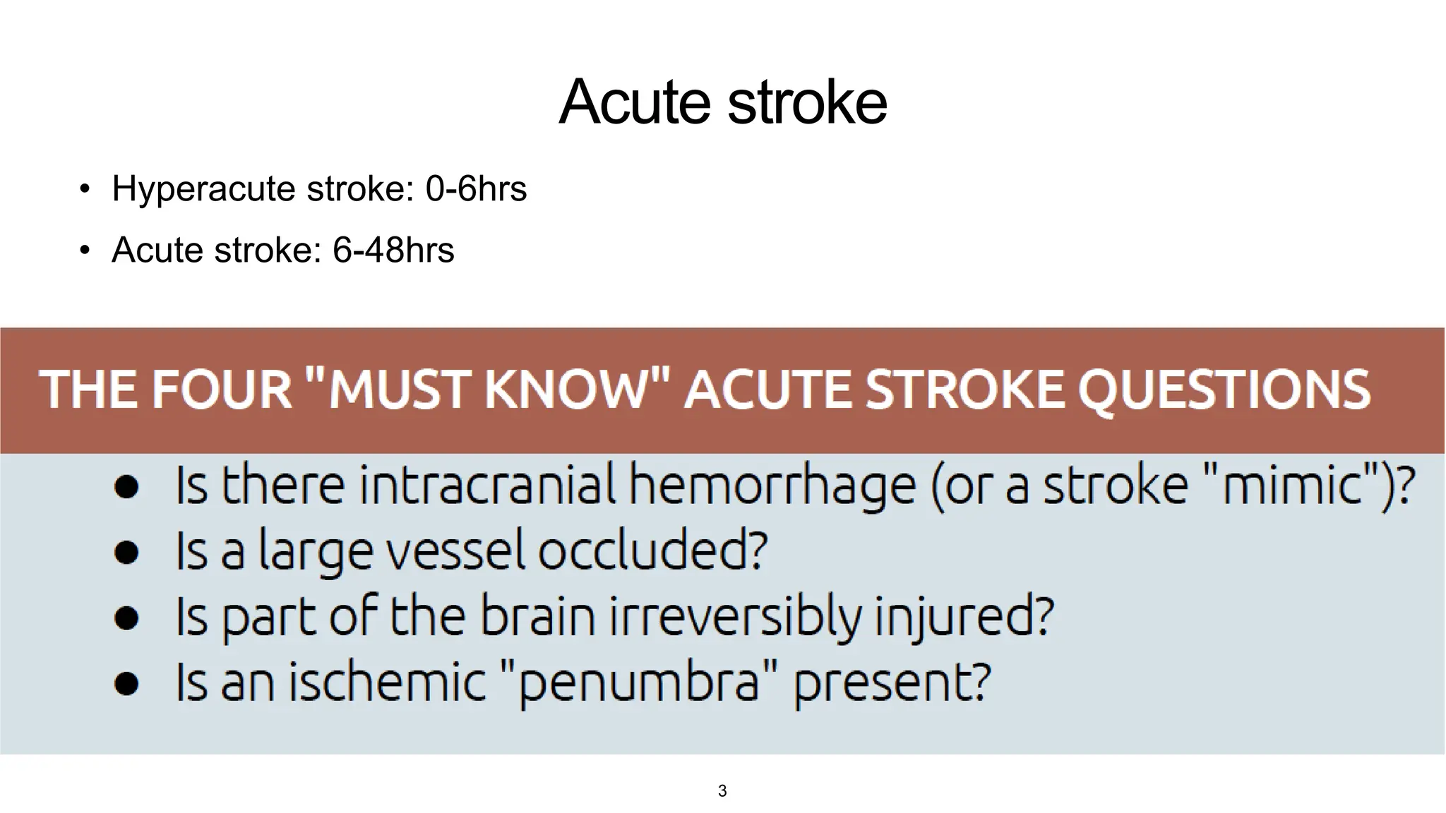
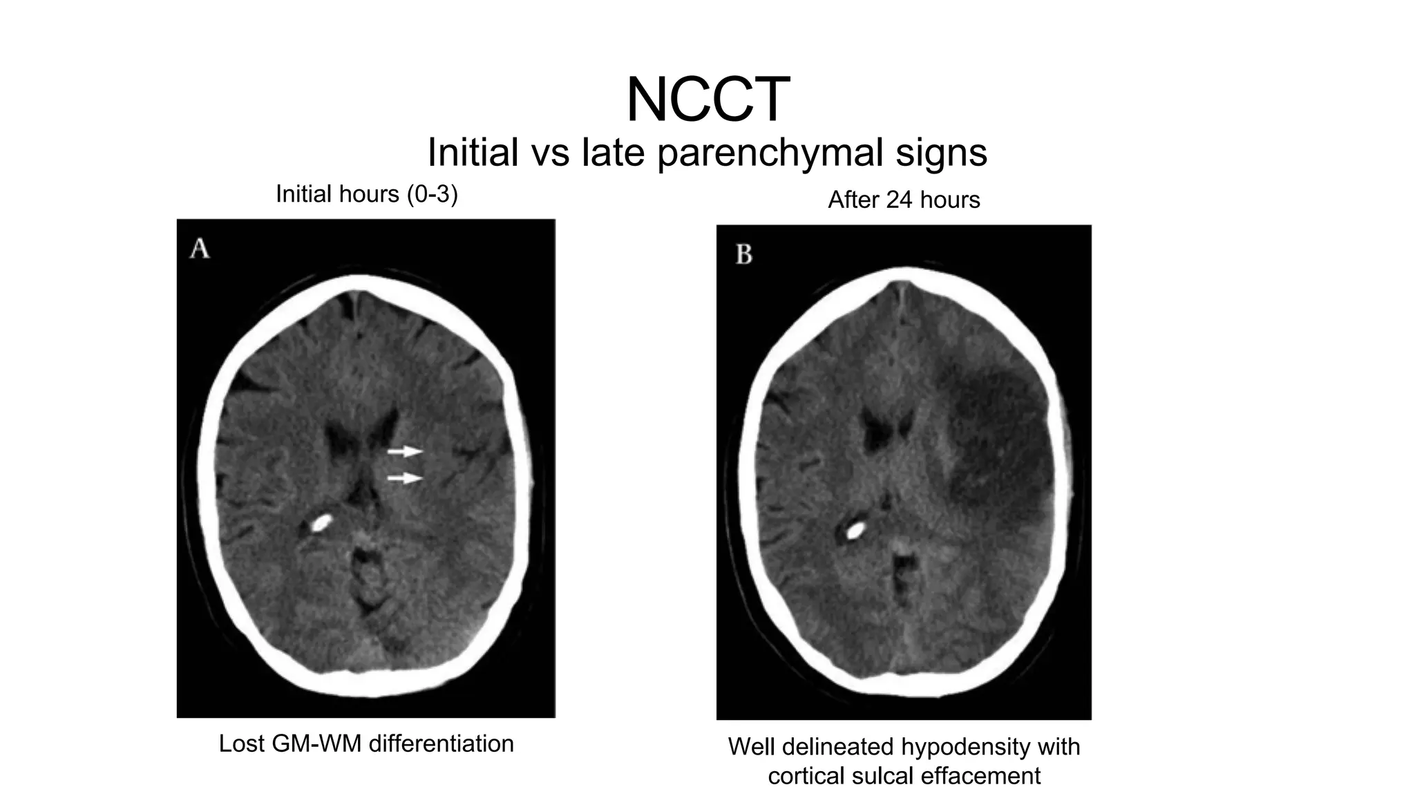
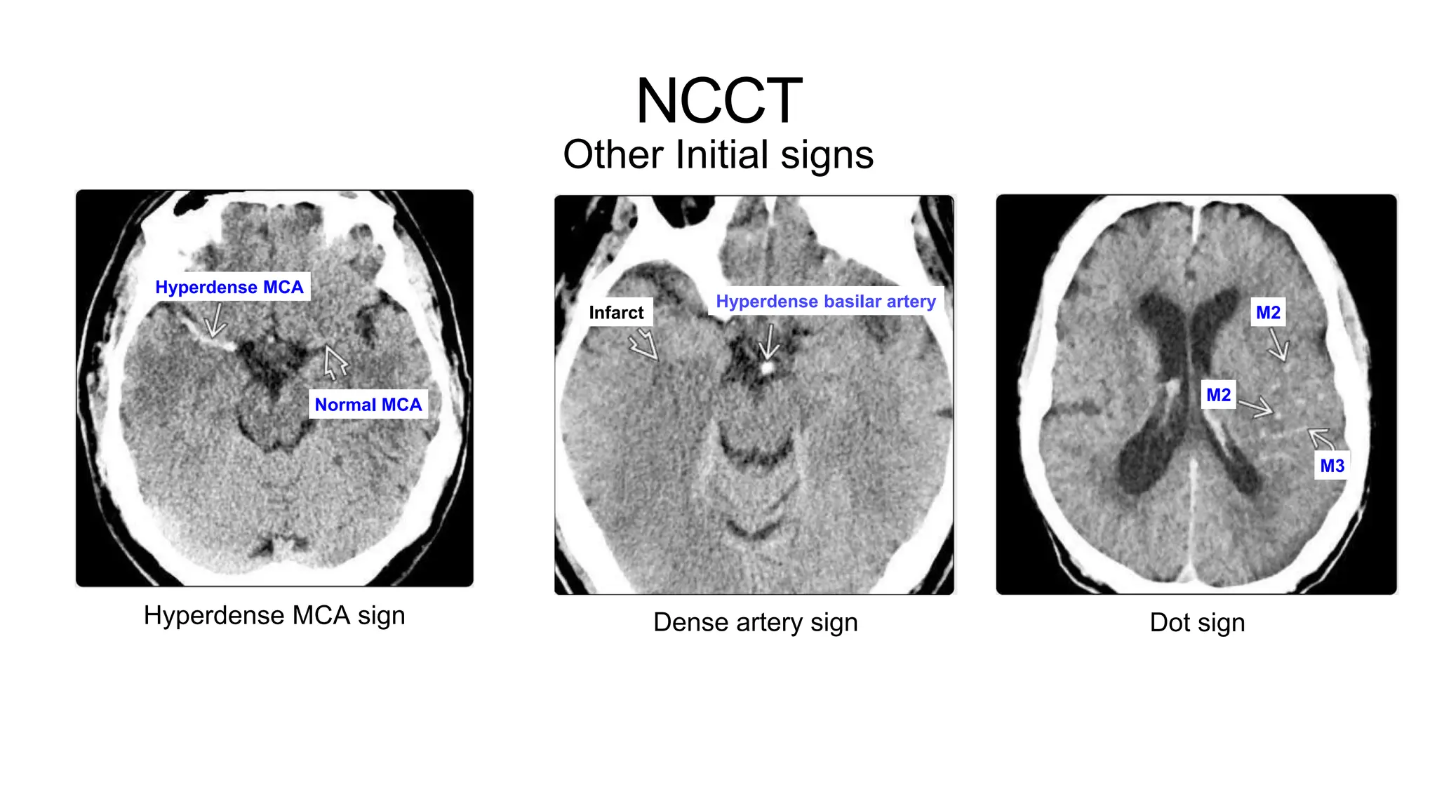
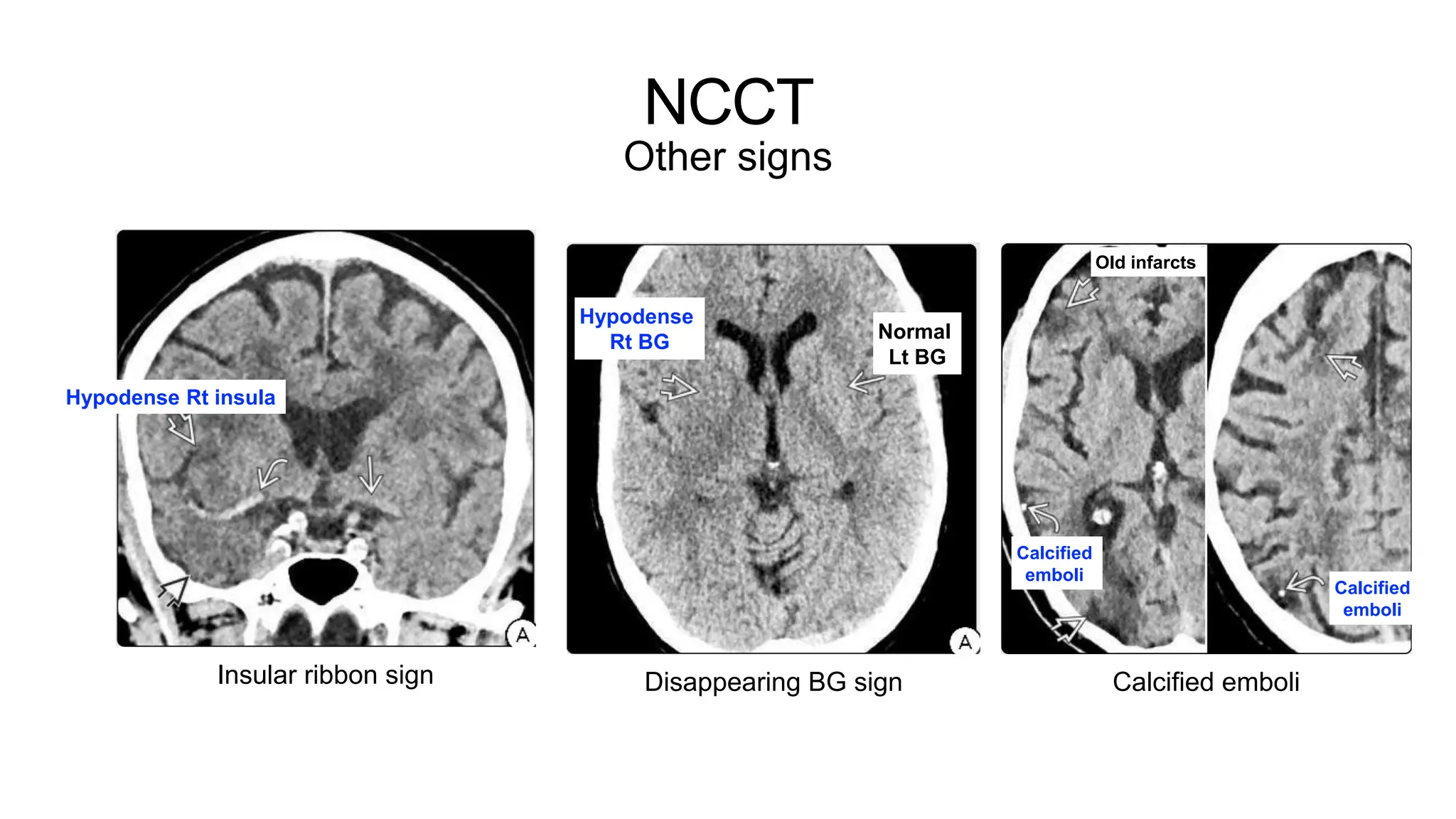
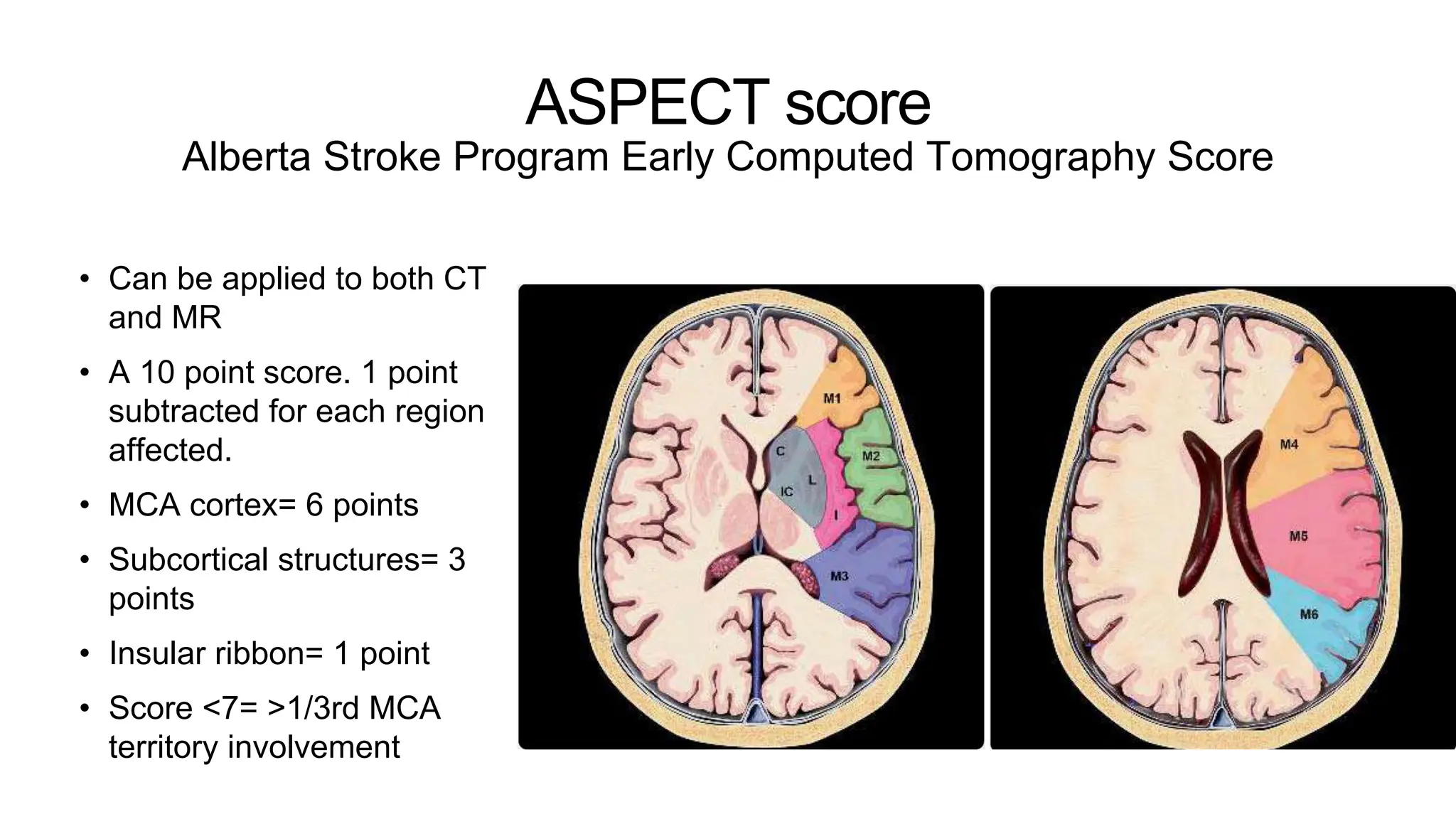
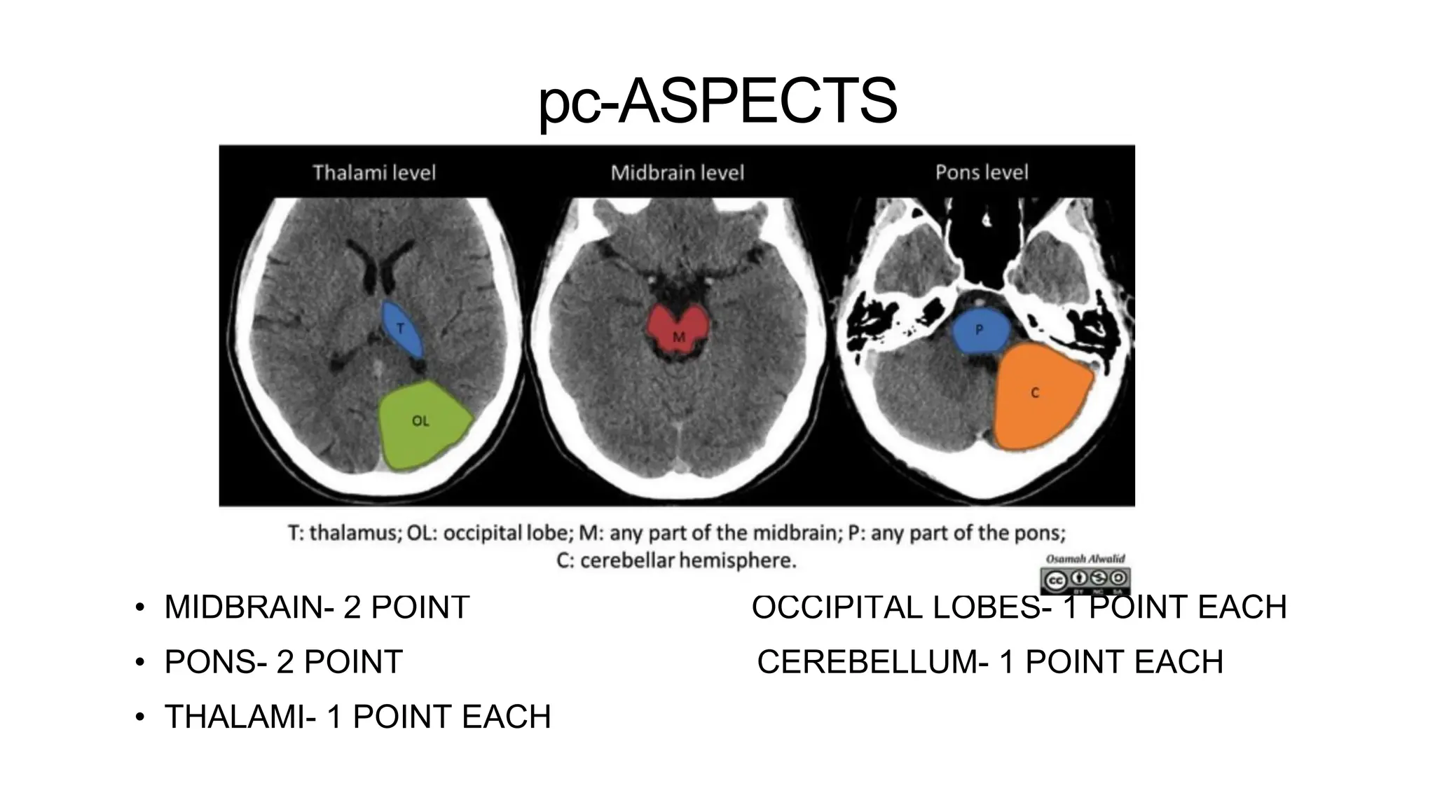
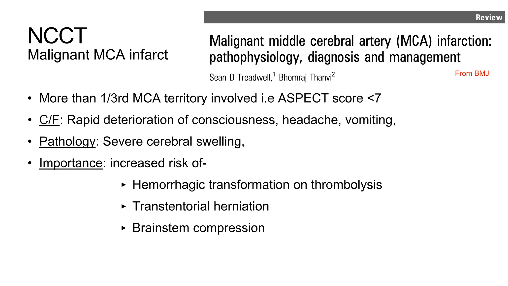
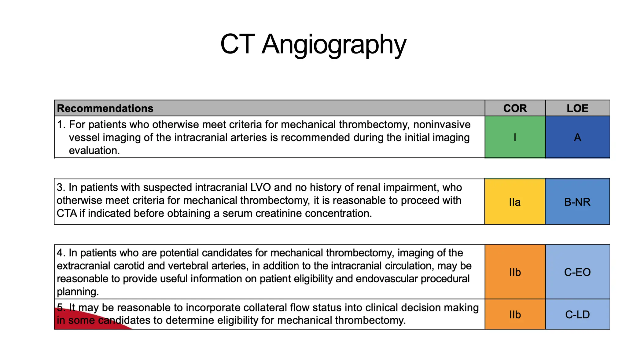
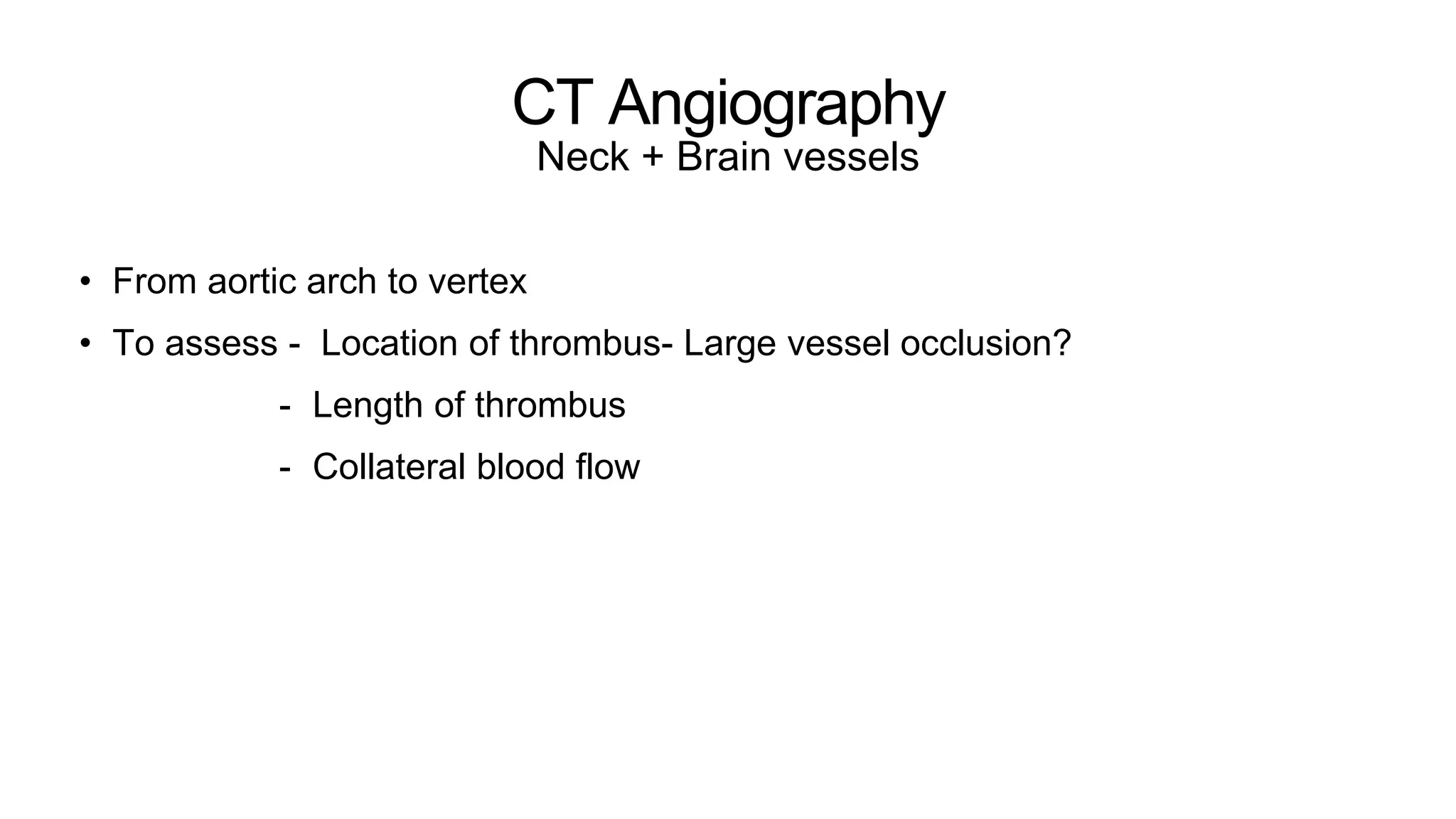
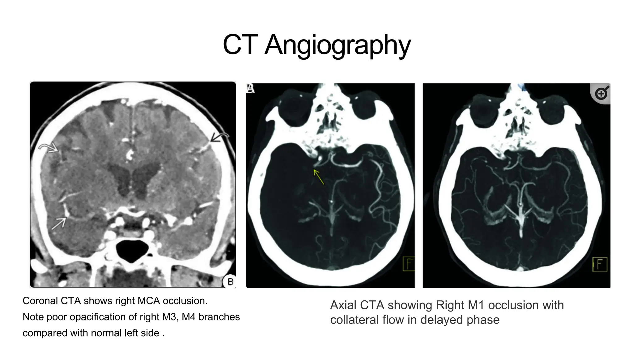
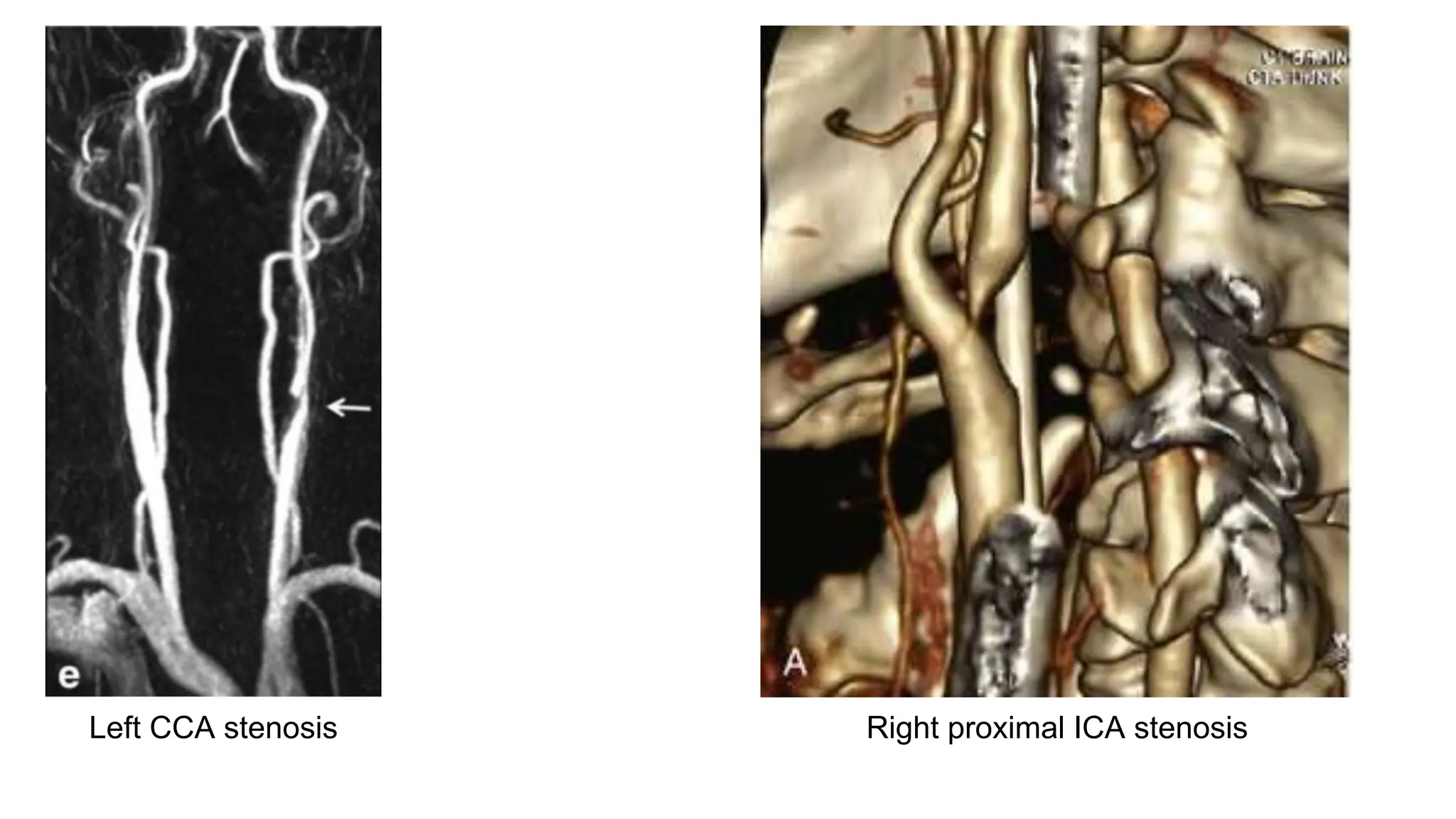
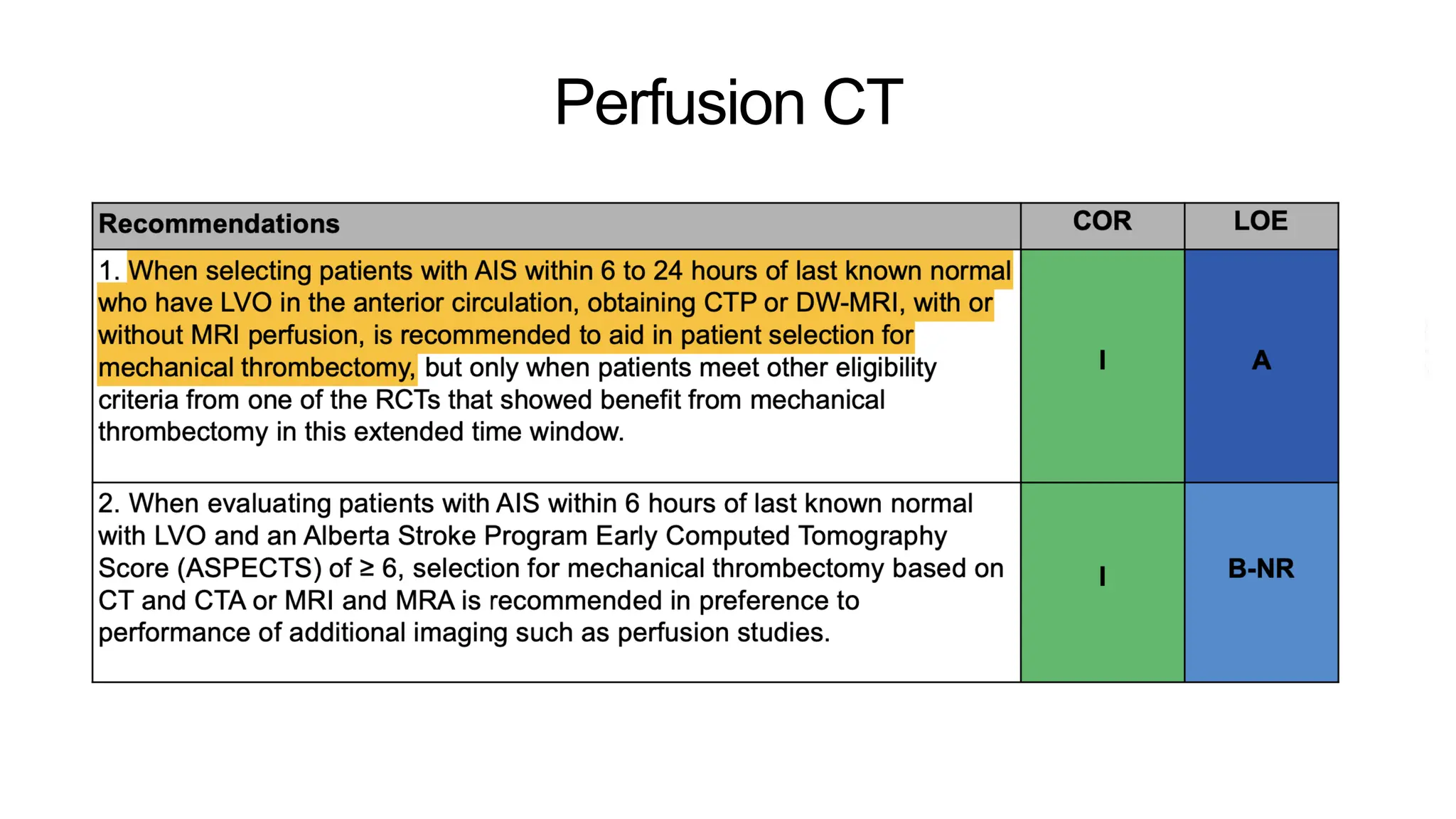
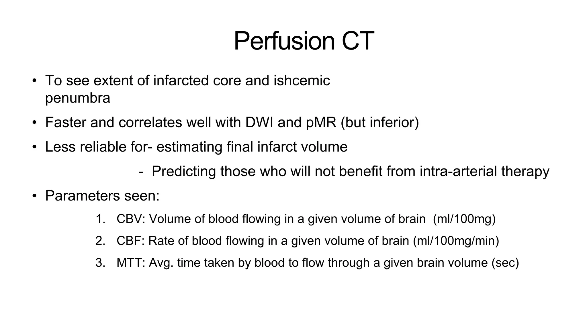
Presenter: Dr. Vipul Phogat
1
20% cases- wrongly diagnosed clinically!!!!!!!!!
To decide treatment
2
Acute stroke
• Hyperacute stroke: 0-6hrs
• Acute stroke: 6-48hrs
3
Acute stroke CT protocol
• A multimodal protocol including
1. NCCT brain
2. CT angio neck + brain vessels ( not MR angio- motion artefacts)
3. Perfusion CT
• All can be done within 15 mins with helical acquisition with separate contrast
boluses.
NCCT
Initial vs late parenchymal signs
Initial hours (0-3)
Lost GM-WM differentiation
After 24 hours
Well delineated hypodensity with
cortical sulcal effacement
NCCT
Other Initial signs
Hyperdense MCA sign Dense artery sign
Hyperdense MCA
Normal MCA
Hyperdense basilar artery
Infarct
Dot sign
M2
M2
M3
NCCT
Other signs
Insular ribbon sign
Hypodense Rt insula
Disappearing BG sign
Hypodense
Rt BG Normal
Lt BG
Calcified emboli
Old infarcts
Calcified
emboli
Calcified
emboli
ASPECT score
Alberta Stroke Program Early Computed Tomography Score
• Can be applied to both CT
and MR
• A 10 point score. 1 point
subtracted for each region
affected.
• MCA cortex= 6 points
• Subcortical structures= 3
points
• Insular ribbon= 1 point
• Score <7= >1/3rd MCA
territory involvement
pc-ASPECTS
• MIDBRAIN- 2 POINT OCCIPITAL LOBES- 1 POINT EACH
• PONS- 2 POINT CEREBELLUM- 1 POINT EACH
• THALAMI- 1 POINT EACH
NCCT
Malignant MCA infarct
• More than 1/3rd MCA territory involved i.e ASPECT score <7
• C/F: Rapid deterioration of consciousness, headache, vomiting,
• Pathology: Severe cerebral swelling,
• Importance: increased risk of-
‣ Hemorrhagic transformation on thrombolysis
‣ Transtentorial herniation
‣ Brainstem compression
From BMJ
CT Angiography
CT Angiography
Neck + Brain vessels
• From aortic arch to vertex
• To assess - Location of thrombus- Large vessel occlusion?
- Length of thrombus
- Collateral blood flow
CT Angiography
Coronal CTA shows right MCA occlusion.
Note poor opacification of right M3, M4 branches
compared with normal left side .
Axial CTA showing Right M1 occlusion with
collateral flow in delayed phase
Left CCA stenosis Right proximal ICA stenosis
Perfusion CT
Perfusion CT
• To see extent of infarcted core and ishcemic
penumbra
• Faster and correlates well with DWI and pMR (but inferior)
• Less reliable for- estimating final infarct volume
- Predicting those who will not benefit from intra-arterial therapy
• Parameters seen:
1. CBV: Volume of blood flowing in a given volume of brain (ml/100mg)
2. CBF: Rate of blood flowing in a given volume of brain (ml/100mg/min)
3. MTT: Avg. time taken by blood to flow through a given brain volume (sec)
pCT
Normal
CBV
CBF
CBV
MTT
Normal perfusion parameters
are:
• Gray matter
◦ CBF: 60 mL/100 g/min
◦ CBV: 4 mL/100 g
◦ MTT: 4 s
• White matter
◦ CBF: 25 mL/100 g/min
◦ CBV: 2 mL/100 g
◦ MTT: 4.8 s
pCT
Stroke
CBF CBV TTP
MTT
CBF 10-25ml/100mg/min-
Ichemia (Blue)
CBF <10ml/100mg/min-
infarction (purple-black)
pCT
Ischemic penumbra
• CBV/CBF mismatch
CBV CBF
CBV/MTT mismatch
CBV MTT
pCT
Acute crossed cerebellar diaschisis
CBF TPP
TPP
MR in stroke
• Time consuming, so expediated rapid stroke protocol:
‣ Fast FLAIR
‣ T2
‣ DWI
‣ pMR
• Superior to CT in detecting small vessel and brainstem stroke.
• Better delineation of ischemic penumbra in cases of CT/CTA and clinical
mismatch
MRI
DWI-ADC
• Positive in ~95% hyperacute infarcts
• Diffusion restriction and ADC fall
• Within minutes
• Due to cytotoxic edema
• DTI: even more sensitive, especially for
pontine and medullary lesions.
• DWI negative strokes: lacunar strokes,
brainstem lesions, rapid recanalisation,
transient/fluctuating hypoperfusion.
MRI
FLAIR
‣ 0-4 hrs- 30% to 50% cases show
hyperintensity and cortical swelling.
‣ >7hr- nearly all strokes positive
‣ T2: Positive after 12-24 hours.
‣ Intra-arterial hyperintensity = slow/retrograde
flow, an early sign.
MRI
FLAIR-DWI mismatch
• DWI+ and FLAIR-
• Occurs in early stroke
• A quick indicator of
viable ischemic
penumbra
• Eligibility for
thrombolysis
MRI
T2-GRE
• Blooming thrombus sign
• Succeptibility vessel sign
MRI
T1-C+
• Intravascular
enhancement due to
slow flow in patent non-
thrombosed vessels
• No parenchymal
enhancement in acute
stroke
Perfusion MRI
• DWI reflects the densely ischemic
core
• pMR shows the ischemic
penumbra also
• DWI-PWI mismatch: Ratio >2.6
provides 90% sensitivity and 83%
specificity
Stroke mimics
False hyperdense vessel sign on CT
In a case of HSV encephalitis, due to
relative hypodensity of surrounding
parenchyma
Stroke mimics
False hyperdense vessel sign on CT
Calcified Right MCA. Attenuation values close to bone
Stroke mimics
False hyperdense vessel sign on CT
Diffuse hyperdense vessels due to raised hematocrit in a case of polycythemia vera
Stroke mimics
DWI
Case of Rt sided weakness with left
cortical and thalamic DWI restriction
(non-vascular pattern) with sparing of
WM, in a case of status epilepticus
DWI restriction in Hypoglcemia in a neonate
Primary CNS lymphoma.
Left frontal periventricular lesion showing
prominent diffusion restriction,
presenting with hyperintensity on DWI (a),
low ADC value (b) and
mild hyperintensity on T2WI (c), all typical
features of this type of hypercellular tumour.
T1WI post gadolinium (d) shows
homogeneous and intense contrast
enhancement

Imaging in acute ischemic stroke cases.pptx

  • 1.
  • 2.
    20% cases- wronglydiagnosed clinically!!!!!!!!! To decide treatment 2
  • 3.
    Acute stroke • Hyperacutestroke: 0-6hrs • Acute stroke: 6-48hrs 3
  • 4.
    Acute stroke CTprotocol • A multimodal protocol including 1. NCCT brain 2. CT angio neck + brain vessels ( not MR angio- motion artefacts) 3. Perfusion CT • All can be done within 15 mins with helical acquisition with separate contrast boluses.
  • 5.
    NCCT Initial vs lateparenchymal signs Initial hours (0-3) Lost GM-WM differentiation After 24 hours Well delineated hypodensity with cortical sulcal effacement
  • 6.
    NCCT Other Initial signs HyperdenseMCA sign Dense artery sign Hyperdense MCA Normal MCA Hyperdense basilar artery Infarct Dot sign M2 M2 M3
  • 7.
    NCCT Other signs Insular ribbonsign Hypodense Rt insula Disappearing BG sign Hypodense Rt BG Normal Lt BG Calcified emboli Old infarcts Calcified emboli Calcified emboli
  • 8.
    ASPECT score Alberta StrokeProgram Early Computed Tomography Score • Can be applied to both CT and MR • A 10 point score. 1 point subtracted for each region affected. • MCA cortex= 6 points • Subcortical structures= 3 points • Insular ribbon= 1 point • Score <7= >1/3rd MCA territory involvement
  • 9.
    pc-ASPECTS • MIDBRAIN- 2POINT OCCIPITAL LOBES- 1 POINT EACH • PONS- 2 POINT CEREBELLUM- 1 POINT EACH • THALAMI- 1 POINT EACH
  • 10.
    NCCT Malignant MCA infarct •More than 1/3rd MCA territory involved i.e ASPECT score <7 • C/F: Rapid deterioration of consciousness, headache, vomiting, • Pathology: Severe cerebral swelling, • Importance: increased risk of- ‣ Hemorrhagic transformation on thrombolysis ‣ Transtentorial herniation ‣ Brainstem compression From BMJ
  • 11.
  • 12.
    CT Angiography Neck +Brain vessels • From aortic arch to vertex • To assess - Location of thrombus- Large vessel occlusion? - Length of thrombus - Collateral blood flow
  • 13.
    CT Angiography Coronal CTAshows right MCA occlusion. Note poor opacification of right M3, M4 branches compared with normal left side . Axial CTA showing Right M1 occlusion with collateral flow in delayed phase
  • 14.
    Left CCA stenosisRight proximal ICA stenosis
  • 15.
  • 16.
    Perfusion CT • Tosee extent of infarcted core and ishcemic penumbra • Faster and correlates well with DWI and pMR (but inferior) • Less reliable for- estimating final infarct volume - Predicting those who will not benefit from intra-arterial therapy • Parameters seen: 1. CBV: Volume of blood flowing in a given volume of brain (ml/100mg) 2. CBF: Rate of blood flowing in a given volume of brain (ml/100mg/min) 3. MTT: Avg. time taken by blood to flow through a given brain volume (sec)
  • 17.
    pCT Normal CBV CBF CBV MTT Normal perfusion parameters are: •Gray matter ◦ CBF: 60 mL/100 g/min ◦ CBV: 4 mL/100 g ◦ MTT: 4 s • White matter ◦ CBF: 25 mL/100 g/min ◦ CBV: 2 mL/100 g ◦ MTT: 4.8 s
  • 18.
    pCT Stroke CBF CBV TTP MTT CBF10-25ml/100mg/min- Ichemia (Blue) CBF <10ml/100mg/min- infarction (purple-black)
  • 19.
    pCT Ischemic penumbra • CBV/CBFmismatch CBV CBF CBV/MTT mismatch CBV MTT
  • 20.
    pCT Acute crossed cerebellardiaschisis CBF TPP TPP
  • 21.
    MR in stroke •Time consuming, so expediated rapid stroke protocol: ‣ Fast FLAIR ‣ T2 ‣ DWI ‣ pMR • Superior to CT in detecting small vessel and brainstem stroke. • Better delineation of ischemic penumbra in cases of CT/CTA and clinical mismatch
  • 22.
    MRI DWI-ADC • Positive in~95% hyperacute infarcts • Diffusion restriction and ADC fall • Within minutes • Due to cytotoxic edema • DTI: even more sensitive, especially for pontine and medullary lesions. • DWI negative strokes: lacunar strokes, brainstem lesions, rapid recanalisation, transient/fluctuating hypoperfusion.
  • 23.
    MRI FLAIR ‣ 0-4 hrs-30% to 50% cases show hyperintensity and cortical swelling. ‣ >7hr- nearly all strokes positive ‣ T2: Positive after 12-24 hours. ‣ Intra-arterial hyperintensity = slow/retrograde flow, an early sign.
  • 24.
    MRI FLAIR-DWI mismatch • DWI+and FLAIR- • Occurs in early stroke • A quick indicator of viable ischemic penumbra • Eligibility for thrombolysis
  • 25.
    MRI T2-GRE • Blooming thrombussign • Succeptibility vessel sign
  • 26.
    MRI T1-C+ • Intravascular enhancement dueto slow flow in patent non- thrombosed vessels • No parenchymal enhancement in acute stroke
  • 27.
    Perfusion MRI • DWIreflects the densely ischemic core • pMR shows the ischemic penumbra also • DWI-PWI mismatch: Ratio >2.6 provides 90% sensitivity and 83% specificity
  • 28.
    Stroke mimics False hyperdensevessel sign on CT In a case of HSV encephalitis, due to relative hypodensity of surrounding parenchyma
  • 29.
    Stroke mimics False hyperdensevessel sign on CT Calcified Right MCA. Attenuation values close to bone
  • 30.
    Stroke mimics False hyperdensevessel sign on CT Diffuse hyperdense vessels due to raised hematocrit in a case of polycythemia vera
  • 31.
    Stroke mimics DWI Case ofRt sided weakness with left cortical and thalamic DWI restriction (non-vascular pattern) with sparing of WM, in a case of status epilepticus DWI restriction in Hypoglcemia in a neonate
  • 35.
    Primary CNS lymphoma. Leftfrontal periventricular lesion showing prominent diffusion restriction, presenting with hyperintensity on DWI (a), low ADC value (b) and mild hyperintensity on T2WI (c), all typical features of this type of hypercellular tumour. T1WI post gadolinium (d) shows homogeneous and intense contrast enhancement

Editor's Notes

  • #6 2. Blurring of GM-WM interface can be seen in 50-70% cases within first 3 hrs. 3. Well delineation of hypodensity due to CT after 24 hrs.
  • #7 1.Most specific, least sensitive
  • #8 3.Calcified emboli from an at-risk ulcerated atheromatous plaque in carotid. Carries 50% risk of repeat ichaemic stroke
  • #17 Techniques: Slow-Infusion/Whole-Brain Technique First-Pass Bolus-Tracking Methodology- Usually one of the slices passes through the basal ganglia, so that portions of the middle, anterior, and posterior cerebral distributions are evaluated. Central volume principal: CBV= CBF/MTT
  • #19 An MCA territory infarct core volume of 70-100 mL is highly specific for poor clinical outcome, independent of associated penumbra and regardless of subsequent recanalization
  • #20 Prolonged MTT over 145% that extends beyond the core infarct area (so-called CBV/MTT mismatch) also characterizes the ischemic penumbra.
  • #21 An important ancillary finding in patients with large MCA infarcts is reduced perfusion in the opposite cerebellar hemisphere. Between 15-20% of large MCA infarcts cause hypoperfusion with reduced CBF in the contralateral cerebellum, a phenomenon called crossed cerebellar diaschisis
  • #23 Very small (lacunar) infarcts, brainstem lesions, clot lysis with recanalization, and moderately reduced or fluctuating hypoperfusion that is not severe enough to restrict water movement have all been cited as possible reasons for DWI-negative acute strokes.