Download to read offline


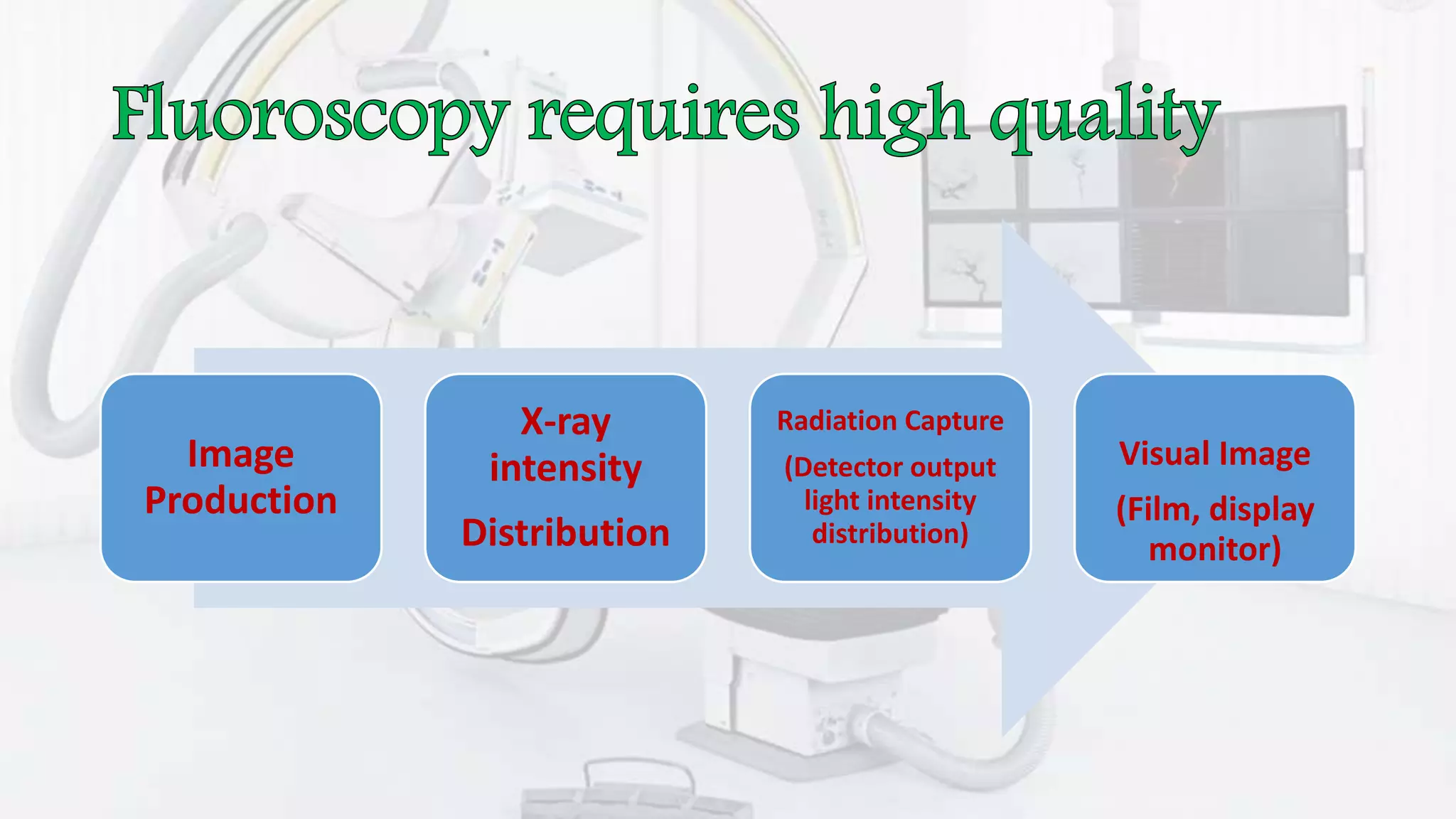


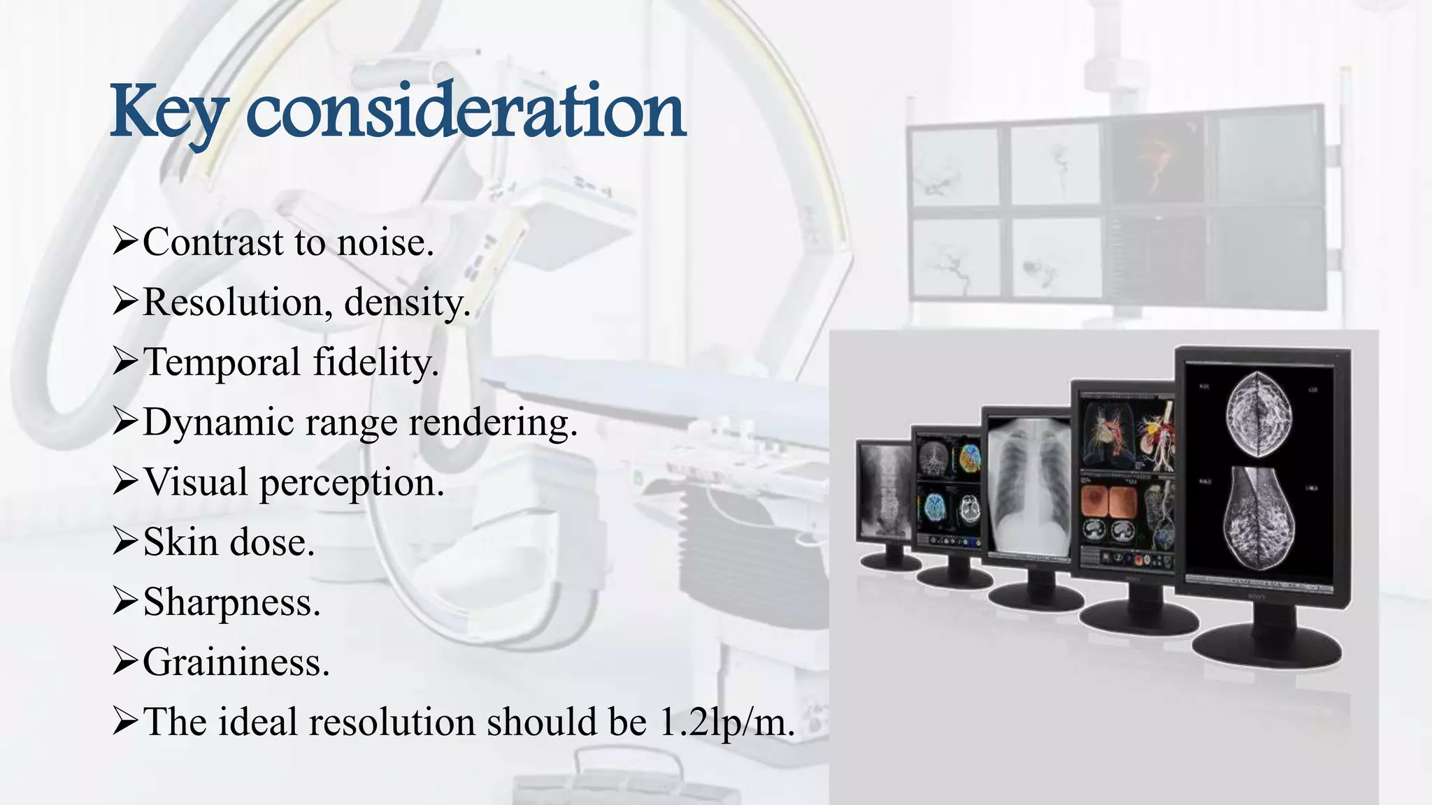


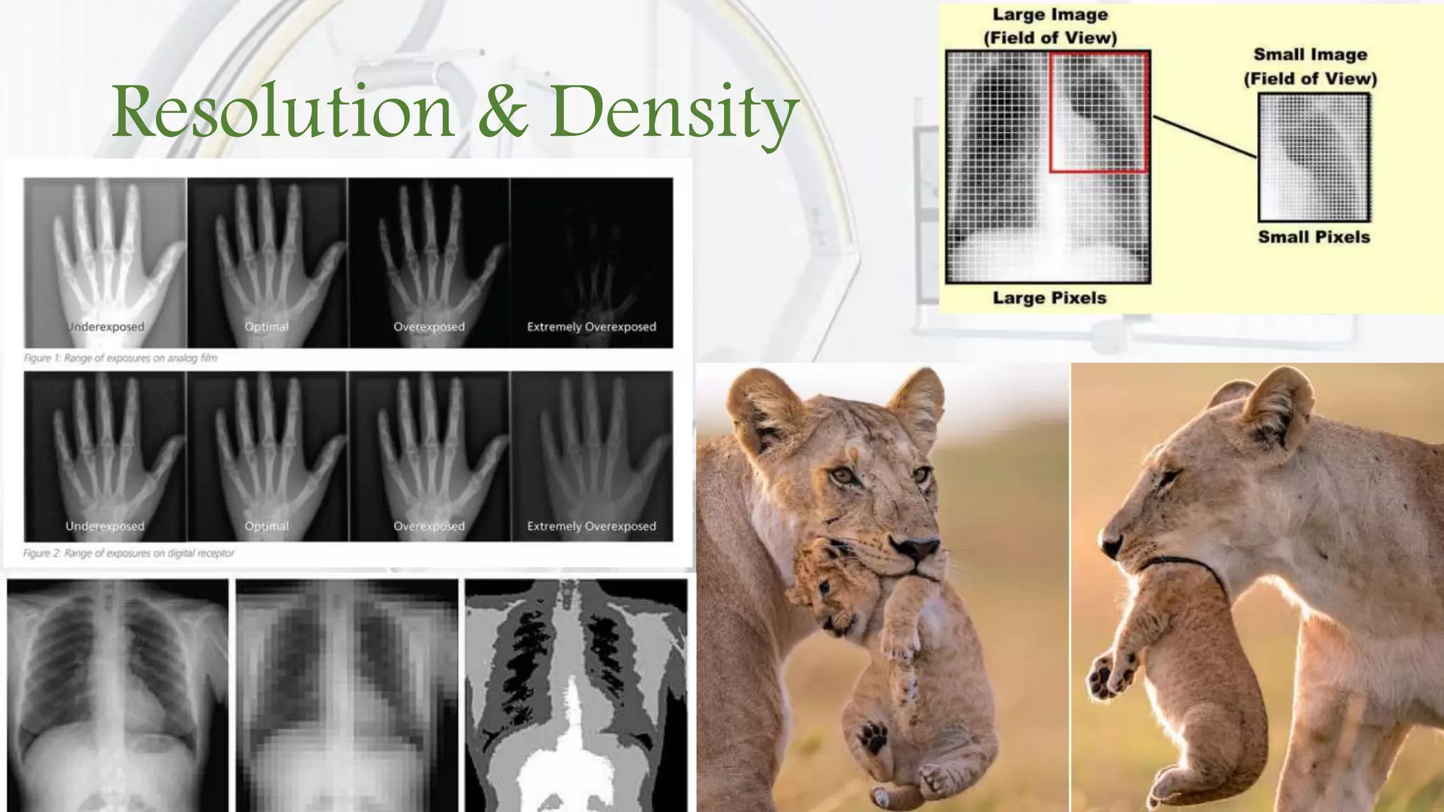


The document discusses factors that influence medical image quality during fluoroscopy. It states that image quality depends on both intrinsic characteristics of the imaging system as well as the observer. Key factors that determine statistical image quality include the number of x-ray photons absorbed and noise from photon variation. Common image distortions in fluoroscopy include veiling glare, vignetting, blooming, and pincushion or S-shaped distortions. The ideal resolution is 1.2 line pairs per millimeter and image quality considerations encompass contrast, resolution, temporal fidelity, dynamic range, and visual perception.