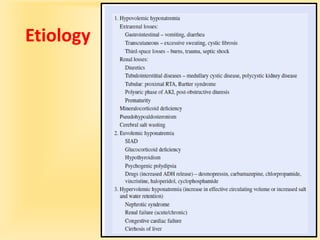
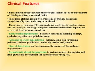
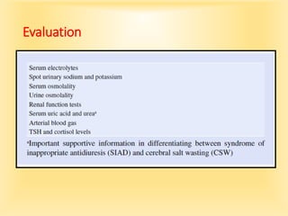
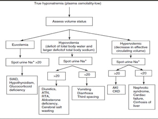
Hyponatremia
Hyponatremia
•Hyponatremia is defined as serum Na +
<135 mEq/l. Mild hyponatremia (serum
Na <135 mEq/l) occurs in 25 % of
hospitalized children, while moderate
hyponatremia (Serum Na + <130 mEq/l)
is seen in 1 % of hospitalized children
• Both total body sodium and TBW determine the serum sodium
concentration.
• Hyponatremia exists when the ratio of water to sodium is increased.
• This condition can occur with low, normal, or high levels of body
sodium. Similarly, body water can be low, normal, or high.
• hypovolemic hyponatremia, (lost sodium).
• The water balance may be positive or negative, but sodium loss has
been higher than water loss.
• The pathogenesis of the hyponatremia is usually a combination of
sodium loss and water retention to compensate for the volume
depletion.
• The patient has a pathologic increase in fluid loss, and this fluid
contains sodium. Most fluid that is lost has a lower sodium
concentration than that of plasma.
• The volume depletion stimulates ADH synthesis, resulting in renal
water retention.
• Volume depletion also decreases the GFR and enhances water
resorption in the proximal tubule, thereby reducing water delivery
to the collecting duct.
• Renal salt wasting occurs in hereditary kidney diseases, such as
juvenile nephronophthisis and autosomal recessive polycystic
kidney disease.
• Acquired tubulointerstitial nephritis, usually secondary to either
medications or infections, may cause salt wasting, along with other
evidence of tubular dysfunction.
• CNS injury may produce cerebral salt wasting, which is
theoretically caused by the production of a natriuretic peptide that
causes renal salt wasting.
• In type II renal tubular acidosis (RTA), usually associated with
Fanconi syndrome , there is increased excretion of sodium and
bicarbonate in the urine.
• Patients with Fanconi syndrome also have glycosuria,
aminoaciduria, and hypophosphatemia because of renal phosphate
wasting .
• Aldosterone is necessary for renal sodium retention and for the
excretion of potassium and acid.
• In congenital adrenal hyperplasia caused by 21-hydroxylase
deficiency, the block of aldosterone production results in
hyponatremia, hyperkalemia, and metabolic acidosis.
• In pseudohypoaldosteronism, aldosterone levels are elevated, but
there is no response because of either a defective sodium channel or a
deficiency of aldosterone receptors.
• A lack of tubular response to aldosterone may occur in children with
urinary tract obstruction, especially during an acute urinary tract
infection.
• In hypervolemic hyponatremia, there is an excess of
TBW and sodium, although the increase in water is greater
than the increase in sodium.
• In most of the conditions that cause hypervolemic
hyponatremia, there is a decrease in the effective blood
volume, resulting from third space fluid loss, vasodilation,
or poor cardiac output.
• The regulatory systems sense a decrease in effective blood
volume and attempt to retain water and sodium to correct
the problem.
• ADH causes renal water retention, and the kidney, under
the influence of aldosterone and other intrarenal
mechanisms, retains sodium. The patient’s sodium
concentration decreases because water intake exceeds
sodium intake and ADH prevents the normal loss of excess
water.
• Patients with hyponatremia and no evidence of volume
overload or volume depletion have euvolemic
hyponatremia.
• These patients typically have an excess of TBW and a slight
decrease in total body sodium. Some of these patients
have an increase in weight, implying that they are volume-
overloaded. Nevertheless, from a clinical standpoint, they
usually appear normal or have subtle signs of fluid
overload.
Etiology
Clinical Features
• The symptoms depend not only on the level of sodium but also on the rapidity
of development (acute versus chronic).
• Sometimes, children present with symptoms of primary disease and
recognition of hyponatremia may be incidental.
• The primary symptoms of hyponatremia are mostly due to cerebral edema,
and the severity of neurologic symptoms correlates well with the rapidity and
severity of the drop in serum sodium:
– Early or mild hyponatremia – headache, nausea and vomiting, lethargy,
confusion, agitation, and gait disturbances
– Advanced or severe hyponatremia – seizures, coma, non-cardiogenic
pulmonary edema, papilledema, and rarely cardiac arrhythmias
• Signs of dehydration may be exaggerated in presence of hypovolemic
hyponatremia.
• Asymptomatic chronic hyponatremia in preterm neonates is associated with
poor growth and development and sensorineural hearing loss.
Evaluation
Approach to Hyponatremia
Treatment
• It is important to ensure that the patient has associated
hypoosmolality. The treatment of hypertonic and
pseudohyponatremia is directed at the underlying disorder.
• While treating hyponatremia, three factors should be taken
into consideration:
severity and duration of hyponatremia , neurological
symptoms , and volume status of the child.
• Asymptomatic hyponatremia is usually chronic (>48 h
duration), while symptomatic hyponatremia is acute (<48 h
duration).
• If the child is in shock or volume depleted, treat with isotonic
saline in sufficient amounts to restore the intravascular
volume before correcting serum sodium.
• Acute hyponatremia, especially those with
hyponatremic encephalopathy, requires early
recognition and treatment .
• Children with chronic hyponatremia are at significant
risk for developing cerebral pontine demyelination, if
hyponatremia is treated aggressively.
• Asymptomatic hyponatremia (chronic hyponatremia)
should be corrected gradually, and recommended safe
limits for the correction of hyponatremia is <10
mmol/l in 24 h or <20 mmol/l in 48 h .
Management of Symptomatic Hyponatremia
• Goal : 5–6 mmol/l increase in serum sodium (SNa) in fi rst 1–2 h.
• End point : Resolution of neurological symptoms or acute rise in
SNa of 5 mmol/l in first 4–6 h.
• Dosage : 2 ml/kg of 3 % NaCl over 10 min (maximum 100 ml).
Repeat bolus 1–2 times as needed until symptoms improve.
• Monitoring : SNa q 2–4 h, signs of fluid overload, urine output,
acid base status,correction in fi rst 48 h should not exceed 15–20
mmol/l.
Management of Asymptomatic Hyponatremia
(Chronichyponatremia)
•Treat the underlying cause Fluid restriction (½ to 2/3
maintenance fluids/day) Oral salt supplementation
•Furosemide (to increase free water loss) + 0.45 %
normal saline (to replace sodium loss in the urine)
•Demeclocycline
•V2-receptor antagonists (vaptans) could be used to
treat euvolemic or hypervolemic hyponatremia that
do not respond to fluid restriction.
Syndrome of Inappropriate Antidiuresis
• Syndrome of inappropriate antidiuresis (SIAD) is characterized by
clinical euvolemia,
low plasma osmolality, and inappropriately concentrated urine, with
normal renal, adrenal, and thyroid function.
• The term SIAD has replaced syndrome of inappropriate antidiuretic
hormone
secretion (SIADH) because not all patients with the syndrome have
inappropriately
elevated circulating levels of arginine vasopressin (ADH).
• The degree of water retention that leads to hyponatremia is
determined by both the fluid intake and the severity of the
impairment in water excretion.
• ADH regulation is impaired in SIAD, and four different
patterns have been described.
Type A – unregulated release of ADH that varies widely with
no relation to the plasma osmolality.
Type B – constant release of ADH with little or no variation
in ADH levels.
Type C – resetting of the osmostat, plasma sodium
concentration is normally
regulated at a lower level (between 125 and 135 mmol/l).
Type D – ADH level is undetectable and is secondary to
activating mutations in V2 vasopressin receptor.
• In SIAD, there is an initial phase of water retention and
hyponatremia followed by partial escape from antidiuresis.
Diagnostic Criteria (Bartter and Schwartz 1967)
Essential criteria
Hyponatremia(serum Na <135 mmol/l)
Decrease extracellular fluid effective osmolality (<270
mOsm/kg H 2 O)
Inappropriate urinary concentration (>100 mOsm/kg H
2 O)
Clinical euvolemia
• Elevated urinary Na (>20 mmol/l) concentration under
conditions of normal salt and water intake
• Absence of adrenal, thyroid, pituitary, or renal insuf fi
ciency or diuretic use Supplemental criteria
• Plasma vasopressin level inappropriately elevated relative
to plasma osmolality
• (Any detectable AVP level when serum osmolality is <270
mOsm/kg H 2 O itself denotes inappropriate elevation.)
• Abnormal water load test – inability to excrete at least 90
% of a 20 ml/kg water load in 4 h and/or failure to dilute
urine osmolality to <100 mOsm/kg H 2 O
• No significant correction of serum sodium with volume
expansion but improvement after fluid restriction
Etiology
CNS disorders – infection, trauma, hypoxic ischemic
encephalopathy, Guillain–Barre syndrome, cerebral
malformations, intracranial hemorrhage, and malignancy
(primary or secondary)
Pulmonary disorders – infections, malignancy, cystic fi brosis,
and positive pressure ventilation
Postsurgery – anesthetic or premedication induced, abdominal,
cardiothoracic and neurosurgery procedures, pain
Drugs – desmopressin, carbamazepine, chlorpropamide,
vincristine, haloperidol,cyclophosphamide
Miscellaneous – acute intermittent porphyria, leukemia,
lymphoma
Evaluation and Differential Diagnosis
SIAD may be difficult to distinguish from cerebral salt wasting (CSW),
a syndrome characterized by hyponatremia and extracellular fluid
depletion due to inappropriate urinary sodium wasting in patients with
intracranial bleeding or following neurosurgical procedures. It is
postulated that unregulated release of brain natriuretic peptide (BNP)
results in impaired renal tubular sodium reabsorption. CSW tends to
be transient and usually resolves within 3–4 weeks.
The primary feature that differentiates cerebral salt wasting from
SIAD is extracellular fluid volume depletion, but clinical assessment of
volume status is imprecise.
It is important to distinguish SIAD from CSW because fluid restriction
is the mainstay of treatment in SIAD while volume repletion with
isotonic saline is the definitive treatment in CSW.
Treatment
• Correct the underlying cause.
• Fluid restriction with appropriate sodium-containing fl uids to avoid
worsening of hyponatremia is the mainstay of therapy. Fluid
restriction to less than 2/3 of maintenance and decreased to ½
maintenance or lower if no improvement in 4–6 h.
• Use 0.45 sodium chloride solution with 5 % dextrose, if intravenous
fluids are indicated.
• Severe symptomatic patients with SIAD often initially require
administration of 3 % NaCl.
• The use of furosemide with 0.45 or 0.9 N sodium chloride solution to
replace urinary losses of sodium can be successful in some children.
• Demeclocycline may be used in those who do not respond to fl uid
restriction.
The dose in children >8 years is 6–12 mg/kg/day divided into 2–3 doses
orally.
However, it has unpredictable renal clearance, and onset of the
response varies and ranges between 5 and 8 days.
• Osmotic diuretics like urea have been used in adults with refractory
SIAD, but safety and efficacy in children has not been established.
• The efficacy and safety of vasopressin receptor antagonists
(tolvaptan, conivaptan) in children has not been established.

hyponatremia

  • 2.
  • 3.
    Hyponatremia •Hyponatremia is definedas serum Na + <135 mEq/l. Mild hyponatremia (serum Na <135 mEq/l) occurs in 25 % of hospitalized children, while moderate hyponatremia (Serum Na + <130 mEq/l) is seen in 1 % of hospitalized children
  • 4.
    • Both totalbody sodium and TBW determine the serum sodium concentration. • Hyponatremia exists when the ratio of water to sodium is increased. • This condition can occur with low, normal, or high levels of body sodium. Similarly, body water can be low, normal, or high. • hypovolemic hyponatremia, (lost sodium). • The water balance may be positive or negative, but sodium loss has been higher than water loss. • The pathogenesis of the hyponatremia is usually a combination of sodium loss and water retention to compensate for the volume depletion. • The patient has a pathologic increase in fluid loss, and this fluid contains sodium. Most fluid that is lost has a lower sodium concentration than that of plasma. • The volume depletion stimulates ADH synthesis, resulting in renal water retention. • Volume depletion also decreases the GFR and enhances water resorption in the proximal tubule, thereby reducing water delivery to the collecting duct.
  • 5.
    • Renal saltwasting occurs in hereditary kidney diseases, such as juvenile nephronophthisis and autosomal recessive polycystic kidney disease. • Acquired tubulointerstitial nephritis, usually secondary to either medications or infections, may cause salt wasting, along with other evidence of tubular dysfunction. • CNS injury may produce cerebral salt wasting, which is theoretically caused by the production of a natriuretic peptide that causes renal salt wasting. • In type II renal tubular acidosis (RTA), usually associated with Fanconi syndrome , there is increased excretion of sodium and bicarbonate in the urine. • Patients with Fanconi syndrome also have glycosuria, aminoaciduria, and hypophosphatemia because of renal phosphate wasting .
  • 6.
    • Aldosterone isnecessary for renal sodium retention and for the excretion of potassium and acid. • In congenital adrenal hyperplasia caused by 21-hydroxylase deficiency, the block of aldosterone production results in hyponatremia, hyperkalemia, and metabolic acidosis. • In pseudohypoaldosteronism, aldosterone levels are elevated, but there is no response because of either a defective sodium channel or a deficiency of aldosterone receptors. • A lack of tubular response to aldosterone may occur in children with urinary tract obstruction, especially during an acute urinary tract infection.
  • 7.
    • In hypervolemichyponatremia, there is an excess of TBW and sodium, although the increase in water is greater than the increase in sodium. • In most of the conditions that cause hypervolemic hyponatremia, there is a decrease in the effective blood volume, resulting from third space fluid loss, vasodilation, or poor cardiac output. • The regulatory systems sense a decrease in effective blood volume and attempt to retain water and sodium to correct the problem. • ADH causes renal water retention, and the kidney, under the influence of aldosterone and other intrarenal mechanisms, retains sodium. The patient’s sodium concentration decreases because water intake exceeds sodium intake and ADH prevents the normal loss of excess water.
  • 8.
    • Patients withhyponatremia and no evidence of volume overload or volume depletion have euvolemic hyponatremia. • These patients typically have an excess of TBW and a slight decrease in total body sodium. Some of these patients have an increase in weight, implying that they are volume- overloaded. Nevertheless, from a clinical standpoint, they usually appear normal or have subtle signs of fluid overload.
  • 9.
  • 10.
    Clinical Features • Thesymptoms depend not only on the level of sodium but also on the rapidity of development (acute versus chronic). • Sometimes, children present with symptoms of primary disease and recognition of hyponatremia may be incidental. • The primary symptoms of hyponatremia are mostly due to cerebral edema, and the severity of neurologic symptoms correlates well with the rapidity and severity of the drop in serum sodium: – Early or mild hyponatremia – headache, nausea and vomiting, lethargy, confusion, agitation, and gait disturbances – Advanced or severe hyponatremia – seizures, coma, non-cardiogenic pulmonary edema, papilledema, and rarely cardiac arrhythmias • Signs of dehydration may be exaggerated in presence of hypovolemic hyponatremia. • Asymptomatic chronic hyponatremia in preterm neonates is associated with poor growth and development and sensorineural hearing loss.
  • 11.
  • 12.
  • 14.
    Treatment • It isimportant to ensure that the patient has associated hypoosmolality. The treatment of hypertonic and pseudohyponatremia is directed at the underlying disorder. • While treating hyponatremia, three factors should be taken into consideration: severity and duration of hyponatremia , neurological symptoms , and volume status of the child. • Asymptomatic hyponatremia is usually chronic (>48 h duration), while symptomatic hyponatremia is acute (<48 h duration). • If the child is in shock or volume depleted, treat with isotonic saline in sufficient amounts to restore the intravascular volume before correcting serum sodium.
  • 15.
    • Acute hyponatremia,especially those with hyponatremic encephalopathy, requires early recognition and treatment . • Children with chronic hyponatremia are at significant risk for developing cerebral pontine demyelination, if hyponatremia is treated aggressively. • Asymptomatic hyponatremia (chronic hyponatremia) should be corrected gradually, and recommended safe limits for the correction of hyponatremia is <10 mmol/l in 24 h or <20 mmol/l in 48 h .
  • 16.
    Management of SymptomaticHyponatremia • Goal : 5–6 mmol/l increase in serum sodium (SNa) in fi rst 1–2 h. • End point : Resolution of neurological symptoms or acute rise in SNa of 5 mmol/l in first 4–6 h. • Dosage : 2 ml/kg of 3 % NaCl over 10 min (maximum 100 ml). Repeat bolus 1–2 times as needed until symptoms improve. • Monitoring : SNa q 2–4 h, signs of fluid overload, urine output, acid base status,correction in fi rst 48 h should not exceed 15–20 mmol/l.
  • 17.
    Management of AsymptomaticHyponatremia (Chronichyponatremia) •Treat the underlying cause Fluid restriction (½ to 2/3 maintenance fluids/day) Oral salt supplementation •Furosemide (to increase free water loss) + 0.45 % normal saline (to replace sodium loss in the urine) •Demeclocycline •V2-receptor antagonists (vaptans) could be used to treat euvolemic or hypervolemic hyponatremia that do not respond to fluid restriction.
  • 19.
    Syndrome of InappropriateAntidiuresis • Syndrome of inappropriate antidiuresis (SIAD) is characterized by clinical euvolemia, low plasma osmolality, and inappropriately concentrated urine, with normal renal, adrenal, and thyroid function. • The term SIAD has replaced syndrome of inappropriate antidiuretic hormone secretion (SIADH) because not all patients with the syndrome have inappropriately elevated circulating levels of arginine vasopressin (ADH). • The degree of water retention that leads to hyponatremia is determined by both the fluid intake and the severity of the impairment in water excretion.
  • 20.
    • ADH regulationis impaired in SIAD, and four different patterns have been described. Type A – unregulated release of ADH that varies widely with no relation to the plasma osmolality. Type B – constant release of ADH with little or no variation in ADH levels. Type C – resetting of the osmostat, plasma sodium concentration is normally regulated at a lower level (between 125 and 135 mmol/l). Type D – ADH level is undetectable and is secondary to activating mutations in V2 vasopressin receptor. • In SIAD, there is an initial phase of water retention and hyponatremia followed by partial escape from antidiuresis.
  • 21.
    Diagnostic Criteria (Bartterand Schwartz 1967) Essential criteria Hyponatremia(serum Na <135 mmol/l) Decrease extracellular fluid effective osmolality (<270 mOsm/kg H 2 O) Inappropriate urinary concentration (>100 mOsm/kg H 2 O)
  • 22.
    Clinical euvolemia • Elevatedurinary Na (>20 mmol/l) concentration under conditions of normal salt and water intake • Absence of adrenal, thyroid, pituitary, or renal insuf fi ciency or diuretic use Supplemental criteria • Plasma vasopressin level inappropriately elevated relative to plasma osmolality • (Any detectable AVP level when serum osmolality is <270 mOsm/kg H 2 O itself denotes inappropriate elevation.) • Abnormal water load test – inability to excrete at least 90 % of a 20 ml/kg water load in 4 h and/or failure to dilute urine osmolality to <100 mOsm/kg H 2 O • No significant correction of serum sodium with volume expansion but improvement after fluid restriction
  • 23.
    Etiology CNS disorders –infection, trauma, hypoxic ischemic encephalopathy, Guillain–Barre syndrome, cerebral malformations, intracranial hemorrhage, and malignancy (primary or secondary) Pulmonary disorders – infections, malignancy, cystic fi brosis, and positive pressure ventilation Postsurgery – anesthetic or premedication induced, abdominal, cardiothoracic and neurosurgery procedures, pain Drugs – desmopressin, carbamazepine, chlorpropamide, vincristine, haloperidol,cyclophosphamide Miscellaneous – acute intermittent porphyria, leukemia, lymphoma
  • 24.
    Evaluation and DifferentialDiagnosis SIAD may be difficult to distinguish from cerebral salt wasting (CSW), a syndrome characterized by hyponatremia and extracellular fluid depletion due to inappropriate urinary sodium wasting in patients with intracranial bleeding or following neurosurgical procedures. It is postulated that unregulated release of brain natriuretic peptide (BNP) results in impaired renal tubular sodium reabsorption. CSW tends to be transient and usually resolves within 3–4 weeks. The primary feature that differentiates cerebral salt wasting from SIAD is extracellular fluid volume depletion, but clinical assessment of volume status is imprecise. It is important to distinguish SIAD from CSW because fluid restriction is the mainstay of treatment in SIAD while volume repletion with isotonic saline is the definitive treatment in CSW.
  • 26.
    Treatment • Correct theunderlying cause. • Fluid restriction with appropriate sodium-containing fl uids to avoid worsening of hyponatremia is the mainstay of therapy. Fluid restriction to less than 2/3 of maintenance and decreased to ½ maintenance or lower if no improvement in 4–6 h. • Use 0.45 sodium chloride solution with 5 % dextrose, if intravenous fluids are indicated. • Severe symptomatic patients with SIAD often initially require administration of 3 % NaCl. • The use of furosemide with 0.45 or 0.9 N sodium chloride solution to replace urinary losses of sodium can be successful in some children.
  • 27.
    • Demeclocycline maybe used in those who do not respond to fl uid restriction. The dose in children >8 years is 6–12 mg/kg/day divided into 2–3 doses orally. However, it has unpredictable renal clearance, and onset of the response varies and ranges between 5 and 8 days. • Osmotic diuretics like urea have been used in adults with refractory SIAD, but safety and efficacy in children has not been established. • The efficacy and safety of vasopressin receptor antagonists (tolvaptan, conivaptan) in children has not been established.