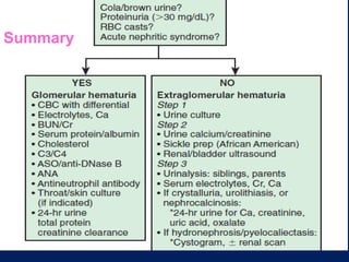
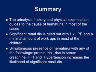
Hematuria in Children
Ali Derakhshan MD
Shiraz University of Medical sciences
Shiraz Nephro-Urology research Center
Shiraz - IRAN
Introduction
◼ A common cause for referral of children to
pediatricians and pediatric nephrologists
◼ Prvalence of Hematuria:1% in girls and
0.5% in boys
Hematuria
Hematuria
Transient phenomenon
of little significance
Transient phenomenon
of little significance
Sign of serious
renal disease
Sign of serious
renal disease
Microscopic Hematuria
• Definition > 5 RBC/HPF
• > 5 RBC/ μL
•Dipstick detects >3 RBC/ μL of urine
•False Negative (formalin ,Ascorbic acid)
•False positive (urine PH>9, oxidizing detergents
•Screening U/A at 5 years and during 2nd decade
•AAP: no recommendation for screening UA
• Definition > 5 RBC/HPF
• > 5 RBC/ μL
•Dipstick detects >3 RBC/ μL of urine
•False Negative (formalin ,Ascorbic acid)
•False positive (urine PH>9, oxidizing detergents
•Screening U/A at 5 years and during 2nd decade
•AAP: no recommendation for screening UA
Causes of Hematuria
Causes of Hematuria
• Glomerular disease
• Lesions along the urinary tract
Conditions unrelated to kidney and urinary tract
disease: following exercise, Febrile disorders, Gastroenteritis
with dehydration, Contamination from external genitalia
Clinical Presentation of
Hematuria in Children
◼ Microscopic hematuria incidental detection could
be isolated or non-isolated
◼ Microscopic hematuria with symptoms
◼ Microscopic hematuria with intermittent gross
hematuria with or without symptoms
◼ Gross hematuria ( intermittent or continuous)with
or without symptoms
Causes Of Discolored Urine
Red or pink urine
◼ Heme (+) : RBC (hematuria), Hburia, Myoglobinuria
◼ Heme (-) :
Drugs: Chloroquine , Deferoxamine , ibuprofen, metronidazole,
rifampin, phenazopyridine, salicylates, iron , sulphasalazine,
Phenothiazines, phenolphthalein, Metabolites: Urates,
porphyrins,
Dyes(veg./fruits) :Beets, black berries, food coloring
Dark brown or black urine
◼ Metabolites: Bile pigments, Meth.Hb, Alanine, Resorcinol,,
Alkaptonuria(Homogenistic acid), Melanin, thymol, tyrosinosis
Glomerular vs Extraglomerular
Hematuria
Urinary
finding
Glomerular Extraglom-
erular
Red cell casts May be present Absent
Red cell
morphology
Dysmorphic Uniform
Proteinuria May be present Absent
Clots Absent May be
present
Color Cola ,Red or
brown
Red
◼ Almost always associated
with glomerulonephritis or
vasculitis Virtually exclude
extra-renal causes of
bleeding
Clinical Approach to
Hematuria
◼ Not to miss serious conditions
◼ Avoid unnecessary and expensive tests
◼ First decision :watchful follow up ,need for
rapid work ups and/or referral to a subspecialist
◼ Reassure the family when appropriate
◼ Guidelines for further studies according to
changes in the course of illness
◼ Careful history
History
◼ Timing of hematuria and associated symptoms
◼ Trauma, exercise, stone passage, URTI, skin
infection and medications
◼ Dysuria, frequency, urgency, back pain, skin
rash, joint symptoms and edema
◼ Sickle cell disease or trait
◼ Family History: hematuria, stone, hypertension,
renal failure,Tx , deafness & coagulopathy
◼ Menarche
Purposeful Physical Examination
◼ General P/E , Edema , BP , Pallor
◼ Hypertension and edema determines how
urgent the diagnostic evaluation be done
◼ Rash , impetigo
◼ Ecchymosis , petechia , hemangioma
◼ Abdominal mass(tumors, hydronephrosis)
◼ Flank tenderness (pyelonephritis , stone)
◼ Inspection of External genitalia
◼ Growth parameters
◼ State of hearing
Purposeful Urinalysis
◼ Look for :
Shape of RBC
RBC cast
Proteinuria
Pyuria
Crystals
◼ Repeat UA for persistence of Hematuria
Child with Lower Urinary Symptoms
◼ Hematuria with dysuria, frequency, urgency,
flank or abdominal pain is suggestive for : UTI,
hypercalciuria or nephrolithiasis
◼ UTI : In 1/3 of cases associated with hematuria
usually microscopic
◼ Hemorrhagic cystitis by adenovirus type 11 and
21 (5days GH then 2-3days micro-hematuria)
◼ Schistosomiasis
Child with Clinical Features of
Glomerulonephritis
◼ Acute nephritic syndrome: edema, ↑BP, oliguria,
hematuria, ↓renal function
◼ Work up for different types of glomerulonephritis
according to the History: ASO, C3,C4,ANA,
Anti-dsDNA, ANCA, Serum IgA.
Asymptomatic Child : Incidental
Microscopic Hematuria in U/A
◼ In routine check up or in a child with febrile illness or any
other presentation
◼ Repeat U/A after resolution of current illness
◼ Repeat U/A twice within1- 2 weeks (+ in all)
◼ UA of siblings and Parents
◼ US ,BUN,Cr,Electrolytes, UCa/Cr ratio, U uric acid/Cr ratio
◼ Color doppler US of renal vessels
◼ Common causes of persistent micro-hematuria: Benign
familial hematuria (TBMN), idiopathic hypercalciuria, IgA
nephropathy and Alport syndrome
Hypercalciuria
◼ Noted in 1981.
◼ Increase urinary excretion calcium with normal
serum calcium levels.
◼ Ca/Cr ratio>0.21mg/mg *or >4mg/kg/day
◼ Measurement of urinary calcium excretion has
become the standard part of the evaluation of
hematuria in children.
◼ Had family history of renal stones.
◼ Rx: hydrochlorothiazide,do not restrict Ca
* >7 years old
Causes of Hematuria
Glomerular:
◼ Post infectious GN
◼ IgA-Nephropathy
◼ Henoch –Schonlein
nephritis
◼ Hereditary nephritis
◼ Benign familial hematuria
◼ MPGN
◼ Lupus nephritis
◼ Vasculitis associated GN
◼ Others
Non glomerular:
◼ Nephrocalcinosis,stone,
hypercalciuria
◼ Interstitial Nephritis, UTI
◼ Renal vein Thrombosis
◼ Sickle cell Dis.
◼ Vascular Malformations :
Hemangioma and AVF,
nutcracker syndrome
◼ Wilm’s Tumor
◼ Renal cell carcinoma
◼ Cystic disease
◼ Trauma
Follow up of Isolated
Hematuria
◼ Permanent disappearance of hematuria
◼ Change in nature with new symptoms or
signs: further work ups
◼ persists and needs regular follow up with
UA every 3-6MO, serum Cr and eventually
may need a kidney Biopsy in case of
persistence for one or 2 years
◼ When a cause is found treatment
accordingly
Indications of Renal Biopsy
◼ In follow up of Hematuria if any of the following:
- Proteinuria
-Hypertension
-Renal isufficiency
◼ Persistent low C3 following PSAGN
◼ Systemic disease with proteinuria: SLE, HSP,
ANCA+vasculitis
◼ Family history suggestive of Alport syndrome
◼ Recurrent gross hematuria of unknown etiology
◼ Keeping in mind that all modalities of tissue study
including light, EM, IF must be available
◼ Important Consideration For Family Anxiety,
Insurance.
aaaaappaaaaaaaaaaa
Summary
Summary
◼ The urinalysis, history and physical examination
guides to the cause of hematuria in most of the
cases
◼ Significant renal dis.is ruled out with Hx , PE and a
minimal amount of work ups in most of the
children
◼ Simultaneous presence of hematuria with any of
the followings: proteinuria , rise in serum
creatinine, FTT and Hypertension increases the
likelihood of significant renal dis.
THANK YOU

Hematuria

  • 1.
    Hematuria in Children AliDerakhshan MD Shiraz University of Medical sciences Shiraz Nephro-Urology research Center Shiraz - IRAN
  • 2.
    Introduction ◼ A commoncause for referral of children to pediatricians and pediatric nephrologists ◼ Prvalence of Hematuria:1% in girls and 0.5% in boys
  • 3.
    Hematuria Hematuria Transient phenomenon of littlesignificance Transient phenomenon of little significance Sign of serious renal disease Sign of serious renal disease
  • 4.
    Microscopic Hematuria Microscopic Hematuria •Definition > 5 RBC/HPF • > 5 RBC/ μL •Dipstick detects >3 RBC/ μL of urine •False Negative (formalin ,Ascorbic acid) •False positive (urine PH>9, oxidizing detergents •Screening U/A at 5 years and during 2nd decade •AAP: no recommendation for screening UA • Definition > 5 RBC/HPF • > 5 RBC/ μL •Dipstick detects >3 RBC/ μL of urine •False Negative (formalin ,Ascorbic acid) •False positive (urine PH>9, oxidizing detergents •Screening U/A at 5 years and during 2nd decade •AAP: no recommendation for screening UA
  • 5.
    Causes of Hematuria Causesof Hematuria • Glomerular disease • Lesions along the urinary tract Conditions unrelated to kidney and urinary tract disease: following exercise, Febrile disorders, Gastroenteritis with dehydration, Contamination from external genitalia
  • 6.
    Clinical Presentation of Hematuriain Children ◼ Microscopic hematuria incidental detection could be isolated or non-isolated ◼ Microscopic hematuria with symptoms ◼ Microscopic hematuria with intermittent gross hematuria with or without symptoms ◼ Gross hematuria ( intermittent or continuous)with or without symptoms
  • 7.
    Causes Of DiscoloredUrine Red or pink urine ◼ Heme (+) : RBC (hematuria), Hburia, Myoglobinuria ◼ Heme (-) : Drugs: Chloroquine , Deferoxamine , ibuprofen, metronidazole, rifampin, phenazopyridine, salicylates, iron , sulphasalazine, Phenothiazines, phenolphthalein, Metabolites: Urates, porphyrins, Dyes(veg./fruits) :Beets, black berries, food coloring Dark brown or black urine ◼ Metabolites: Bile pigments, Meth.Hb, Alanine, Resorcinol,, Alkaptonuria(Homogenistic acid), Melanin, thymol, tyrosinosis
  • 8.
    Glomerular vs Extraglomerular Hematuria Urinary finding GlomerularExtraglom- erular Red cell casts May be present Absent Red cell morphology Dysmorphic Uniform Proteinuria May be present Absent Clots Absent May be present Color Cola ,Red or brown Red ◼ Almost always associated with glomerulonephritis or vasculitis Virtually exclude extra-renal causes of bleeding
  • 9.
    Clinical Approach to Hematuria ◼Not to miss serious conditions ◼ Avoid unnecessary and expensive tests ◼ First decision :watchful follow up ,need for rapid work ups and/or referral to a subspecialist ◼ Reassure the family when appropriate ◼ Guidelines for further studies according to changes in the course of illness ◼ Careful history
  • 10.
    History ◼ Timing ofhematuria and associated symptoms ◼ Trauma, exercise, stone passage, URTI, skin infection and medications ◼ Dysuria, frequency, urgency, back pain, skin rash, joint symptoms and edema ◼ Sickle cell disease or trait ◼ Family History: hematuria, stone, hypertension, renal failure,Tx , deafness & coagulopathy ◼ Menarche
  • 11.
    Purposeful Physical Examination ◼General P/E , Edema , BP , Pallor ◼ Hypertension and edema determines how urgent the diagnostic evaluation be done ◼ Rash , impetigo ◼ Ecchymosis , petechia , hemangioma ◼ Abdominal mass(tumors, hydronephrosis) ◼ Flank tenderness (pyelonephritis , stone) ◼ Inspection of External genitalia ◼ Growth parameters ◼ State of hearing
  • 12.
    Purposeful Urinalysis ◼ Lookfor : Shape of RBC RBC cast Proteinuria Pyuria Crystals ◼ Repeat UA for persistence of Hematuria
  • 13.
    Child with LowerUrinary Symptoms ◼ Hematuria with dysuria, frequency, urgency, flank or abdominal pain is suggestive for : UTI, hypercalciuria or nephrolithiasis ◼ UTI : In 1/3 of cases associated with hematuria usually microscopic ◼ Hemorrhagic cystitis by adenovirus type 11 and 21 (5days GH then 2-3days micro-hematuria) ◼ Schistosomiasis
  • 14.
    Child with ClinicalFeatures of Glomerulonephritis ◼ Acute nephritic syndrome: edema, ↑BP, oliguria, hematuria, ↓renal function ◼ Work up for different types of glomerulonephritis according to the History: ASO, C3,C4,ANA, Anti-dsDNA, ANCA, Serum IgA.
  • 15.
    Asymptomatic Child :Incidental Microscopic Hematuria in U/A ◼ In routine check up or in a child with febrile illness or any other presentation ◼ Repeat U/A after resolution of current illness ◼ Repeat U/A twice within1- 2 weeks (+ in all) ◼ UA of siblings and Parents ◼ US ,BUN,Cr,Electrolytes, UCa/Cr ratio, U uric acid/Cr ratio ◼ Color doppler US of renal vessels ◼ Common causes of persistent micro-hematuria: Benign familial hematuria (TBMN), idiopathic hypercalciuria, IgA nephropathy and Alport syndrome
  • 16.
    Hypercalciuria ◼ Noted in1981. ◼ Increase urinary excretion calcium with normal serum calcium levels. ◼ Ca/Cr ratio>0.21mg/mg *or >4mg/kg/day ◼ Measurement of urinary calcium excretion has become the standard part of the evaluation of hematuria in children. ◼ Had family history of renal stones. ◼ Rx: hydrochlorothiazide,do not restrict Ca * >7 years old
  • 18.
    Causes of Hematuria Glomerular: ◼Post infectious GN ◼ IgA-Nephropathy ◼ Henoch –Schonlein nephritis ◼ Hereditary nephritis ◼ Benign familial hematuria ◼ MPGN ◼ Lupus nephritis ◼ Vasculitis associated GN ◼ Others Non glomerular: ◼ Nephrocalcinosis,stone, hypercalciuria ◼ Interstitial Nephritis, UTI ◼ Renal vein Thrombosis ◼ Sickle cell Dis. ◼ Vascular Malformations : Hemangioma and AVF, nutcracker syndrome ◼ Wilm’s Tumor ◼ Renal cell carcinoma ◼ Cystic disease ◼ Trauma
  • 19.
    Follow up ofIsolated Hematuria ◼ Permanent disappearance of hematuria ◼ Change in nature with new symptoms or signs: further work ups ◼ persists and needs regular follow up with UA every 3-6MO, serum Cr and eventually may need a kidney Biopsy in case of persistence for one or 2 years ◼ When a cause is found treatment accordingly
  • 20.
    Indications of RenalBiopsy ◼ In follow up of Hematuria if any of the following: - Proteinuria -Hypertension -Renal isufficiency ◼ Persistent low C3 following PSAGN ◼ Systemic disease with proteinuria: SLE, HSP, ANCA+vasculitis ◼ Family history suggestive of Alport syndrome ◼ Recurrent gross hematuria of unknown etiology ◼ Keeping in mind that all modalities of tissue study including light, EM, IF must be available ◼ Important Consideration For Family Anxiety, Insurance.
  • 21.
  • 22.
    Summary ◼ The urinalysis,history and physical examination guides to the cause of hematuria in most of the cases ◼ Significant renal dis.is ruled out with Hx , PE and a minimal amount of work ups in most of the children ◼ Simultaneous presence of hematuria with any of the followings: proteinuria , rise in serum creatinine, FTT and Hypertension increases the likelihood of significant renal dis.
  • 23.