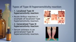
HYPERSENSITIVITY
By:- Raj Sonali Harsh
What is Hypersensitivity?
›It is an immunological state in which the
immune system “over react” to foreign
antigen such that the immune response
itself is more harmful than the antigen.
Types of Hypersensitivity
› Type I:- allergic reaction (“immediate hypersensitivity”)
› Type II:- cytotoxic reaction
› Type III:- immune complex reaction
› Type IV:- delayed cell-mediated reaction (DTH)
Type I Hypersensitivity
 Type I hypersensitivity is an allergic reaction provoked by re-
exposure to a specific antigen.
 Exposure may be by ingestion, inhalation, injection, or direct
contact.
 The reaction is mediated by IgE antibodies and produced by the
immediate release of histamine, tryptase, arachidonate and
derivatives by basophils and mast cells..
 This causes an inflammatory response leading to an immediate
(within seconds to minutes) reaction.
 The reaction may be either local or systemic. Symptoms vary from
mild irritation to sudden death from anaphylactic shock.
 Treatment usually involves epinephrine, antihistamines, and
corticosteroids
Some examples:
 Allergic asthma
 Allergic conjunctivitis
 Allergic rhinitis ("hay fever")
 Anaphylaxis
 Angioedema
 Urticaria (hives)
Pathologic Lesions
Vascular dilation, edema,
smooth muscle
contraction, mucus
production, tissue injury,
inflammation
Mechanism:
Type II - antibody-dependent
 In type II hypersensitivity, the antibodies produced by the
immune response bind to antigens on the patient's own cell
surfaces.
 The antigens recognized in this way may either be intrinsic
("self" antigen, innately part of the patient's cells) or extrinsic
(absorbed onto the cells during exposure to some foreign
antigen) possibly as part of infection with a pathogen
 IgG and IgM antibodies bind to these antigens to form complexes
that activate the classical pathway of complement activation for
eliminating cells presenting foreign antigens (which are usually, but
not in this case, pathogens).
 As a result mediators of acute inflammation are generated at the
site and membrane attack complexes cause cell lysis and death.
The reaction takes hours to a day.
Examples
 Autoimmune haemolytic anaemia
 Pernicious anemia
 Immune thrombocytopenia
 Transfusion reactions
 Hashimoto's thyroiditis
 Graves' disease
 Myasthenia gravis
 Farmer's Lung
 Hemolytic disease of the newborn
Pathologic
lesions:-
Phagocytosis and lysis of
cells; inflammation; in
some diseases, functional
derangements without
cell or tissue injury
Mechanism:-
Lab Diagnosis
Diagnostic tests include detection of circulating antibody against the tissues involved and the
presence of antibody and complement in the lesion (biopsy) by immunofluorescence
Type III - immune complex
In type III hypersensitivity:
 soluble immune complexes (aggregations of antigens and
IgG and IgM antibodies) form in the blood and are deposited
in various tissues (typically the skin, kidney and joints)
 This may trigger an immune response according to
the classical pathway of complement activation.
 The reaction takes hours to days to develop
Examples:
 Immune complex
glomerulonephritis
 Rheumatoid arthritis
 Serum sickness
 Subacute bacterial endocarditis
 Symptoms of malaria
 Systemic lupus erythematosus
 Arthus reaction
Pathologic
lesions:-
Inflammation,
necrotizing vasculitis
(fibrinoid necrosis)
Types of Type III hypersensitivity reaction
› 1. Localized Type III
hypersensitivity reaction:
• Acute Arthus reaction is
example of localized Type
hypersensitivity reaction.
› 2. Generalized Type III
hypersensitivity reaction:
• Serum sickness is an
generalized Type III
hypersensitivity reaction.
TYPE IV HYPERSENSITIVITY
 Type IV hypersensitivity is also known as cell mediated
or delayed type hypersensitivity.
 The classical example of this hypersensitivity is
tuberculin (Montoux) reaction
 Reaction peaks 48 hours after the injection of antigen
(PPD or old tuberculin). The lesion is characterized by
induration and erythema
 Type IV hypersensitivity is involved in
the pathogenesis of many autoimmune
and infectious diseases:
 Tuberculosis
 Leprosy
 Blastomycosis
 Histoplasmosis
 Toxoplasmosis
 Leishmaniasis
 Granulomas due to infections and
foreign antigens
Pathologic lesions:-
Perivascular cellular
infiltrates; edema;
granuloma formation; cell
destruction
 Another form of delayed hypersensitivity is contact dermatitis
(poison ivy (figure 6), chemicals, heavy metals, etc.) in which the
lesions are more papular
 Type IV hypersensitivity can be classified into three categories
depending on the time of onset and clinical and histological
presentation
Mechanism:
 The mechanism includes T lymphocytes and monocytes and/or macrophages.
 Cytotoxic T cells (Tc) cause direct damage whereas helper T (TH1) cells
secrete cytokines which activate cytotoxic T cells, recruit and activate
monocytes and macrophages, which cause the bulk of the damage
 The delayed hypersensitivity lesions mainly contain monocytes and a few T
cells.
Diagnosis
 Diagnostic tests in vivo include delayed cutaneous reaction (e.g.
Montoux test )
 In vitro tests for delayed hypersensitivity include mitogenic response,
lympho-cytotoxicity and IL-2 production.
 Corticosteroids & other immunosuppressive agents are used in
treatment.
hypersensitivity

hypersensitivity

  • 1.
  • 2.
    What is Hypersensitivity? ›Itis an immunological state in which the immune system “over react” to foreign antigen such that the immune response itself is more harmful than the antigen.
  • 3.
    Types of Hypersensitivity ›Type I:- allergic reaction (“immediate hypersensitivity”) › Type II:- cytotoxic reaction › Type III:- immune complex reaction › Type IV:- delayed cell-mediated reaction (DTH)
  • 4.
    Type I Hypersensitivity Type I hypersensitivity is an allergic reaction provoked by re- exposure to a specific antigen.  Exposure may be by ingestion, inhalation, injection, or direct contact.  The reaction is mediated by IgE antibodies and produced by the immediate release of histamine, tryptase, arachidonate and derivatives by basophils and mast cells..
  • 5.
     This causesan inflammatory response leading to an immediate (within seconds to minutes) reaction.  The reaction may be either local or systemic. Symptoms vary from mild irritation to sudden death from anaphylactic shock.  Treatment usually involves epinephrine, antihistamines, and corticosteroids
  • 6.
    Some examples:  Allergicasthma  Allergic conjunctivitis  Allergic rhinitis ("hay fever")  Anaphylaxis  Angioedema  Urticaria (hives) Pathologic Lesions Vascular dilation, edema, smooth muscle contraction, mucus production, tissue injury, inflammation
  • 7.
  • 8.
    Type II -antibody-dependent  In type II hypersensitivity, the antibodies produced by the immune response bind to antigens on the patient's own cell surfaces.  The antigens recognized in this way may either be intrinsic ("self" antigen, innately part of the patient's cells) or extrinsic (absorbed onto the cells during exposure to some foreign antigen) possibly as part of infection with a pathogen
  • 9.
     IgG andIgM antibodies bind to these antigens to form complexes that activate the classical pathway of complement activation for eliminating cells presenting foreign antigens (which are usually, but not in this case, pathogens).  As a result mediators of acute inflammation are generated at the site and membrane attack complexes cause cell lysis and death. The reaction takes hours to a day.
  • 10.
    Examples  Autoimmune haemolyticanaemia  Pernicious anemia  Immune thrombocytopenia  Transfusion reactions  Hashimoto's thyroiditis  Graves' disease  Myasthenia gravis  Farmer's Lung  Hemolytic disease of the newborn Pathologic lesions:- Phagocytosis and lysis of cells; inflammation; in some diseases, functional derangements without cell or tissue injury
  • 11.
    Mechanism:- Lab Diagnosis Diagnostic testsinclude detection of circulating antibody against the tissues involved and the presence of antibody and complement in the lesion (biopsy) by immunofluorescence
  • 12.
    Type III -immune complex In type III hypersensitivity:  soluble immune complexes (aggregations of antigens and IgG and IgM antibodies) form in the blood and are deposited in various tissues (typically the skin, kidney and joints)  This may trigger an immune response according to the classical pathway of complement activation.  The reaction takes hours to days to develop
  • 13.
    Examples:  Immune complex glomerulonephritis Rheumatoid arthritis  Serum sickness  Subacute bacterial endocarditis  Symptoms of malaria  Systemic lupus erythematosus  Arthus reaction Pathologic lesions:- Inflammation, necrotizing vasculitis (fibrinoid necrosis)
  • 14.
    Types of TypeIII hypersensitivity reaction › 1. Localized Type III hypersensitivity reaction: • Acute Arthus reaction is example of localized Type hypersensitivity reaction. › 2. Generalized Type III hypersensitivity reaction: • Serum sickness is an generalized Type III hypersensitivity reaction.
  • 15.
    TYPE IV HYPERSENSITIVITY Type IV hypersensitivity is also known as cell mediated or delayed type hypersensitivity.  The classical example of this hypersensitivity is tuberculin (Montoux) reaction  Reaction peaks 48 hours after the injection of antigen (PPD or old tuberculin). The lesion is characterized by induration and erythema
  • 16.
     Type IVhypersensitivity is involved in the pathogenesis of many autoimmune and infectious diseases:  Tuberculosis  Leprosy  Blastomycosis  Histoplasmosis  Toxoplasmosis  Leishmaniasis  Granulomas due to infections and foreign antigens Pathologic lesions:- Perivascular cellular infiltrates; edema; granuloma formation; cell destruction
  • 17.
     Another formof delayed hypersensitivity is contact dermatitis (poison ivy (figure 6), chemicals, heavy metals, etc.) in which the lesions are more papular  Type IV hypersensitivity can be classified into three categories depending on the time of onset and clinical and histological presentation
  • 18.
    Mechanism:  The mechanismincludes T lymphocytes and monocytes and/or macrophages.  Cytotoxic T cells (Tc) cause direct damage whereas helper T (TH1) cells secrete cytokines which activate cytotoxic T cells, recruit and activate monocytes and macrophages, which cause the bulk of the damage  The delayed hypersensitivity lesions mainly contain monocytes and a few T cells. Diagnosis  Diagnostic tests in vivo include delayed cutaneous reaction (e.g. Montoux test )  In vitro tests for delayed hypersensitivity include mitogenic response, lympho-cytotoxicity and IL-2 production.  Corticosteroids & other immunosuppressive agents are used in treatment.