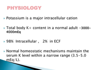
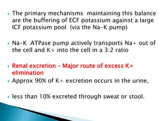
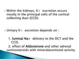
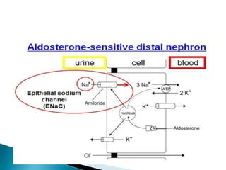
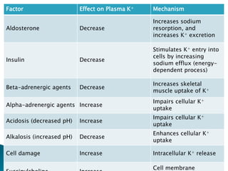
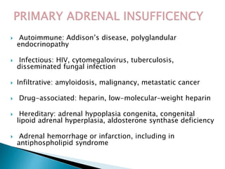
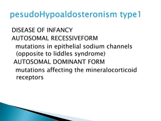
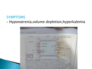
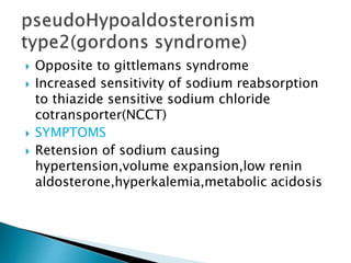
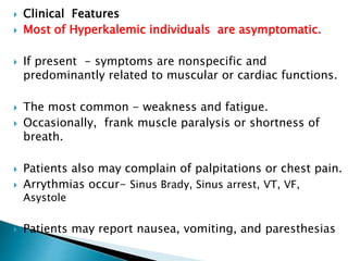
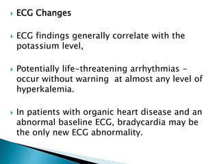
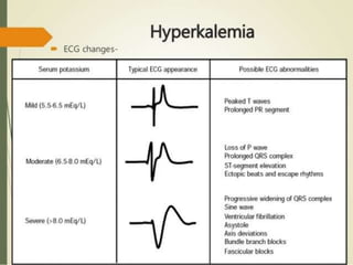
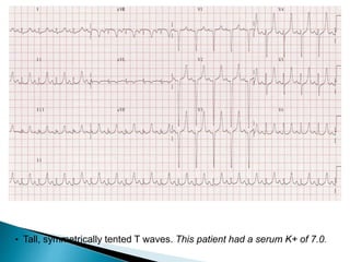
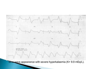
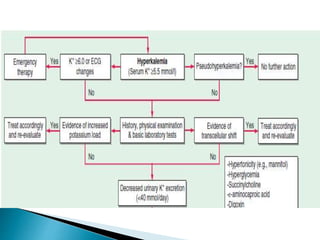
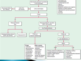
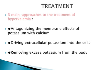
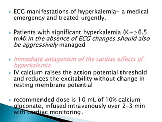
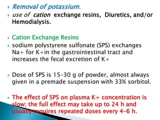
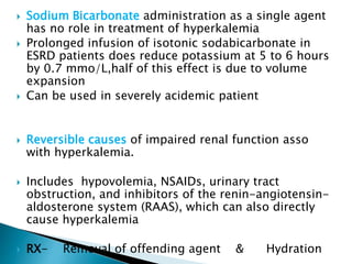
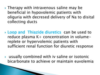
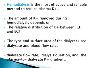
Dr. RAGHU NANDHINI
PHYSIOLOGY
 Potassium is a major intracellular cation
 Total body K+ content in a normal adult -3000-
4000mEq
 98% Intracellular , 2% in ECF
 Normal homeostatic mechanisms maintain the
serum K level within a narrow range (3.5-5.0
mEq/L).
 The primary mechanisms maintaining this balance
are the buffering of ECF potassium against a large
ICF potassium pool (via the Na-K pump)
 Na-K ATPase pump actively transports Na+ out of
the cell and K+ into the cell in a 3:2 ratio
 Renal excretion – Major route of excess K+
elimination
 Approx 90% of K+ excretion occurs in the urine,
 less than 10% excreted through sweat or stool.
 Within the kidneys, K+ excretion occurs
mostly in the principal cells of the cortical
collecting duct (CCD).
 Urinary K+ excretion depends on :
1. luminal Na+ delivery to the DCT and the
CCD,
2. effect of Aldosterone and other adrenal
corticosteroids with mineralocorticoid activity.
Factor Effect on Plasma K+ Mechanism
Aldosterone Decrease
Increases sodium
resorption, and
increases K+ excretion
Insulin Decrease
Stimulates K+ entry into
cells by increasing
sodium efflux (energy-
dependent process)
Beta-adrenergic agents Decrease
Increases skeletal
muscle uptake of K+
Alpha-adrenergic agents Increase
Impairs cellular K+
uptake
Acidosis (decreased pH) Increase
Impairs cellular K+
uptake
Alkalosis (increased pH) Decrease
Enhances cellular K+
uptake
Cell damage Increase Intracellular K+ release
Cell membrane
HYPERKALEMIA
 Defined as a plasma potassium level of >5.5
mEq/L
 Severe hyperkalemia when serum potssium
Levels are >6.0meq/L
decrease in renal excretion is the most
frequent cause
 Artificial increase in serum potassium during
or after venipuncture
 Mainely occurs due to marked increase in
muscle activity durin venipuncture
 Marked increase in cellular
elements(thrombocytosis,leucocytosis,erythro
cytosis)
 Cooling of blood followin venipuncture is
another cause
 Genetic causes causing increase in passive
potassium permeability for erythrocytes
II. Intra- to extracellular shift
 Acidosis – Uptake of H+, efflux of K+
NAGMA
 Hyperosmolality; hypertonic dextrose,
mannitol, - Solvent Drag effect
 β2-Adrenergic antagonists (noncardioselective
agents)
Suppresses catecholamine stimulated renin release-
in turn aldosterone synthesis
 Digoxin and related glycosides (yellow
oleander, foxglove, bufadienolide)- Inhibits
Na/K ATPase
 Hyperkalemic periodic paralysis- Episodic attack
of muscle weakness asso with Hyper k+. Na Muscle
channelopathy
 Lysine, arginine, and ε-aminocaproic acid
(structurally similar, positively charged)
 Succinylcholine; depolarises Muscle cells,
Efflux of K+ through AChRs . Contraindicated in
thermal trauma, neuromuscular injury, disuse atrophy,
mucositis, or prolonged immobilization- upregulated
AChRs
 Rapid tumor lysis / Rhabdomyolysis
 III. Inadequate excretion
 A. Inhibition of the renin-angiotensin-
aldosterone axis;
(↑ risk of hyperkalemia when these drugs
are used in combination
 Angiotensin-converting enzyme (ACE)
inhibitors
 Renin inhibitors; aliskiren
(in combination with ACE inhibitors or
angiotensin receptor blockers [ARBs])
 Angiotensin receptor blockers (ARBs)
 Blockade of the mineralocorticoid receptor:
- spironolactone, eplerenone,
 Blockade of the epithelial sodium channel
(ENaC): amiloride, triamterene, trimethoprim,
pentamidine, nafamostat
B. Decreased distal delivery
 Congestive heart failure
 Volume depletion
C. Hyporeninemic hypoaldosteronism
 Tubulointerstitial diseases:
SLE, sickle cell anemia, obstructive uropathy
 Diabetes, diabetic nephropathy
 Drugs: nonsteroidal anti-inflammatory drugs
(NSAIDs), cyclooxygenase 2 (COX2) inhibitors,
β-blockers, cyclosporine, tacrolimus
 Chronic kidney disease, advanced age
 Pseudohypoaldosteronism type II: defects in
WNK1 or WNK4 kinases, Kelch-like 3 (KLHL3),
or Cullin 3 (CUL3)
In The above said conditions –most Pt will
be volume expanded- secondary increse in
circulating ANP that inhibit both Renal renin
release and adrenal aldosterone release
D. Renal resistance to mineralocorticoid
 Tubulointerstitial diseases:
SLE, amyloidosis, sickle cell anemia,
obstructive uropathy, post–acute tubular
necrosis
 Hereditary:
pseudohypoaldosteronism type I; defects
in the mineralocorticoid receptor or the
epithelial sodium channel (ENaC)
E. Advanced renal insufficiency
 Chronic kidney disease
 Acute oliguric kidney disease
 Autoimmune: Addison’s disease, polyglandular
endocrinopathy
 Infectious: HIV, cytomegalovirus, tuberculosis,
disseminated fungal infection
 Infiltrative: amyloidosis, malignancy, metastatic cancer
 Drug-associated: heparin, low-molecular-weight heparin
 Hereditary: adrenal hypoplasia congenita, congenital
lipoid adrenal hyperplasia, aldosterone synthase deficiency
 Adrenal hemorrhage or infarction, including in
antiphospholipid syndrome
DISEASE OF INFANCY
AUTOSOMAL RECESSIVEFORM
mutations in epithelial sodium channels
(opposite to liddles syndrome)
AUTOSOMAL DOMINANT FORM
mutations affecting the mineralocorticoid
receptors
SYMPTOMS
 Hyponatrenia,volume depletion,hyperkalemia
 Opposite to gittlemans syndrome
 Increased sensitivity of sodium reabsorption
to thiazide sensitive sodium chloride
cotransporter(NCCT)
 SYMPTOMS
 Retension of sodium causing
hypertension,volume expansion,low renin
aldosterone,hyperkalemia,metabolic acidosis
 Clinical Features
 Most of Hyperkalemic individuals are asymptomatic.
 If present - symptoms are nonspecific and
predominantly related to muscular or cardiac functions.
 The most common - weakness and fatigue.
 Occasionally, frank muscle paralysis or shortness of
breath.
 Patients also may complain of palpitations or chest pain.
 Arrythmias occur- Sinus Brady, Sinus arrest, VT, VF,
Asystole
 Patients may report nausea, vomiting, and paresthesias
 ECG Changes
 ECG findings generally correlate with the
potassium level,
 Potentially life-threatening arrhythmias -
occur without warning at almost any level of
hyperkalemia.
 In patients with organic heart disease and an
abnormal baseline ECG, bradycardia may be
the only new ECG abnormality.
• Tall, symmetrically tented T waves. This patient had a serum K+ of 7.0.
Sine wave appearance with severe hyperkalaemia (K+ 9.9 mEq/L).
DIAGNOSTIC APPROACH TO
HYPERKALEMIA
 Tests In Evaluation of Hyperkalemia
 History on medications,diet,risk factors for
kidney failure,reduction in urine output,blood
pressure,volume status.
 BUN,creatinine,serum osmolarity,
 Serum Electrolytes- including Mg, Ca
 Urine potassium, sodium, and osmolality
 Complete blood count (CBC)
 ECG
Trans-tubular potassium gradient (TTKG)
 TTKG is an index reflecting the conservation
of potassium in the cortical collecting ducts
(CCD) of the kidneys.
 It is useful in diagnosing the causes of
hyperkalemia or hypokalemia.
 TTKG estimates the ratio of potassium in the
lumen of the CCD to that in the peritubular
capillaries.
 TTKG= Urine K/ Serum K x serum
Osm/Urine osm
 3 main approaches to the treatment of
hyperkalemia :
 ●Antagonizing the membrane effects of
potassium with calcium
 ●Driving extracellular potassium into the cells
 ●Removing excess potassium from the body
 ECG manifestations of hyperkalemia- a medical
emergency and treated urgently.
 Patients with significant hyperkalemia (K+≥6.5
mM) in the absence of ECG changes should also
be aggressively managed
 Immediate antagonism of the cardiac effects of
hyperkalemia
 IV calcium raises the action potential threshold
and reduces the excitability without change in
resting membrane potential
 recommended dose is 10 mL of 10% calcium
gluconate, infused intravenously over 2–3 min
with cardiac monitoring.
 The effect of infusion strats after 1to 3 min
and lasts for 30 to 60 min
 Dose should be repeated if there is no change
in ECG findings and they recur after intial
improvement
 Rapid reduction in plasma K+ concentration by
redistribution into cells.
 Insulin lowers plasma K+ concentration by
shifting K+in to cells
 Can be given as constant infusion or bolus
regimen
 Infusion regimen:10 units of regular insulin in
500ml of 10%dextroseover 60 min
 Bolous regimen: used in emergency conditons
recommended dose is 10 units of regular
insulin iv followed by 50 ml of 50%dextrose
 Effect begin 10 to 20 min,peaks at 30 to 60 min
and lasts for 4 to 6 hours
 In hyperkalemic patients with glucose
concentrations>200mg/dl insulin should be
given without glucose with blood glucose
monitering
 In almost all the patients plasma potassium
drops by0.5 to 1.2mmol/L after treatment
 Combined treatment with Beta 2 agonists in
addition to providing a synergistic effect with
insulin in lowering plasma potassium,may reduce
incidence of hypoglycemia
 β2-agonists, most commonly albuterol, are effective
but underused agents for the acute management of
hyperkalemia.
 However 20% of patients with end stage renal
disease(ESRD) are resistant to B2 agonists
 reaches peak at about 90 min lasts for 2-6 hoursThe
recommended dose for inhaled albuterol is 10 to 20 mg
in 4 ml of normal saline inhaled over 10 min
 Effect starts at 30 min
 Hyperglycemia is a side effect along with tachycardia
 Should be used with caution in hyperkalemia with
cardiac disease
 Removal of potassium.
 use of cation exchange resins, Diuretics, and/or
Hemodialysis.
 Cation Exchange Resins
 sodium polystyrene sulfonate (SPS) exchanges
Na+ for K+in the gastrointestinal tract and
increases the fecal excretion of K+
 Dose of SPS is 15–30 g of powder, almost always
given in a premade suspension with 33% sorbitol.
 The effect of SPS on plasma K+ concentration is
slow; the full effect may take up to 24 h and
usually requires repeated doses every 4–6 h.
 Sodium Bicarbonate administration as a single agent
has no role in treatment of hyperkalemia
 Prolonged infusion of isotonic sodabicarbonate in
ESRD patients does reduce potassium at 5 to 6 hours
by 0.7 mmo/L,half of this effect is due to volume
expansion
 Can be used in severely acidemic patient
 Reversible causes of impaired renal function asso
with hyperkalemia.
 Includes hypovolemia, NSAIDs, urinary tract
obstruction, and inhibitors of the renin-angiotensin-
aldosterone system (RAAS), which can also directly
cause hyperkalemia
 RX- Removal of offending agent & Hydration
 Therapy with intravenous saline may be
beneficial in hypovolemic patients with
oliguria with decresed delivery of Na to disital
collecting ducts
 Loop and Thiazide diuretics can be used to
reduce plasma K+ concentration in volume-
replete or hypervolemic patients with
sufficient renal function for diuretic response
 usually combined with iv saline or isotonic
bicarbonate to achieve or maintain euvolemia
 Hemodialysis is the most effective and reliable
method to reduce plasma K+ .
 The amount of K+ removed during
hemodialysis depends on
 The relative distribution of K+ between ICF
and ECF
 The type and surface area of the dialyzer used,
 dialysate and blood flow rates,
 dialysate flow rate, dialysis duration, and the
plasma-to- dialysate K+ gradient.
Hyperkalemia 160108171542

Hyperkalemia 160108171542

  • 1.
  • 2.
    PHYSIOLOGY  Potassium isa major intracellular cation  Total body K+ content in a normal adult -3000- 4000mEq  98% Intracellular , 2% in ECF  Normal homeostatic mechanisms maintain the serum K level within a narrow range (3.5-5.0 mEq/L).
  • 3.
     The primarymechanisms maintaining this balance are the buffering of ECF potassium against a large ICF potassium pool (via the Na-K pump)  Na-K ATPase pump actively transports Na+ out of the cell and K+ into the cell in a 3:2 ratio  Renal excretion – Major route of excess K+ elimination  Approx 90% of K+ excretion occurs in the urine,  less than 10% excreted through sweat or stool.
  • 4.
     Within thekidneys, K+ excretion occurs mostly in the principal cells of the cortical collecting duct (CCD).  Urinary K+ excretion depends on : 1. luminal Na+ delivery to the DCT and the CCD, 2. effect of Aldosterone and other adrenal corticosteroids with mineralocorticoid activity.
  • 6.
    Factor Effect onPlasma K+ Mechanism Aldosterone Decrease Increases sodium resorption, and increases K+ excretion Insulin Decrease Stimulates K+ entry into cells by increasing sodium efflux (energy- dependent process) Beta-adrenergic agents Decrease Increases skeletal muscle uptake of K+ Alpha-adrenergic agents Increase Impairs cellular K+ uptake Acidosis (decreased pH) Increase Impairs cellular K+ uptake Alkalosis (increased pH) Decrease Enhances cellular K+ uptake Cell damage Increase Intracellular K+ release Cell membrane
  • 7.
    HYPERKALEMIA  Defined asa plasma potassium level of >5.5 mEq/L  Severe hyperkalemia when serum potssium Levels are >6.0meq/L decrease in renal excretion is the most frequent cause
  • 8.
     Artificial increasein serum potassium during or after venipuncture  Mainely occurs due to marked increase in muscle activity durin venipuncture  Marked increase in cellular elements(thrombocytosis,leucocytosis,erythro cytosis)  Cooling of blood followin venipuncture is another cause  Genetic causes causing increase in passive potassium permeability for erythrocytes
  • 9.
    II. Intra- toextracellular shift  Acidosis – Uptake of H+, efflux of K+ NAGMA  Hyperosmolality; hypertonic dextrose, mannitol, - Solvent Drag effect  β2-Adrenergic antagonists (noncardioselective agents) Suppresses catecholamine stimulated renin release- in turn aldosterone synthesis  Digoxin and related glycosides (yellow oleander, foxglove, bufadienolide)- Inhibits Na/K ATPase
  • 10.
     Hyperkalemic periodicparalysis- Episodic attack of muscle weakness asso with Hyper k+. Na Muscle channelopathy  Lysine, arginine, and ε-aminocaproic acid (structurally similar, positively charged)  Succinylcholine; depolarises Muscle cells, Efflux of K+ through AChRs . Contraindicated in thermal trauma, neuromuscular injury, disuse atrophy, mucositis, or prolonged immobilization- upregulated AChRs  Rapid tumor lysis / Rhabdomyolysis
  • 11.
     III. Inadequateexcretion  A. Inhibition of the renin-angiotensin- aldosterone axis; (↑ risk of hyperkalemia when these drugs are used in combination  Angiotensin-converting enzyme (ACE) inhibitors  Renin inhibitors; aliskiren (in combination with ACE inhibitors or angiotensin receptor blockers [ARBs])
  • 12.
     Angiotensin receptorblockers (ARBs)  Blockade of the mineralocorticoid receptor: - spironolactone, eplerenone,  Blockade of the epithelial sodium channel (ENaC): amiloride, triamterene, trimethoprim, pentamidine, nafamostat B. Decreased distal delivery  Congestive heart failure  Volume depletion
  • 13.
    C. Hyporeninemic hypoaldosteronism Tubulointerstitial diseases: SLE, sickle cell anemia, obstructive uropathy  Diabetes, diabetic nephropathy  Drugs: nonsteroidal anti-inflammatory drugs (NSAIDs), cyclooxygenase 2 (COX2) inhibitors, β-blockers, cyclosporine, tacrolimus
  • 14.
     Chronic kidneydisease, advanced age  Pseudohypoaldosteronism type II: defects in WNK1 or WNK4 kinases, Kelch-like 3 (KLHL3), or Cullin 3 (CUL3) In The above said conditions –most Pt will be volume expanded- secondary increse in circulating ANP that inhibit both Renal renin release and adrenal aldosterone release
  • 15.
    D. Renal resistanceto mineralocorticoid  Tubulointerstitial diseases: SLE, amyloidosis, sickle cell anemia, obstructive uropathy, post–acute tubular necrosis  Hereditary: pseudohypoaldosteronism type I; defects in the mineralocorticoid receptor or the epithelial sodium channel (ENaC) E. Advanced renal insufficiency  Chronic kidney disease  Acute oliguric kidney disease
  • 16.
     Autoimmune: Addison’sdisease, polyglandular endocrinopathy  Infectious: HIV, cytomegalovirus, tuberculosis, disseminated fungal infection  Infiltrative: amyloidosis, malignancy, metastatic cancer  Drug-associated: heparin, low-molecular-weight heparin  Hereditary: adrenal hypoplasia congenita, congenital lipoid adrenal hyperplasia, aldosterone synthase deficiency  Adrenal hemorrhage or infarction, including in antiphospholipid syndrome
  • 17.
    DISEASE OF INFANCY AUTOSOMALRECESSIVEFORM mutations in epithelial sodium channels (opposite to liddles syndrome) AUTOSOMAL DOMINANT FORM mutations affecting the mineralocorticoid receptors
  • 18.
  • 19.
     Opposite togittlemans syndrome  Increased sensitivity of sodium reabsorption to thiazide sensitive sodium chloride cotransporter(NCCT)  SYMPTOMS  Retension of sodium causing hypertension,volume expansion,low renin aldosterone,hyperkalemia,metabolic acidosis
  • 20.
     Clinical Features Most of Hyperkalemic individuals are asymptomatic.  If present - symptoms are nonspecific and predominantly related to muscular or cardiac functions.  The most common - weakness and fatigue.  Occasionally, frank muscle paralysis or shortness of breath.  Patients also may complain of palpitations or chest pain.  Arrythmias occur- Sinus Brady, Sinus arrest, VT, VF, Asystole  Patients may report nausea, vomiting, and paresthesias
  • 21.
     ECG Changes ECG findings generally correlate with the potassium level,  Potentially life-threatening arrhythmias - occur without warning at almost any level of hyperkalemia.  In patients with organic heart disease and an abnormal baseline ECG, bradycardia may be the only new ECG abnormality.
  • 23.
    • Tall, symmetricallytented T waves. This patient had a serum K+ of 7.0.
  • 24.
    Sine wave appearancewith severe hyperkalaemia (K+ 9.9 mEq/L).
  • 25.
  • 26.
     Tests InEvaluation of Hyperkalemia  History on medications,diet,risk factors for kidney failure,reduction in urine output,blood pressure,volume status.  BUN,creatinine,serum osmolarity,  Serum Electrolytes- including Mg, Ca  Urine potassium, sodium, and osmolality  Complete blood count (CBC)  ECG
  • 27.
    Trans-tubular potassium gradient(TTKG)  TTKG is an index reflecting the conservation of potassium in the cortical collecting ducts (CCD) of the kidneys.  It is useful in diagnosing the causes of hyperkalemia or hypokalemia.  TTKG estimates the ratio of potassium in the lumen of the CCD to that in the peritubular capillaries.  TTKG= Urine K/ Serum K x serum Osm/Urine osm
  • 30.
     3 mainapproaches to the treatment of hyperkalemia :  ●Antagonizing the membrane effects of potassium with calcium  ●Driving extracellular potassium into the cells  ●Removing excess potassium from the body
  • 31.
     ECG manifestationsof hyperkalemia- a medical emergency and treated urgently.  Patients with significant hyperkalemia (K+≥6.5 mM) in the absence of ECG changes should also be aggressively managed  Immediate antagonism of the cardiac effects of hyperkalemia  IV calcium raises the action potential threshold and reduces the excitability without change in resting membrane potential  recommended dose is 10 mL of 10% calcium gluconate, infused intravenously over 2–3 min with cardiac monitoring.
  • 32.
     The effectof infusion strats after 1to 3 min and lasts for 30 to 60 min  Dose should be repeated if there is no change in ECG findings and they recur after intial improvement
  • 33.
     Rapid reductionin plasma K+ concentration by redistribution into cells.  Insulin lowers plasma K+ concentration by shifting K+in to cells  Can be given as constant infusion or bolus regimen  Infusion regimen:10 units of regular insulin in 500ml of 10%dextroseover 60 min  Bolous regimen: used in emergency conditons recommended dose is 10 units of regular insulin iv followed by 50 ml of 50%dextrose
  • 34.
     Effect begin10 to 20 min,peaks at 30 to 60 min and lasts for 4 to 6 hours  In hyperkalemic patients with glucose concentrations>200mg/dl insulin should be given without glucose with blood glucose monitering  In almost all the patients plasma potassium drops by0.5 to 1.2mmol/L after treatment  Combined treatment with Beta 2 agonists in addition to providing a synergistic effect with insulin in lowering plasma potassium,may reduce incidence of hypoglycemia
  • 35.
     β2-agonists, mostcommonly albuterol, are effective but underused agents for the acute management of hyperkalemia.  However 20% of patients with end stage renal disease(ESRD) are resistant to B2 agonists  reaches peak at about 90 min lasts for 2-6 hoursThe recommended dose for inhaled albuterol is 10 to 20 mg in 4 ml of normal saline inhaled over 10 min  Effect starts at 30 min  Hyperglycemia is a side effect along with tachycardia  Should be used with caution in hyperkalemia with cardiac disease
  • 36.
     Removal ofpotassium.  use of cation exchange resins, Diuretics, and/or Hemodialysis.  Cation Exchange Resins  sodium polystyrene sulfonate (SPS) exchanges Na+ for K+in the gastrointestinal tract and increases the fecal excretion of K+  Dose of SPS is 15–30 g of powder, almost always given in a premade suspension with 33% sorbitol.  The effect of SPS on plasma K+ concentration is slow; the full effect may take up to 24 h and usually requires repeated doses every 4–6 h.
  • 37.
     Sodium Bicarbonateadministration as a single agent has no role in treatment of hyperkalemia  Prolonged infusion of isotonic sodabicarbonate in ESRD patients does reduce potassium at 5 to 6 hours by 0.7 mmo/L,half of this effect is due to volume expansion  Can be used in severely acidemic patient  Reversible causes of impaired renal function asso with hyperkalemia.  Includes hypovolemia, NSAIDs, urinary tract obstruction, and inhibitors of the renin-angiotensin- aldosterone system (RAAS), which can also directly cause hyperkalemia  RX- Removal of offending agent & Hydration
  • 38.
     Therapy withintravenous saline may be beneficial in hypovolemic patients with oliguria with decresed delivery of Na to disital collecting ducts  Loop and Thiazide diuretics can be used to reduce plasma K+ concentration in volume- replete or hypervolemic patients with sufficient renal function for diuretic response  usually combined with iv saline or isotonic bicarbonate to achieve or maintain euvolemia
  • 39.
     Hemodialysis isthe most effective and reliable method to reduce plasma K+ .  The amount of K+ removed during hemodialysis depends on  The relative distribution of K+ between ICF and ECF  The type and surface area of the dialyzer used,  dialysate and blood flow rates,  dialysate flow rate, dialysis duration, and the plasma-to- dialysate K+ gradient.