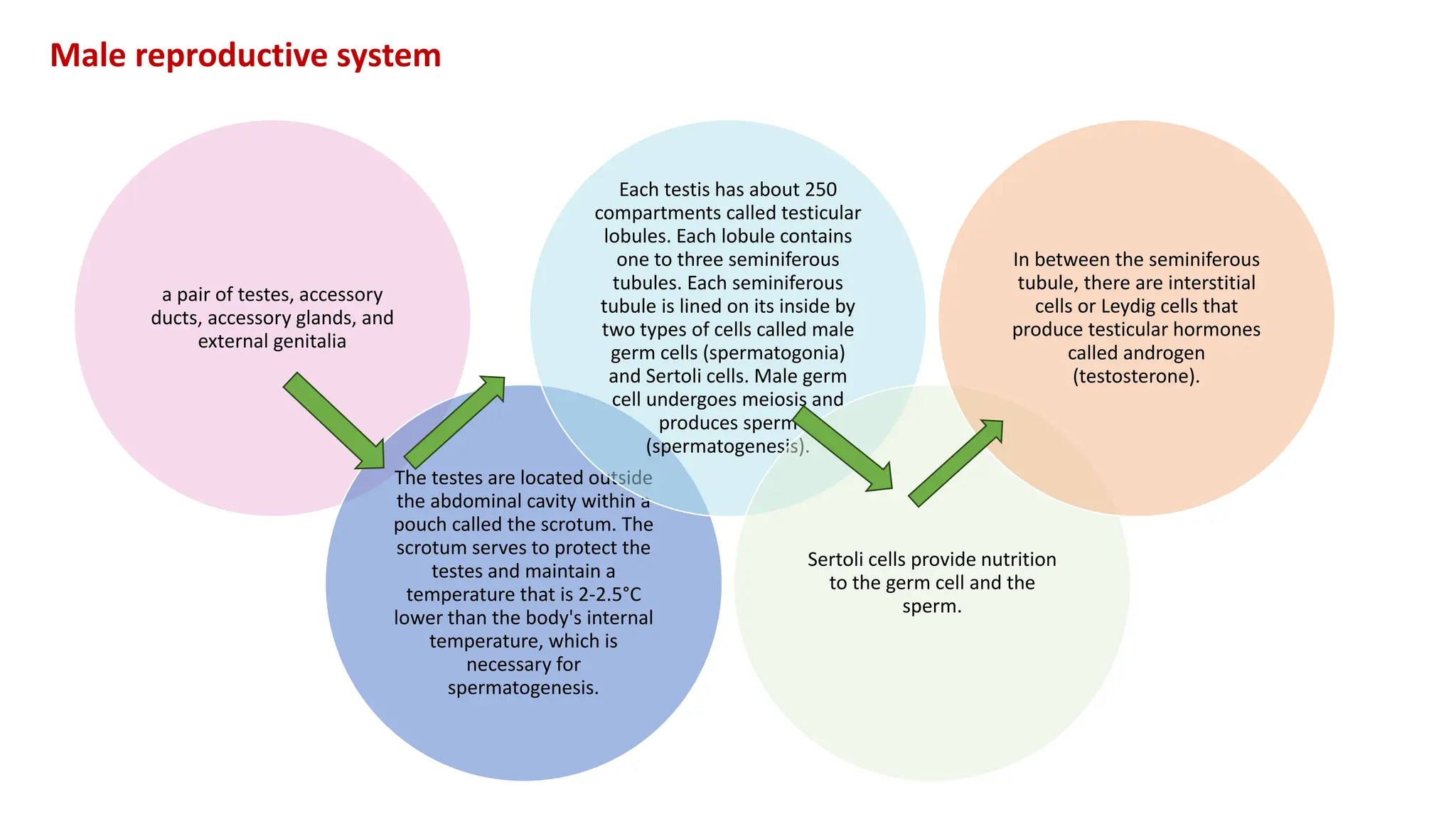
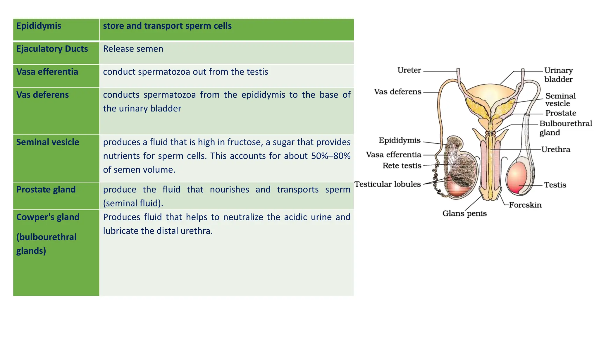
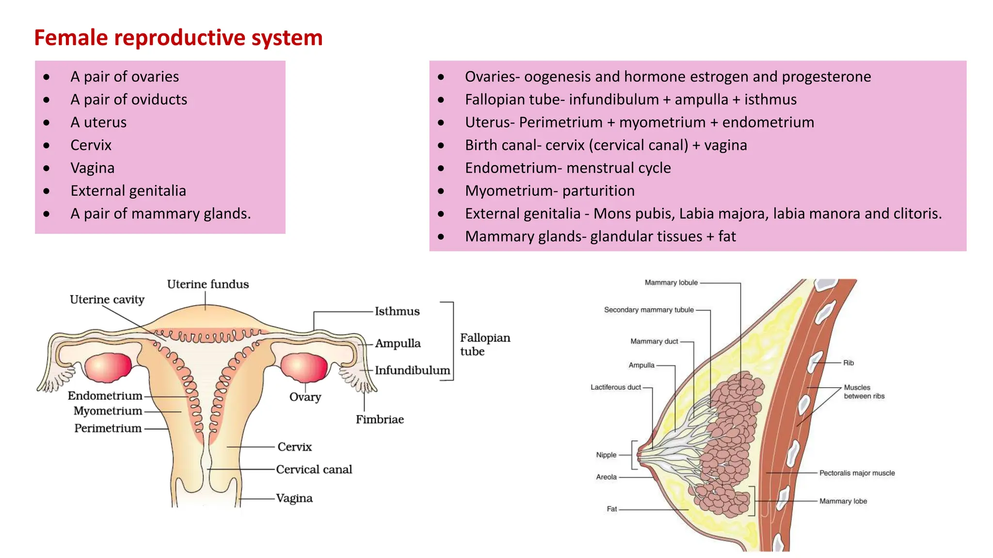
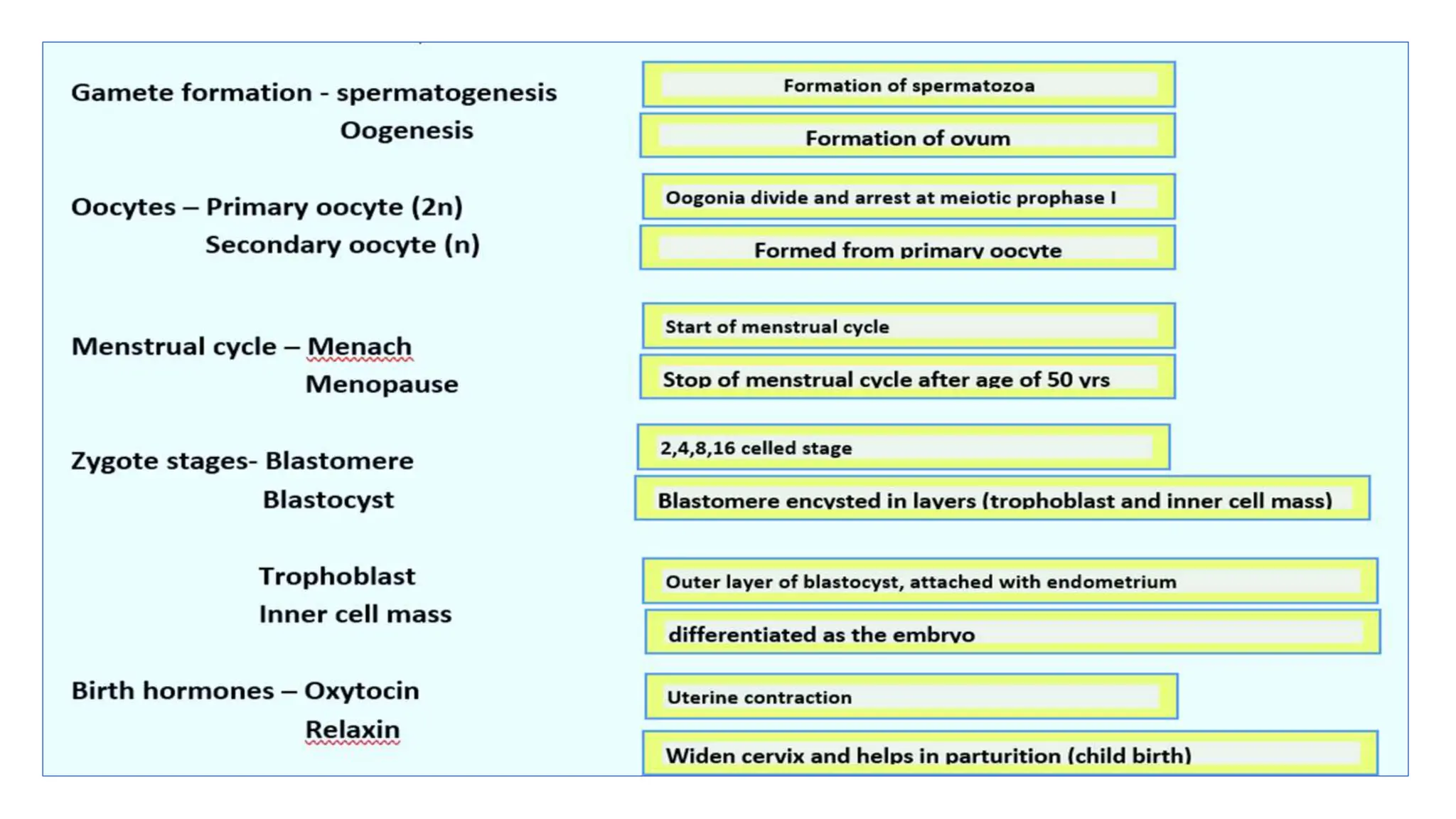
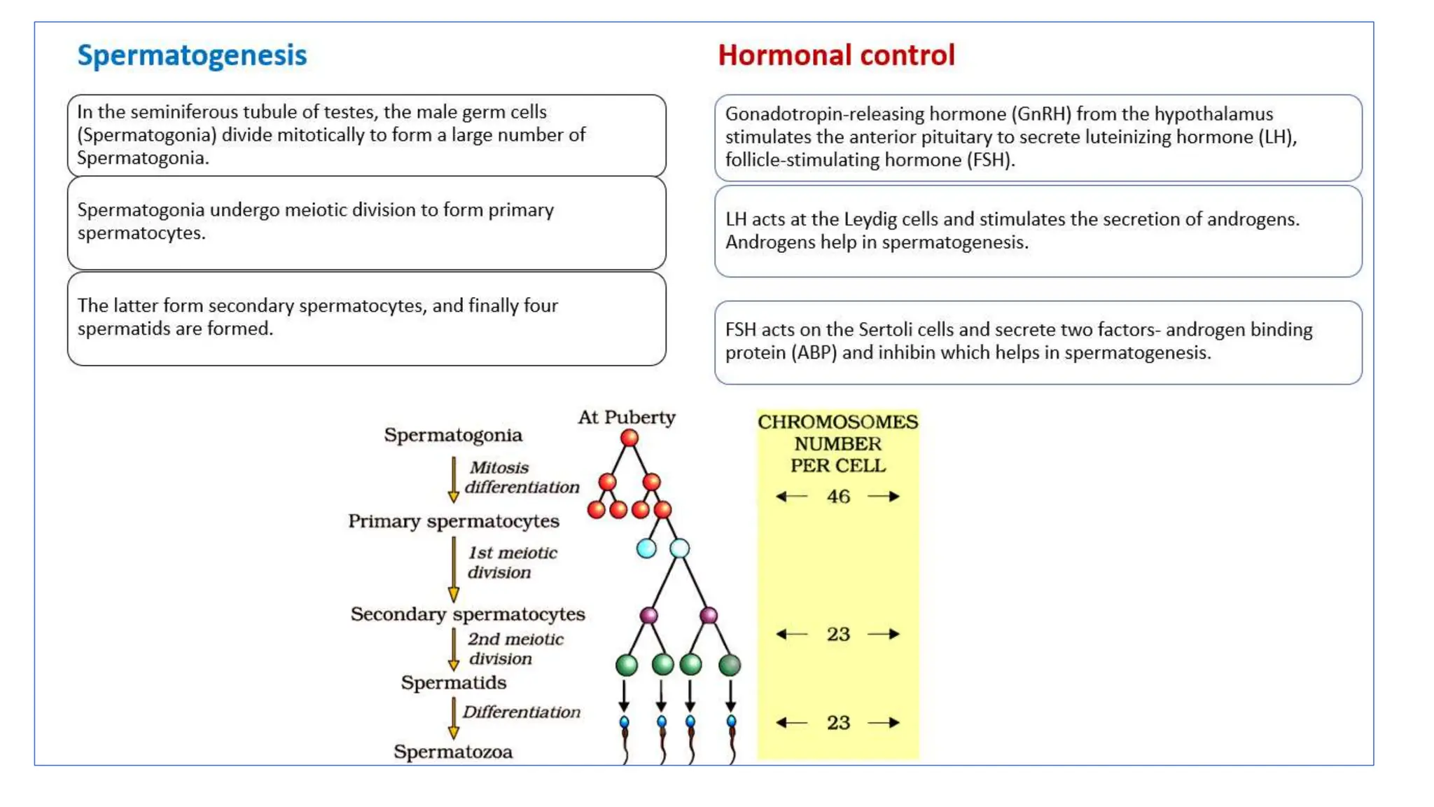
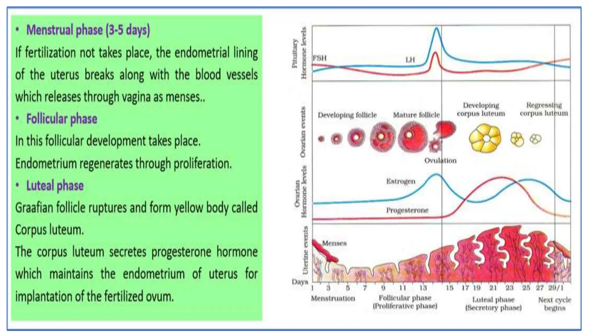
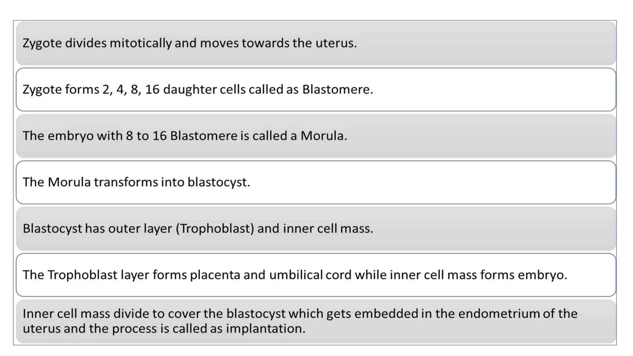
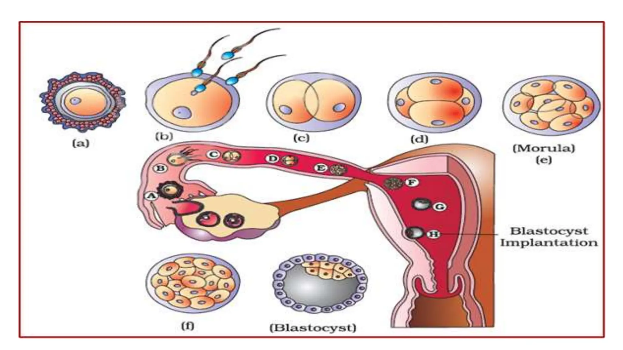
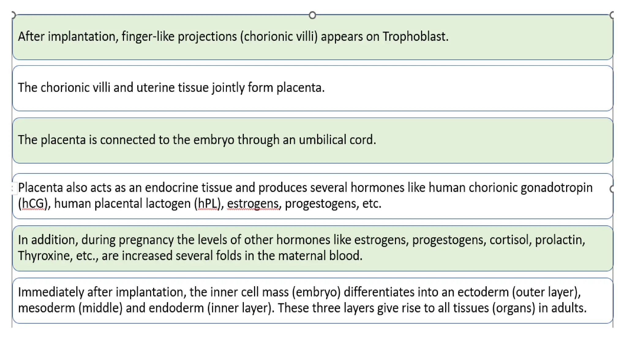
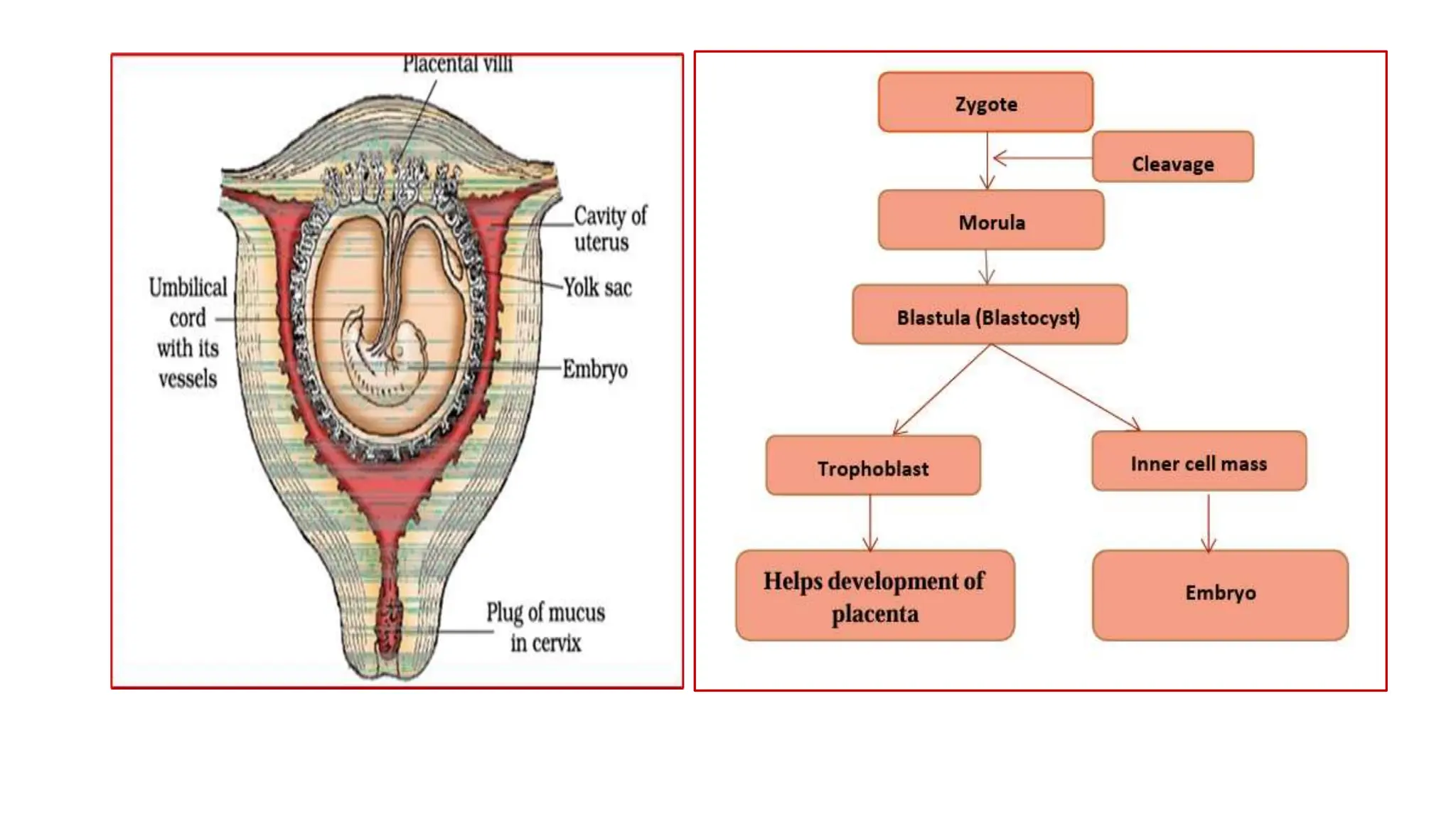
HUMAN
REPRODUCTIO
N
Dr. R. K. Upadhyay
Male reproductive system
a pair of testes, accessory
ducts, accessory glands, and
external genitalia
The testes are located outside
the abdominal cavity within a
pouch called the scrotum. The
scrotum serves to protect the
testes and maintain a
temperature that is 2-2.5°C
lower than the body's internal
temperature, which is
necessary for
spermatogenesis.
Each testis has about 250
compartments called testicular
lobules. Each lobule contains
one to three seminiferous
tubules. Each seminiferous
tubule is lined on its inside by
two types of cells called male
germ cells (spermatogonia)
and Sertoli cells. Male germ
cell undergoes meiosis and
produces sperm
(spermatogenesis).
Sertoli cells provide nutrition
to the germ cell and the
sperm.
In between the seminiferous
tubule, there are interstitial
cells or Leydig cells that
produce testicular hormones
called androgen
(testosterone).
Epididymis store and transport sperm cells
Ejaculatory Ducts Release semen
Vasa efferentia conduct spermatozoa out from the testis
Vas deferens conducts spermatozoa from the epididymis to the base of
the urinary bladder
Seminal vesicle produces a fluid that is high in fructose, a sugar that provides
nutrients for sperm cells. This accounts for about 50%–80%
of semen volume.
Prostate gland produce the fluid that nourishes and transports sperm
(seminal fluid).
Cowper's gland
(bulbourethral
glands)
Produces fluid that helps to neutralize the acidic urine and
lubricate the distal urethra.
Female reproductive system
 Ovaries- oogenesis and hormone estrogen and progesterone
 Fallopian tube- infundibulum + ampulla + isthmus
 Uterus- Perimetrium + myometrium + endometrium
 Birth canal- cervix (cervical canal) + vagina
 Endometrium- menstrual cycle
 Myometrium- parturition
 External genitalia - Mons pubis, Labia majora, labia manora and clitoris.
 Mammary glands- glandular tissues + fat
 A pair of ovaries
 A pair of oviducts
 A uterus
 Cervix
 Vagina
 External genitalia
 A pair of mammary glands.
One month Heart is formed
Two month Development of limbs and digits
12 weeks (first trimester) Most of the major organ systems are formed, for
example, the limbs and external Genital organs are
well developed.
Fifth month First movements and appearance of hair on the head
After 24 weeks (end of second
trimester)
The body is covered with fine hair, eye-lids separate,
and eyelashes are formed.
9 month Foetus is fully developed
Scrotum It is a pouch-like structure outside the abdominal cavity in which the testes is situated. It helps in maintaining the low temperature of the testes (2–2.5o C lower than the normal
internal body temperature) necessary for spermatogenesis.
Somniferous tubule In each testicular lobule 1- 3 highly coiled seminiferous tubules in which sperms are produced.
Sertoli cells Sertoli cells provide nutrition to the male germ cells.
Leydig cell Present in interstitial space (regions outside the seminiferous tubules). It secretes testicular hormones called androgens
Urethral meatus It is the external opening of the penis.
Seminal plasma Secretion of the male accessory glands (seminal vesicles, prostate and bulbourethral gland) constitute the seminal plasma.
Male accessory glands Paired seminal vesicles, prostate and paired bulbourethral glands
Lactiferous duct Through which milk is sucked out.
Spermatogonia Immature diploid male germ cells (Spermatogonia) produce sperms by spermatogenesis. These are present on the inside wall of the seminiferous tubule.
Acrosome Cap-like structure on sperm head. it is enzymes that help fertilization of the ovum.
Semen The seminal plasma along with the sperms constitutes the semen.
Oogonia Gamete mother cells (oogonia) are formed within each fetal ovary which later develops into the egg.
Graafian follicle Mature follicles formed during oogenesis. When it own ruptures releases the secondary oocyte (ovum) and reaming part forms the carpus luteum.
Zona pellucida The membrane on secondary oocyte, prevent polyspermy
Carpus luteum After ovulation remaining parts of the Graafian follicle transform in the corpus luteum. It secretes progesterone hormone, which is essential for the maintenance of the
endometrium.
Polar body It is a small haploid cell that is formed during oogenesis. It doesn’t have the ability to be fertilized.
Cleavage The mitotic division in the zygote is called cleavage.
Placenta The chorionic villi and uterine tissue become interdigitated with each other and jointly form a structural and functional unit called the placenta
Chorionic villi After implantation, finger-like projections appear on the trophoblast called chorionic villi which are surrounded by the uterine tissue and maternal blood.
Stem cell Inner cell mass contains certain cells called stem cells which have the potency to give rise to all the tissues and organs.
Implantation Implantation is the process in which the mammalian embryo (blastocyst) becomes attached to the endometrium of the uterus.
Fetus ejection reflex Mild uterine contractions are generated by the placenta when the fetus is fully developed, at the time of parturition.

HUMAN REPRODUCTION .pptx class XII Biology

  • 1.
  • 2.
    Male reproductive system apair of testes, accessory ducts, accessory glands, and external genitalia The testes are located outside the abdominal cavity within a pouch called the scrotum. The scrotum serves to protect the testes and maintain a temperature that is 2-2.5°C lower than the body's internal temperature, which is necessary for spermatogenesis. Each testis has about 250 compartments called testicular lobules. Each lobule contains one to three seminiferous tubules. Each seminiferous tubule is lined on its inside by two types of cells called male germ cells (spermatogonia) and Sertoli cells. Male germ cell undergoes meiosis and produces sperm (spermatogenesis). Sertoli cells provide nutrition to the germ cell and the sperm. In between the seminiferous tubule, there are interstitial cells or Leydig cells that produce testicular hormones called androgen (testosterone).
  • 3.
    Epididymis store andtransport sperm cells Ejaculatory Ducts Release semen Vasa efferentia conduct spermatozoa out from the testis Vas deferens conducts spermatozoa from the epididymis to the base of the urinary bladder Seminal vesicle produces a fluid that is high in fructose, a sugar that provides nutrients for sperm cells. This accounts for about 50%–80% of semen volume. Prostate gland produce the fluid that nourishes and transports sperm (seminal fluid). Cowper's gland (bulbourethral glands) Produces fluid that helps to neutralize the acidic urine and lubricate the distal urethra.
  • 4.
    Female reproductive system Ovaries- oogenesis and hormone estrogen and progesterone  Fallopian tube- infundibulum + ampulla + isthmus  Uterus- Perimetrium + myometrium + endometrium  Birth canal- cervix (cervical canal) + vagina  Endometrium- menstrual cycle  Myometrium- parturition  External genitalia - Mons pubis, Labia majora, labia manora and clitoris.  Mammary glands- glandular tissues + fat  A pair of ovaries  A pair of oviducts  A uterus  Cervix  Vagina  External genitalia  A pair of mammary glands.
  • 14.
    One month Heartis formed Two month Development of limbs and digits 12 weeks (first trimester) Most of the major organ systems are formed, for example, the limbs and external Genital organs are well developed. Fifth month First movements and appearance of hair on the head After 24 weeks (end of second trimester) The body is covered with fine hair, eye-lids separate, and eyelashes are formed. 9 month Foetus is fully developed
  • 15.
    Scrotum It isa pouch-like structure outside the abdominal cavity in which the testes is situated. It helps in maintaining the low temperature of the testes (2–2.5o C lower than the normal internal body temperature) necessary for spermatogenesis. Somniferous tubule In each testicular lobule 1- 3 highly coiled seminiferous tubules in which sperms are produced. Sertoli cells Sertoli cells provide nutrition to the male germ cells. Leydig cell Present in interstitial space (regions outside the seminiferous tubules). It secretes testicular hormones called androgens Urethral meatus It is the external opening of the penis. Seminal plasma Secretion of the male accessory glands (seminal vesicles, prostate and bulbourethral gland) constitute the seminal plasma. Male accessory glands Paired seminal vesicles, prostate and paired bulbourethral glands Lactiferous duct Through which milk is sucked out. Spermatogonia Immature diploid male germ cells (Spermatogonia) produce sperms by spermatogenesis. These are present on the inside wall of the seminiferous tubule. Acrosome Cap-like structure on sperm head. it is enzymes that help fertilization of the ovum. Semen The seminal plasma along with the sperms constitutes the semen. Oogonia Gamete mother cells (oogonia) are formed within each fetal ovary which later develops into the egg. Graafian follicle Mature follicles formed during oogenesis. When it own ruptures releases the secondary oocyte (ovum) and reaming part forms the carpus luteum. Zona pellucida The membrane on secondary oocyte, prevent polyspermy Carpus luteum After ovulation remaining parts of the Graafian follicle transform in the corpus luteum. It secretes progesterone hormone, which is essential for the maintenance of the endometrium. Polar body It is a small haploid cell that is formed during oogenesis. It doesn’t have the ability to be fertilized. Cleavage The mitotic division in the zygote is called cleavage. Placenta The chorionic villi and uterine tissue become interdigitated with each other and jointly form a structural and functional unit called the placenta Chorionic villi After implantation, finger-like projections appear on the trophoblast called chorionic villi which are surrounded by the uterine tissue and maternal blood. Stem cell Inner cell mass contains certain cells called stem cells which have the potency to give rise to all the tissues and organs. Implantation Implantation is the process in which the mammalian embryo (blastocyst) becomes attached to the endometrium of the uterus. Fetus ejection reflex Mild uterine contractions are generated by the placenta when the fetus is fully developed, at the time of parturition.