The reproductive systemis responsible for producing, storing,
and transporting gametes (sperm in males, eggs in females)
and facilitating reproduction. It is divided into male and
female reproductive systems, each with specialized
structures and functions.
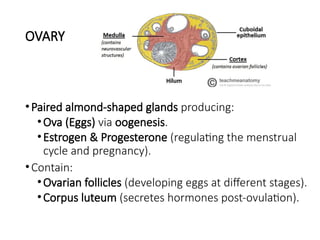
OVARY
•Paired almond-shaped glandsproducing:
•Ova (Eggs) via oogenesis.
•Estrogen & Progesterone (regulating the menstrual
cycle and pregnancy).
•Contain:
•Ovarian follicles (developing eggs at different stages).
•Corpus luteum (secretes hormones post-ovulation).
7.
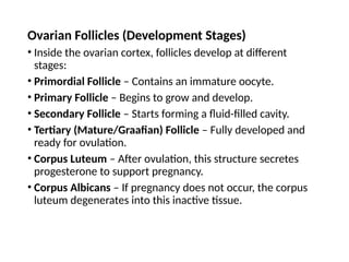
Ovarian Follicles (DevelopmentStages)
• Inside the ovarian cortex, follicles develop at different
stages:
• Primordial Follicle – Contains an immature oocyte.
• Primary Follicle – Begins to grow and develop.
• Secondary Follicle – Starts forming a fluid-filled cavity.
• Tertiary (Mature/Graafian) Follicle – Fully developed and
ready for ovulation.
• Corpus Luteum – After ovulation, this structure secretes
progesterone to support pregnancy.
• Corpus Albicans – If pregnancy does not occur, the corpus
luteum degenerates into this inactive tissue.
8.
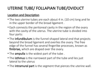
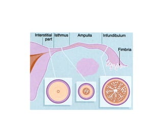
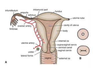
UTERINE TUBE/ FOLLAPIANTUBE/OVIDUCT
Location and Description
• The two uterine tubes are each about 4 in. (10 cm) long and lie
in the upper border of the broad ligament .
• Each connects the peritoneal cavity in the region of the ovary
with the cavity of the uterus. The uterine tube is divided into
four parts:
• The infundibulum is the funnel-shaped lateral end that projects
beyond the broad ligament and overlies the ovary. The free
edge of the funnel has several fingerlike processes, known as
fimbriae, which are draped over the ovary.
• The ampulla is the widest part of the tube .
• The isthmus is the narrowest part of the tube and lies just
lateral to the uterus
• The intramural part is the segment that pierces the uterine wall.
10.
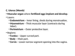
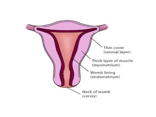
C. Uterus (Womb)
•Muscular organ where fertilized eggs implant and develop.
• Layers:
• Endometrium – Inner lining, sheds during menstruation.
• Myometrium – Thick muscular layer (contracts during
labor).
• Perimetrium – Outer protective layer.
• Regions:
• Fundus – Upper curved part.
• Body – Main part.
• Cervix – Lower narrow segment opening into the vagina.
11.
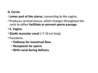
D. Cervix
• Lowerpart of the uterus, connecting to the vagina.
• Produces cervical mucus, which changes throughout the
cycle to either facilitate or prevent sperm passage.
• E. Vagina
• Elastic muscular canal (~7-10 cm long).
• Functions:
• Pathway for menstrual flow.
• Receptacle for sperm.
• Birth canal during delivery.
16.
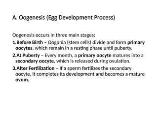
A. Oogenesis (EggDevelopment Process)
Oogenesis occurs in three main stages:
1.Before Birth – Oogonia (stem cells) divide and form primary
oocytes, which remain in a resting phase until puberty.
2.At Puberty – Every month, a primary oocyte matures into a
secondary oocyte, which is released during ovulation.
3.After Fertilization – If a sperm fertilizes the secondary
oocyte, it completes its development and becomes a mature
ovum.
17.
B. Ovulation
• Occursmid-cycle (around day 14) in a 28-day menstrual
cycle.
• A mature follicle ruptures and releases a secondary oocyte
into the fallopian tube.
• If fertilization does not occur, the oocyte degenerates within
24 hours.
External Male ReproductiveOrgans
• Penis
• Glans Penis
• Shaft
• Prepuce (Foreskin)
• Scrotum
• Contains the testicles and regulates temperature for
sperm production.
20.
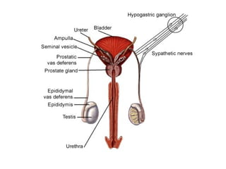
Internal Male ReproductiveOrgans
A. Primary Reproductive Organs (Gonads)
• Testes (Testicles)
B. Accessory Glands
• Seminal Vesicles
• Prostate Gland
• Bulbourethral (Cowper’s) Glands
C. Duct System (Sperm Transport Pathway)
• Epididymis
• Vas Deferens (Ductus Deferens)
• Ejaculatory Duct
• Urethra
21.
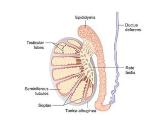
1. Testes (Testicles)
•The testes are the primary reproductive organs (gonads) of the
male. They are located inside the scrotum and are responsible for:
• Sperm production (spermatogenesis)
• Testosterone secretion
• Structure of the Testes
• Each testis is made up of:
• Seminiferous Tubules – Coiled structures where sperm are produced.
• Sertoli Cells – Support and nourish developing sperm.
• Leydig Cells – Produce testosterone, which regulates sperm
production and secondary sexual characteristics.
24.
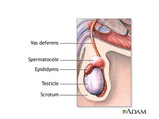
2. Epididymis
• Along, coiled tube located on the back of each testis, where sperm
mature and gain the ability to swim.
• Head of Epididymis – Collects sperm from the testes.
• Body of Epididymis – Site of sperm maturation.
• Tail of Epididymis – Stores mature sperm until ejaculation.
25.
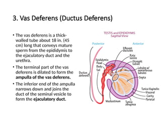
3. Vas Deferens(Ductus Deferens)
• The vas deferens is a thick-
walled tube about 18 in. (45
cm) long that conveys mature
sperm from the epididymis to
the ejaculatory duct and the
urethra.
• The terminal part of the vas
deferens is dilated to form the
ampulla of the vas deferens.
• The inferior end of the ampulla
narrows down and joins the
duct of the seminal vesicle to
form the ejaculatory duct.
26.
4. Ejaculatory Duct
•Formed by the fusion of the vas deferens and the seminal vesicle
duct.
• Passes through the prostate gland and opens into the urethra.
• Transports semen (sperm + fluids) during ejaculation.
27.
5. Urethra
• Ashared passage for urine and semen, extending from the bladder
to the tip of the penis.
• Prostatic Urethra – Passes through the prostate gland.
• Membranous Urethra – Passes through the pelvic floor muscles.
• Penile (Spongy) Urethra – Passes through the penis and ends at the
external opening.
28.
6. Accessory Glands(Semen Production)
A. Seminal Vesicles
• Paired glands located behind the bladder.
• Produce seminal fluid rich in fructose, which nourishes
sperm.
• Secretes prostaglandins, which help sperm move through
the female reproductive tract.
B. Prostate Gland
• A walnut-sized gland located below the bladder.
• Produces prostatic fluid, which makes semen alkaline to
neutralize acidic vaginal pH.
• Enhances sperm motility and survival.
29.
6. Accessory Glands(Semen Production)
C. Bulbourethral (Cowper’s) Glands
• Small, pea-sized glands near the base of the penis.
• Secretes pre-ejaculate fluid to lubricate the urethra and clear
residual urine before ejaculation.
31.
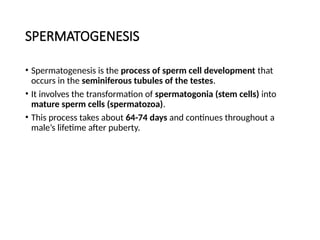
SPERMATOGENESIS
• Spermatogenesis isthe process of sperm cell development that
occurs in the seminiferous tubules of the testes.
• It involves the transformation of spermatogonia (stem cells) into
mature sperm cells (spermatozoa).
• This process takes about 64-74 days and continues throughout a
male’s lifetime after puberty.
32.
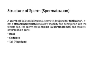
Structure of Sperm(Spermatozoon)
A sperm cell is a specialized male gamete designed for fertilization. It
has a streamlined structure to allow mobility and penetration into the
female egg. The sperm cell is haploid (23 chromosomes) and consists
of three main parts:
• Head
• Midpiece
• Tail (Flagellum)


![Reproductive%20 System[1]](https://cdn.slidesharecdn.com/ss_thumbnails/reproductive20system1-1220708198883512-8-thumbnail.jpg?width=640&height=640&fit=bounds)


