Downloaded 234 times


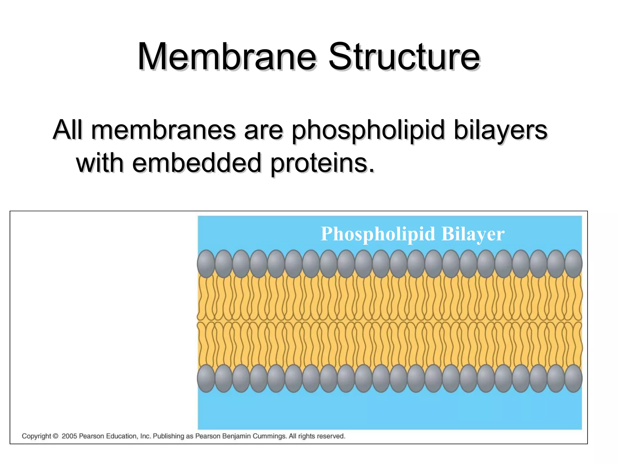


The document discusses cell membrane structure and function. It describes the fluid mosaic model of the plasma membrane, which is composed of a phospholipid bilayer with embedded proteins. Membranes organize cellular chemical activities and form boundaries that exhibit selective permeability. Membranes also compartmentalize reactions through internal organelles. Transport across membranes can occur passively through diffusion or facilitated diffusion, or actively through proteins that require energy.