Downloaded 599 times
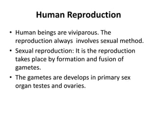
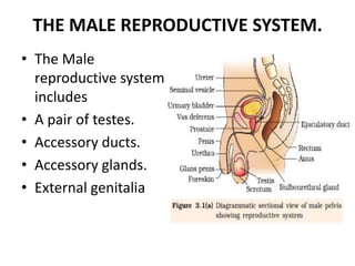
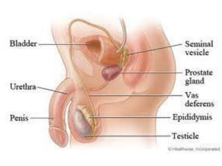
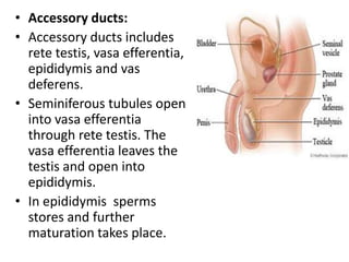
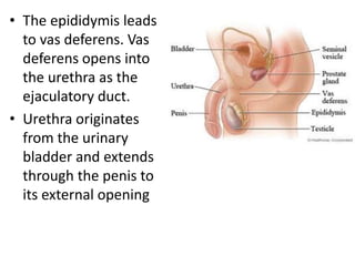
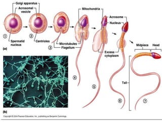
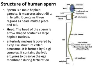
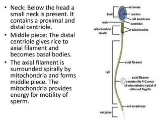
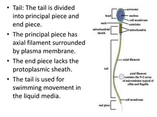

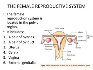
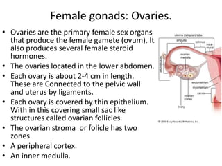
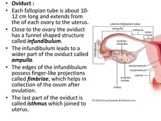
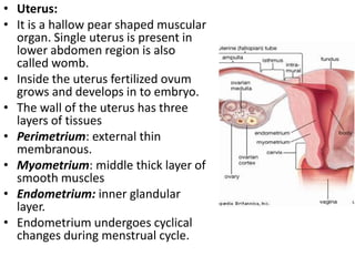
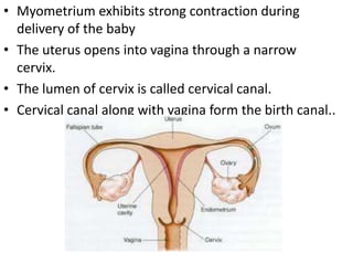
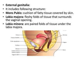
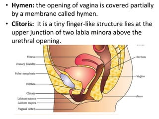
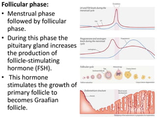
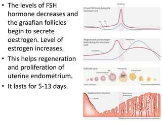
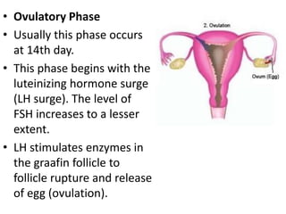
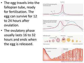
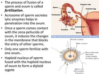


The document summarizes human reproduction. It describes that human reproduction involves sexual reproduction through the formation and fusion of male and female gametes in the testes and ovaries. It then provides details on the male reproductive system including the testes, accessory ducts, glands, and external genitalia. Spermatogenesis and sperm structure are explained. The female reproductive system including ovaries, oviducts, uterus, cervix, vagina and external genitalia are outlined. The menstrual cycle and mammary glands are also summarized.