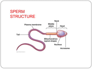
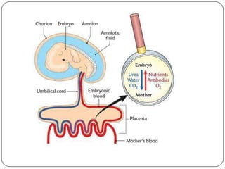
Chapter-3
NCERT
By- Punya Mohanty
Class –XII
HUMAN REPRODUCTION
THE MALE REPRODUCTIVE SYSTEM.
 Located in the pelvis
region.
 Male reproductive system
includes
 A pair of testes.
 Accessory ducts.
 Accessory glands.
 External genitalia
Testes:
 Located outside the abdominal cavity within a pouch called scrotum.
 Scrotum provides low temperature required for spermatogenesis.
 Each testis is about 4 to 5 cm length and 2 to 3 cm width.
 Each testis has about 250 compartments called testicular lobules.
 Each lobule contains one to three seminiferous tubules.
 Seminiferous tubules lined by male germ cells and Sertoli cells.
 Male germ cell undergoes meiosis and produce sperm.
 Sertoli cells provide nutrition to the germ cell and the sperm.
 In between the seminiferous tubule there is interstitial cell or Leydig
cell.
 Leydig cells produce testicular hormones
called androgen (testosterone).
Accessory ducts:
 Includes rete testis, vasa efferentia, epididymis and vas
deferens.
 Seminiferous tubules open into vasa efferentia through rete
testis.
 The vasa efferentia leaves the testis and open into epididymis.
 The epididymis leads to vas deferens that ascends to the
abdomen through inguinal canal and loops over the urinary
bladder.
 Vas deferens receives a duct from seminal vesicle and opens
into the urethra as the ejaculatory duct.
 Urethra originates from the urinary bladder and extends
through the penis to its external opening calledurethral
meatus.
Accessory glands:
 Includes
 Paired seminal vesicle
 A prostate gland
 Paired bulbourethral gland.
 Secretion of these glands constitutes the seminal
plasma.
 Seminal plasma rich in fructose, calcium, and
certain enzyme.
 Secretion of bulbo-urethral glands helps in
lubrication of penis.
External genitalia:
 Penis is the external genitalia.
 It is made of special tissue that helps in erection
of the penis to facilitate insemination.
 The enlarged end of penis is called glans penis.
 Glans penis is covered by a loose fold of skin
called foreskin.
THE FEMALE REPRODUCTIVE
SYSTEM
 Located in the pelvic region
of the
female.
 The female reproductive
system includes:
 A pair of ovaries
 A pair of oviduct.
 Uterus
 Cervix
 Vagina
 External genitalia.
 A pair of mammary gland
Ovaries:
 It is the primary female sex organs that produce
the female gamete (ovum).
 It also produces several steroid hormones.
 The ovaries located in the lower abdomen.
 Each ovary is about 2-4 cm in length.
 Connected to the pelvic wall and uterus by
ligaments.
 Each ovary is covered by thin epithelium which
encloses the ovarian stroma
 The ovarian stroma has two zones
 A peripheral cortex.
 An inner medulla.
Oviduct:
 Oviducts, uterus and vagina constitute the female
accessory ducts.
 Each fallopian tube is about 10-12 cm long and extends
from the
periphery of each ovary to the uterus.
 Close to the ovary the oviduct has a funnel shaped
structure called infundibulum?
 The edges of the infundibulum possess finger-like
projections called fimbriae, which helps in collection
of the ovum after ovulation.
 The infundibulum leads to a wider part of the
oviduct called ampulla.
 The last part of the oviduct is called isthmus which
joined to uterus.
Uterus:
 It is single and is called womb.
 It is inverted pear shaped.
 Attached the pelvic wall by ligaments.
 The uterus opens into vagina through a narrow cervix.
 The lumen of cervix is called cervical canal.
 Cervical canal along with vagina form the birth canal.
 The wall of the uterus has three layers of tissues
 Perimetrium: external thin membranous.
 Myometrium: middle thick layer of smooth muscles
 Endometrium: inner glandular layer.
 Endometrium undergoes cyclical changes during
menstrual cycle.
 Myometrium exhibits strong contraction during delivery of
the baby.
External genitalia:
 It includes following structure:
 Mons Pubis: cushion of fatty covered by skin and
pubic hair.
 Labia majora: fleshy folds of tissue which extends
down from the mons pubis and surrounds the
vaginal opening.
 Labia minora: are paired folds of tissue under
the labia majora.
 Hymen: the opening of vagina is often covered
partially by a membrane called hymen.
 Clitoris: a tiny finger-like structure lies at the upper
junction of two labia minora above the urethral
opening.
Mammary glands:
 Mammary gland consists of glandular tissue and fat.
 Glandular tissue of each breast divided into 15-20
mammary
lobes.
 Mammary lobes contain cluster of cells called
alveoli.
 The cells of alveoli secrete milk, stored in the lumen
of alveoli.
 The alveoli open into mammary tubules.
 The tubules of each lobe join to form a mammary
duct.
 Several mammary ducts join to form a wider
mammary ampulla.
 Mammary ampulla connected to lactiferous duct,
GAMETOGENESIS: (formation of
gametes)
 Spermatogenesis:
 Formation of sperm from the germ cell in the testes is spermatogenesis.
 The process begins at puberty.
 Spermatogonia present in the lining of seminiferous tubules undergo
mitotic division to increase their number.
 Each spermatogonium is diploid (2n) which contain 46 chromosomes.
 Innermost layer of spermatogonial becomes larger called primary
spermatocyte.
 Primary spermatocyte undergoes meiosis-I to form two equal haploid (n)
secondary
spermatocytes (n).
 Each secondary spermatocyte undergoes meiosis-II to
form two equal, haploid spermatids.
 Each primary spermatocyte produces four spermatids.
 Spermatids transformed into spermatozoa (sperms) by
the process called spermiogenesis.
 The sperm head embedded in the Sertoli cell.
SPERMATOGENESIS PROCESS
Hormonal control of
spermatogenesis:
 This process is initiated at puberty due to
secretion of gonadotrophins releasing
hormone (GnRH)
 GnRH secreted form hypothalamus and
stimulate anterior
pituitary to secrete two gonadotrophins.
 Luteinizing hormone (LH) and
 Follicle stimulating Hormone (FSH)
 LH acts on Leydig cells and stimulates synthesis
of
androgens.
 Androgen stimulates spermatogenesis.
 FSH acts on Sertoli cells and stimulates
spermatogenesis in other ways.
Structure of sperm:
 Ultrastructure of sperm consists of a head, neck, a middle piece
and a tail.
 Whole body of sperm surrounded by plasma membrane.
 The sperm head contain an elongated haploid nucleus.
 Above the nucleus a cap like structure present called acrosome.
 The acrosome contains enzymes which help in fertilization of
ovum.
 The middle piece contains mitochondria, which provide
energy for movement of tail that facilitate sperm motility.
 Human male ejaculates 200-300 million sperms during coitus.
 60 percent must have normal shape and size and 40 percent of
them must show
vigorous motility.
 Sperm released from seminiferous tubules enters into accessory
ducts.
 On their way fluids from seminal vesicle and prostate gland added
which
SPERM
STRUCTURE
Oogenesis:
 Formation of a mature female gamete or ovum is calledoogenesis.
 Oogenesis starts during embryonic stage, 25th week of the fetal age.
 Germinal epithelium of ovary divided mitotically to produce millions of gamete
mother cell or oogonia.
 No oogonia formed or added after birth.
 Oogonia enters into meiosis-I and proceeds uptodiakinesis of Prophase-I and get
suspended, at this stage calledprimary Oocytes.
 Each primary oocyte surrounded by layers of granulose cells and then
called primary follicle.
 At puberty only 60,000 to 80,000 primary oocytes are left in each ovary.
 After puberty primary follicles get surrounded by more layers of granulosa cells and
a new theca to formsecondary follicles.
 The secondary follicle transformed into tertiary follicle, characterized by a fluid
filled cavity calledantrum.
 The theca layers organized into an inner theca interna and outer theca externa.
 During the growth of primary follicle into tertiary follicle during puberty, the
primary oocyte restarts its first meiotic division and completes it within tertiary
follicle resulting two unequal haploid cells.
 Large haploid cell is called secondary oocyte.
 A tiny cell called first polar body.
CONT
…. The secondary oocyte retains bulk of the nutrient rich cytoplasm of
primary oocyte.
 The tertiary follicle having secondary oocyte further changes
into Graafian follicle.
 The secondary oocyte surrounded by a new
membrane, zonapellucida.
 The secondary oocyte undergoes second meiotic division continued
upto metaphase-II and get suspended until entry of sperm.
 At this stage Graafian follicle releases secondary oocyte from the
ovary by the process called ovulation.
 On entry of a sperm into the secondary oocytes stimulates it to
complete meiosis-II and there is formation of a haploid ovum and
a second polar body (n).
Menstrual cycle:
 Reproductive cycle of female primates is
called menstrual cycle.
 The first menstruation begins at puberty is
called Menarche.
 Menstrual cycle repeated at an average interval of
28/29 days.
 One ovum is released in the middle of each
menstrual cycle.
Menstrual cycle has following phases:
 Menstrual phase:
 1st phase of menstrual cycle.
 Menstrual flow occurs.
 Lasts for 3-5 days.
 Breakdown of endometrial lining and blood vessel.
 Mucus and blood comes out through vagina.
 It occurs only when ovum released but no
fertilization.
 Lack of menstruation is the indication of pregnancy.
Follicular phase:
 Menstrual phase followed by follicular phase.
 Primary follicle becomes Graafian follicle.
 Regeneration and proliferation of uterine
endometrium.
 LH and FSH level increases gradually in follicular
phase.
 Level of estrogen increases as it is secreted from
growing follicle.
 It lasts for 5-13 days.
Ovulatory phase:
 FSH and LH attain peak level in this period (14th
day).
 This is called LH surge, which induces rupture of
Graafian follicle and release of ovum from the ovary
called ovulation.
Luteal phase:
 Remaining part of Graafian follicle transformed into corpus
luteum.
 Coupusluteum produces large amount of progesterone.
 Progesterone maintains the uterine endometrium, and
prepares it for implantation.
 Thickness of uterine endometrium increase in many folds, due
to proliferation.
 If there is fertilization, corpus luteum grows further and
pregnancy continued, menstrual cycle stopped.
 In the absence of fertilization corpus luteum degenerates.
 Disintegration of endometrium leading to menstruation.
 Menstrual cycle ceases around 50 years of age,
called menopause.
FERTILIZATION AND
IMPLANTATION:
 During copulation (coitus) semen is released by the
penis into the vagina is called insemination.
 The motile sperm swim rapidly, pass through cervix,
uterus and finally reach the junction of isthmus and
ampulla(ammpullary-isthmic junction).
 The ovum released from the ovary also transported to
ampullary isthmic junction where fertilization takes
place.
 Fertilization only takes place if both sperm and ovum
reach ampullary – isthmic junction simultaneously.
 The process of fusion of a sperm and ovum is
called fertilization.
 Acrosome of sperm secretes enzymes helps in
penetration into the ovum.
CONT
… Once a sperm comes contact with the zonapellucida of
ovum and induces the changes in the membrane that
blocks the entry of additional sperms.
 That ensures monospermy and prevents polyspermy.
 Only one sperm fertilize with one ovum.
 Entry of sperm into the ovum induces the ovum to
complete its second meiotic division of secondary
oocyte.
 Meiosis-II is also unequal cytokinesis resulting
production of one large ovum (ootid) and one
small second polar body.
 Haploid nucleus of sperm fused with the haploid
nucleus of ovum to form a diploid zygote.
 The zygote undergoes a series of mitotic cell divisions
called cleavage.
 The stages of development are:
Fertilized ovum (zygote)
2-cell stage
4-cell stage
8-cell stage
Morula
Blastula
 Gastrula
Sex determination:
 Sex of a baby has been decided during fertilization and in the
zygote.
 Sex is determined by the sex-chromosomes present in
gametes.
 Human female contain two XX chromosomes.
 Human male contain XY chromosomes.
 All the female gametes produced with only ǮXǯchromosome.
 Sperms produced by male, 50% with ǮXǯand 50 % with ǮYǯ
chromosome.
 After fertilization zygote either carries XX or XY
chromosomes.
 Zygote with XX chromosomes develop into female and with
XY chromosome develops into male
Cleavage:
 Repeated mitotic division of the zygote without growth resulting a
multicellular ball like embryo is calledcleavage.
 Cleavage starts soon after fertilization.
 Daughter cells produced during cleavage are called blastomeres.
 The product of cleavage is called Morula, which is 8 to 16 celled.
 The morula continues to divide and grow and transformed
into blastocyst.
 The blastomeres in blastocyst arranged into an outer layer
called trophoblast and an inner mass of cells attached to trophoblast
called inner cell mass.
 Trophoblast cells attached to the endometrium helps development of
placenta.
 Inner cell mass gets differentiated into the embryo.
 After attachment the uterine cells divide rapidly and cover the
blastocyst.
 Blastocyst completely embedded in the uterine endometrium. This is
called implantation.
Pregnancy and embryonic development:
 After implantation, finger like projections appears
on the trophoblast called chorionic villi.
 Chorionic villi surrounded by uterine tissue and
maternal blood.
 Temporary association between the fetal tissue
(chorionic villi) and maternal tissue (uterine
endometrium) is called placenta.
Function of placenta:
 The embryo connected to the placenta by umbilical
cord, which transports substances to and from the
embryo.
 Facilitate transport of oxygen and nutrient from mother
to embryo.
 Removes CO2 and waste material from the embryo.
 Acts as endocrine gland and produces several hormones
like:
 Human chorionic gonadotrophins (hCG)
 Human placental lactogen (hPL)
 Estrogen.
 Progesterone
 Relaxin produced from the ovary in the later stage of
pregnancy.
Embryonic development:
 After implantation the inner cell mass of blastocyst
differentiated into an outer layer
called ectoderm and an inner layer
called endoderm.
 Mesoderm differentiated in-between ectoderm and
endoderm.
 The inner cell mass thus called stem cells, having
potency to produce all types of cell, tissues and
organs by differentiation.
Organogenesis:
 Formation of different organs in the embryo is
called organogenesis.
 Human pregnancy lasts for 9 months.
 After one month of pregnancy heart is formed in the embryo.
 By the end of 2nd month the foetus develops limbs and digits.
 By the end of 12 weeks (first trimester) most of organ system is
formed (limbs and external genitalia are well developed).
 First movement of foetus and appearance of hairs observed in 5th
month.
 By the end of 24th week (2nd trimesters) the body is covered with
fine hairs, eye-lids separate, and eyelashes are formed.
 By the end of 9 months the foetus is fully developed and is ready for
delivery.
PARTURITION AND LACTATION:
 The period of pregnancy is called gestation period. (9 months).
 Ejection or expulsion or delivery of foetus is called parturition.
 Parturition is due to vigorous contraction of uterine Myometrium.
 The signal of parturition is originated from the fully developed
foetus and the placenta which induces mild contraction of uterus
called fetal ejection reflex.
 Fetal ejection reflex triggers the release of Oxytocin from pituitary.
 Oxytocin induces stronger contraction of uterine endometrium.
 Stimulatory reflex continues stronger contraction leads to
expulsion.
 After delivery the placenta is also expelled out of the uterus.
Lactation:
 The mammary gland of the female more
differentiated during pregnancy,
 Mammary gland starts producing milk towards the
end of the pregnancy.
 Process of milk production in mammary gland is
called lactation.
 Milk produced during initial days of lactation is
called colostrum.
 Colostrum contains several antibodies which
provide immunity to the new born baby.
Thanking
you

Human Reproduction

  • 1.
    Chapter-3 NCERT By- Punya Mohanty Class–XII HUMAN REPRODUCTION
  • 2.
    THE MALE REPRODUCTIVESYSTEM.  Located in the pelvis region.  Male reproductive system includes  A pair of testes.  Accessory ducts.  Accessory glands.  External genitalia
  • 3.
    Testes:  Located outsidethe abdominal cavity within a pouch called scrotum.  Scrotum provides low temperature required for spermatogenesis.  Each testis is about 4 to 5 cm length and 2 to 3 cm width.  Each testis has about 250 compartments called testicular lobules.  Each lobule contains one to three seminiferous tubules.  Seminiferous tubules lined by male germ cells and Sertoli cells.  Male germ cell undergoes meiosis and produce sperm.  Sertoli cells provide nutrition to the germ cell and the sperm.  In between the seminiferous tubule there is interstitial cell or Leydig cell.  Leydig cells produce testicular hormones called androgen (testosterone).
  • 4.
    Accessory ducts:  Includesrete testis, vasa efferentia, epididymis and vas deferens.  Seminiferous tubules open into vasa efferentia through rete testis.  The vasa efferentia leaves the testis and open into epididymis.  The epididymis leads to vas deferens that ascends to the abdomen through inguinal canal and loops over the urinary bladder.  Vas deferens receives a duct from seminal vesicle and opens into the urethra as the ejaculatory duct.  Urethra originates from the urinary bladder and extends through the penis to its external opening calledurethral meatus.
  • 5.
    Accessory glands:  Includes Paired seminal vesicle  A prostate gland  Paired bulbourethral gland.  Secretion of these glands constitutes the seminal plasma.  Seminal plasma rich in fructose, calcium, and certain enzyme.  Secretion of bulbo-urethral glands helps in lubrication of penis.
  • 6.
    External genitalia:  Penisis the external genitalia.  It is made of special tissue that helps in erection of the penis to facilitate insemination.  The enlarged end of penis is called glans penis.  Glans penis is covered by a loose fold of skin called foreskin.
  • 7.
    THE FEMALE REPRODUCTIVE SYSTEM Located in the pelvic region of the female.  The female reproductive system includes:  A pair of ovaries  A pair of oviduct.  Uterus  Cervix  Vagina  External genitalia.  A pair of mammary gland
  • 8.
    Ovaries:  It isthe primary female sex organs that produce the female gamete (ovum).  It also produces several steroid hormones.  The ovaries located in the lower abdomen.  Each ovary is about 2-4 cm in length.  Connected to the pelvic wall and uterus by ligaments.  Each ovary is covered by thin epithelium which encloses the ovarian stroma  The ovarian stroma has two zones  A peripheral cortex.  An inner medulla.
  • 9.
    Oviduct:  Oviducts, uterusand vagina constitute the female accessory ducts.  Each fallopian tube is about 10-12 cm long and extends from the periphery of each ovary to the uterus.  Close to the ovary the oviduct has a funnel shaped structure called infundibulum?  The edges of the infundibulum possess finger-like projections called fimbriae, which helps in collection of the ovum after ovulation.  The infundibulum leads to a wider part of the oviduct called ampulla.  The last part of the oviduct is called isthmus which joined to uterus.
  • 10.
    Uterus:  It issingle and is called womb.  It is inverted pear shaped.  Attached the pelvic wall by ligaments.  The uterus opens into vagina through a narrow cervix.  The lumen of cervix is called cervical canal.  Cervical canal along with vagina form the birth canal.  The wall of the uterus has three layers of tissues  Perimetrium: external thin membranous.  Myometrium: middle thick layer of smooth muscles  Endometrium: inner glandular layer.  Endometrium undergoes cyclical changes during menstrual cycle.  Myometrium exhibits strong contraction during delivery of the baby.
  • 11.
    External genitalia:  Itincludes following structure:  Mons Pubis: cushion of fatty covered by skin and pubic hair.  Labia majora: fleshy folds of tissue which extends down from the mons pubis and surrounds the vaginal opening.  Labia minora: are paired folds of tissue under the labia majora.  Hymen: the opening of vagina is often covered partially by a membrane called hymen.  Clitoris: a tiny finger-like structure lies at the upper junction of two labia minora above the urethral opening.
  • 12.
    Mammary glands:  Mammarygland consists of glandular tissue and fat.  Glandular tissue of each breast divided into 15-20 mammary lobes.  Mammary lobes contain cluster of cells called alveoli.  The cells of alveoli secrete milk, stored in the lumen of alveoli.  The alveoli open into mammary tubules.  The tubules of each lobe join to form a mammary duct.  Several mammary ducts join to form a wider mammary ampulla.  Mammary ampulla connected to lactiferous duct,
  • 13.
    GAMETOGENESIS: (formation of gametes) Spermatogenesis:  Formation of sperm from the germ cell in the testes is spermatogenesis.  The process begins at puberty.  Spermatogonia present in the lining of seminiferous tubules undergo mitotic division to increase their number.  Each spermatogonium is diploid (2n) which contain 46 chromosomes.  Innermost layer of spermatogonial becomes larger called primary spermatocyte.  Primary spermatocyte undergoes meiosis-I to form two equal haploid (n) secondary spermatocytes (n).  Each secondary spermatocyte undergoes meiosis-II to form two equal, haploid spermatids.  Each primary spermatocyte produces four spermatids.  Spermatids transformed into spermatozoa (sperms) by the process called spermiogenesis.  The sperm head embedded in the Sertoli cell.
  • 14.
  • 15.
    Hormonal control of spermatogenesis: This process is initiated at puberty due to secretion of gonadotrophins releasing hormone (GnRH)  GnRH secreted form hypothalamus and stimulate anterior pituitary to secrete two gonadotrophins.  Luteinizing hormone (LH) and  Follicle stimulating Hormone (FSH)  LH acts on Leydig cells and stimulates synthesis of androgens.  Androgen stimulates spermatogenesis.  FSH acts on Sertoli cells and stimulates spermatogenesis in other ways.
  • 16.
    Structure of sperm: Ultrastructure of sperm consists of a head, neck, a middle piece and a tail.  Whole body of sperm surrounded by plasma membrane.  The sperm head contain an elongated haploid nucleus.  Above the nucleus a cap like structure present called acrosome.  The acrosome contains enzymes which help in fertilization of ovum.  The middle piece contains mitochondria, which provide energy for movement of tail that facilitate sperm motility.  Human male ejaculates 200-300 million sperms during coitus.  60 percent must have normal shape and size and 40 percent of them must show vigorous motility.  Sperm released from seminiferous tubules enters into accessory ducts.  On their way fluids from seminal vesicle and prostate gland added which
  • 17.
  • 18.
    Oogenesis:  Formation ofa mature female gamete or ovum is calledoogenesis.  Oogenesis starts during embryonic stage, 25th week of the fetal age.  Germinal epithelium of ovary divided mitotically to produce millions of gamete mother cell or oogonia.  No oogonia formed or added after birth.  Oogonia enters into meiosis-I and proceeds uptodiakinesis of Prophase-I and get suspended, at this stage calledprimary Oocytes.  Each primary oocyte surrounded by layers of granulose cells and then called primary follicle.  At puberty only 60,000 to 80,000 primary oocytes are left in each ovary.  After puberty primary follicles get surrounded by more layers of granulosa cells and a new theca to formsecondary follicles.  The secondary follicle transformed into tertiary follicle, characterized by a fluid filled cavity calledantrum.  The theca layers organized into an inner theca interna and outer theca externa.  During the growth of primary follicle into tertiary follicle during puberty, the primary oocyte restarts its first meiotic division and completes it within tertiary follicle resulting two unequal haploid cells.  Large haploid cell is called secondary oocyte.  A tiny cell called first polar body.
  • 19.
    CONT …. The secondaryoocyte retains bulk of the nutrient rich cytoplasm of primary oocyte.  The tertiary follicle having secondary oocyte further changes into Graafian follicle.  The secondary oocyte surrounded by a new membrane, zonapellucida.  The secondary oocyte undergoes second meiotic division continued upto metaphase-II and get suspended until entry of sperm.  At this stage Graafian follicle releases secondary oocyte from the ovary by the process called ovulation.  On entry of a sperm into the secondary oocytes stimulates it to complete meiosis-II and there is formation of a haploid ovum and a second polar body (n).
  • 20.
    Menstrual cycle:  Reproductivecycle of female primates is called menstrual cycle.  The first menstruation begins at puberty is called Menarche.  Menstrual cycle repeated at an average interval of 28/29 days.  One ovum is released in the middle of each menstrual cycle.
  • 21.
    Menstrual cycle hasfollowing phases:  Menstrual phase:  1st phase of menstrual cycle.  Menstrual flow occurs.  Lasts for 3-5 days.  Breakdown of endometrial lining and blood vessel.  Mucus and blood comes out through vagina.  It occurs only when ovum released but no fertilization.  Lack of menstruation is the indication of pregnancy.
  • 22.
    Follicular phase:  Menstrualphase followed by follicular phase.  Primary follicle becomes Graafian follicle.  Regeneration and proliferation of uterine endometrium.  LH and FSH level increases gradually in follicular phase.  Level of estrogen increases as it is secreted from growing follicle.  It lasts for 5-13 days.
  • 23.
    Ovulatory phase:  FSHand LH attain peak level in this period (14th day).  This is called LH surge, which induces rupture of Graafian follicle and release of ovum from the ovary called ovulation.
  • 24.
    Luteal phase:  Remainingpart of Graafian follicle transformed into corpus luteum.  Coupusluteum produces large amount of progesterone.  Progesterone maintains the uterine endometrium, and prepares it for implantation.  Thickness of uterine endometrium increase in many folds, due to proliferation.  If there is fertilization, corpus luteum grows further and pregnancy continued, menstrual cycle stopped.  In the absence of fertilization corpus luteum degenerates.  Disintegration of endometrium leading to menstruation.  Menstrual cycle ceases around 50 years of age, called menopause.
  • 26.
    FERTILIZATION AND IMPLANTATION:  Duringcopulation (coitus) semen is released by the penis into the vagina is called insemination.  The motile sperm swim rapidly, pass through cervix, uterus and finally reach the junction of isthmus and ampulla(ammpullary-isthmic junction).  The ovum released from the ovary also transported to ampullary isthmic junction where fertilization takes place.  Fertilization only takes place if both sperm and ovum reach ampullary – isthmic junction simultaneously.  The process of fusion of a sperm and ovum is called fertilization.  Acrosome of sperm secretes enzymes helps in penetration into the ovum.
  • 27.
    CONT … Once asperm comes contact with the zonapellucida of ovum and induces the changes in the membrane that blocks the entry of additional sperms.  That ensures monospermy and prevents polyspermy.  Only one sperm fertilize with one ovum.  Entry of sperm into the ovum induces the ovum to complete its second meiotic division of secondary oocyte.  Meiosis-II is also unequal cytokinesis resulting production of one large ovum (ootid) and one small second polar body.  Haploid nucleus of sperm fused with the haploid nucleus of ovum to form a diploid zygote.
  • 30.
     The zygoteundergoes a series of mitotic cell divisions called cleavage.  The stages of development are: Fertilized ovum (zygote) 2-cell stage 4-cell stage 8-cell stage Morula Blastula  Gastrula
  • 33.
    Sex determination:  Sexof a baby has been decided during fertilization and in the zygote.  Sex is determined by the sex-chromosomes present in gametes.  Human female contain two XX chromosomes.  Human male contain XY chromosomes.  All the female gametes produced with only ǮXǯchromosome.  Sperms produced by male, 50% with ǮXǯand 50 % with ǮYǯ chromosome.  After fertilization zygote either carries XX or XY chromosomes.  Zygote with XX chromosomes develop into female and with XY chromosome develops into male
  • 34.
    Cleavage:  Repeated mitoticdivision of the zygote without growth resulting a multicellular ball like embryo is calledcleavage.  Cleavage starts soon after fertilization.  Daughter cells produced during cleavage are called blastomeres.  The product of cleavage is called Morula, which is 8 to 16 celled.  The morula continues to divide and grow and transformed into blastocyst.  The blastomeres in blastocyst arranged into an outer layer called trophoblast and an inner mass of cells attached to trophoblast called inner cell mass.  Trophoblast cells attached to the endometrium helps development of placenta.  Inner cell mass gets differentiated into the embryo.  After attachment the uterine cells divide rapidly and cover the blastocyst.  Blastocyst completely embedded in the uterine endometrium. This is called implantation.
  • 35.
    Pregnancy and embryonicdevelopment:  After implantation, finger like projections appears on the trophoblast called chorionic villi.  Chorionic villi surrounded by uterine tissue and maternal blood.  Temporary association between the fetal tissue (chorionic villi) and maternal tissue (uterine endometrium) is called placenta.
  • 36.
    Function of placenta: The embryo connected to the placenta by umbilical cord, which transports substances to and from the embryo.  Facilitate transport of oxygen and nutrient from mother to embryo.  Removes CO2 and waste material from the embryo.  Acts as endocrine gland and produces several hormones like:  Human chorionic gonadotrophins (hCG)  Human placental lactogen (hPL)  Estrogen.  Progesterone  Relaxin produced from the ovary in the later stage of pregnancy.
  • 38.
    Embryonic development:  Afterimplantation the inner cell mass of blastocyst differentiated into an outer layer called ectoderm and an inner layer called endoderm.  Mesoderm differentiated in-between ectoderm and endoderm.  The inner cell mass thus called stem cells, having potency to produce all types of cell, tissues and organs by differentiation.
  • 39.
    Organogenesis:  Formation ofdifferent organs in the embryo is called organogenesis.  Human pregnancy lasts for 9 months.  After one month of pregnancy heart is formed in the embryo.  By the end of 2nd month the foetus develops limbs and digits.  By the end of 12 weeks (first trimester) most of organ system is formed (limbs and external genitalia are well developed).  First movement of foetus and appearance of hairs observed in 5th month.  By the end of 24th week (2nd trimesters) the body is covered with fine hairs, eye-lids separate, and eyelashes are formed.  By the end of 9 months the foetus is fully developed and is ready for delivery.
  • 42.
    PARTURITION AND LACTATION: The period of pregnancy is called gestation period. (9 months).  Ejection or expulsion or delivery of foetus is called parturition.  Parturition is due to vigorous contraction of uterine Myometrium.  The signal of parturition is originated from the fully developed foetus and the placenta which induces mild contraction of uterus called fetal ejection reflex.  Fetal ejection reflex triggers the release of Oxytocin from pituitary.  Oxytocin induces stronger contraction of uterine endometrium.  Stimulatory reflex continues stronger contraction leads to expulsion.  After delivery the placenta is also expelled out of the uterus.
  • 43.
    Lactation:  The mammarygland of the female more differentiated during pregnancy,  Mammary gland starts producing milk towards the end of the pregnancy.  Process of milk production in mammary gland is called lactation.  Milk produced during initial days of lactation is called colostrum.  Colostrum contains several antibodies which provide immunity to the new born baby.
  • 44.