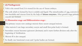
SlideShare a Scribd company logo
STD-12 BIOLOGY
CHAPTER- 3
HUMAN
REPRODUCTION
Sanjay Siddhapura
M.Sc., B. Ed., GSET, GPSC.
INTRODUCTION
• Humans are sexually reproducing and viviparous.
• The reproductive events in humans include formation of gametes (gametogenesis), i.e., sperms
in males and ovum in females.
• Transfer of sperms into the female genital tract (insemination).
• Fusion of male and female gametes (fertilisation) leading to formation of zygote.
• This is followed by formation and development of blastocyst and its attachment to the uterine
wall (implantation).
• Embryonic development (gestation) and delivery of the baby (parturition).
• You have learnt that these reproductive events occur after puberty.
• There are remarkable differences between the reproductive events in the male and in the female,
for example, sperm formation continues even in old men, but formation of ovum ceases in
women around the age of fifty years.
HUMAN REPRODUCTIVE SYSTEM
• Primary sex organs produce gametes and also secrete sex hormones.
• Secondary sex organs are important in reproduction, but do not produce gametes or sex hormones.
• By accessory sex characters both sexes can be distinguished externally but do not directly play any role in
reproduction.
Puberty
• The age of sexual maturity is called
puberty. External or accessory sex
characters first appear in puberty.
• Puberty occurs in girls at the age of
11 to 14 years.
• Puberty occurs in boys at the age of
14 to 16 years. Child
MALE REPRODUCTIVE SYSTEM
• The male reproductive system is located in
the pelvis region.
• Male reproductive system of man consists’
of paired testes, accessory glands, a duct
system, external genitalia and penis.
• The testes are situated outside the
abdominal cavity within a pouch called
scrotum.
• The scrotum helps in maintaining the low
temperature of the testes (2–2.5o C lower
than the normal internal body temperature)
necessary for spermatogenesis.
Testes
• Testes are the primary male sex organs.
• Testis produces spermatozoa and secretes the male sex
hormone testosterone.
• In mammals, the testes are located in the extra-abdominal
scrotal sacs because maturation of sperm needs low
temperature.
• The temperature in scrotum is 2 to 4°C below the
temperature of abdominal cavity.
• In adults, each testis is oval in shape, with a length of
about 4 to 5 cm and a width of about 2 to 3 cm.
• The testis is covered by a dense covering.
• Each testis has about 250 compartments called testicular
lobules.
• Each lobule contains one to three highly coiled
seminiferous tubules in which sperms are produced.
They are approximately 750 seminiferous tubules present.
• Each seminiferous tubule is lined on its inside by two
types of cells called male germ cells (spermatogonia) and
Sertoli cells.
• The male germ cells undergo meiotic divisions finally
leading to sperm formation.
• Sertoli cells provide nutrition to the germ cells.
• The regions outside the seminiferous tubules called
interstitial spaces, contain small blood vessels and
interstitial cells or Leydig cells.
• Leydig cells synthesise and secrete testicular hormones
called androgens. Other immunologically competent cells
are also present.
Rete testis
Vasa efferentia
testicular
• The male sex accessory ducts include rete testis, vasa
efferentia, epididymis and vas deferens.
• All the seminiferous tubules in each testis open into a network
called rete testis leading to fine ductules called vasa efferentia.
• The vasa efferentia leave the testis and open into epididymis
located along the posterior surface of each testis.
• Epididymis
• Several tubes called vasa efferentia arise from rete testis and
conduct sperms into epididymis.
• Epididymis is an irregular, narrow and highly convoluted
tubule found on the inner surface of testis.
• Epididymis has three parts: anterior caput epididymis,
middle corpus epididymis and posterior cauda epididymis.
• Sperms achieve maturity and motility in epididymis.
• Epididymis stores the sperms temporarily.
• Vas deferens
• Vas deferens or sperm duct arises from cauda epididymis,
ascends to abdomen, passes over the urinary bladder and
receive the duct from seminal vesicle behind the urinary
bladder forming ejaculatory duct.
• Ejaculatory duct passes through the prostate to open into
urethra shortly after its origin from urinary bladder.
• The urethra receives rite ducts of prostate and Cowper’s
gland (bulbourethral gland) pass through penis and
opens to outside.
• Seminal vesicles
• Seminal vesicles (Uterus masculinus) are situated at the
junction of vas deferens and prostate gland. They are
narrow, long pouches with muscular tissue on their wall.
• About 70% of the semen is seminal fluid produced by
seminal vesicles.
• Seminal Fluid : Seminal fluid has a pH of about 7.4 and
contains fructose, citrate, ascorbic acid, prostaglandins and
various enzymes.
• The fructose is a source of energy for the spermatozoa.
• Prostate Gland
• Prostate is situated around the first part of the urethra. It contains prostatic utricle and acini and covered by a
capsule.
• Enlargement of prostate results in prostatitis, urination is difficult or impossible.
• Prostatic Fluid: Prostatic secretion forms about 20% of the volume of semen.
• It is slightly acidic, pH 6.5 due to the presence of citric acid.
• Prostatic secretion contains substances important for sperm mobility notably albumin and proteolytic
enzymes fibrinolysin and fibrinogenase.
• Cowper’s glands (bulbourethral glands)
• Cowper’s glands are present in male mammals.
• Cowper’s glands are yellow in colour.
• These are also known as bulbourethral glands, situated beneath bladder and behind the urethra.
• Cowper’s glands open into urethra before entering into penis.
• Cowper’s Fluid : Secretion of cowper’s gland is slightly alkaline and is produced during sexual stimulation.
• Cowper’s gland secretion acts as a lubricant for the glans penis. It also neutralizes any urine in urethra.
• Urethra is a thick-walled muscular duct and it is a
common passage for both urine and semen and called
urinogenital duct.
• Urethra traverse and opens at the tip of the penis as
the male urinogenital aperture.
• Penis
• Penis is the copulatory organ. It is vascular, spongy
and erectile structure.
• It has glans penis at tip covered with a fold of skin
called prepuce.
• Penis contains three longitudinal columns of spongy
erectile tissue which becomes filled with blood during
sexual excitement.
• Surrounding the urinogenital duct is corpus
spongiosum and above it lie two corpora cavernosa.
• Penis serves to transmit sperms into the vaginaof the
female during sexual intercourse.
Urethra
FEMALE REPRODUCTIVE SYSTEM
• Female reproductive system consists of the ovaries, oviducts (fallopian tubes), uterus,
cervix, vagina, accessory genital glands, mammary glands, etc.
Ovary
• The ovary is the primary female sex organ. The ovaries are located one
on each side of the lower abdomen.
• Each ovary is about 2 to 4 cm in length and is connected to
• the pelvic wall and uterus by ligaments.
• Ovary produces ova and secretes female sex hormones viz., estrogens
and progesterone.
• The ovary remains attached to the abdominal wall by a ligament called
mesovarium.
• The ovary is solid, the blood vessels and nerve enter ovary at hilus.
• Ovary is covered by a cubical epithelium called the germinal
epithelium.
• Ovary contains connective tissue, stroma composed of an outer dense
layer called cortex and an inner loose layer known as medulla.
• Cortex contains many small and large, spherical or oval ovarian
follicles. A fully matured follicle is called Graafian follicle.
• Graafian follicle contains a large central follicular cavity
or antrum with a fluid called liquor folliculi.
• The follicle cells lining the cavity are termed membrane
granulosa.
• The oocyte of mammal is enclosed in a perforated
membrane called zone pellucida.
• Next to zona pellucida, cells of inner margin of discus
proligerous (cumulus) present a characteristic radial
arrangement and is termed corona radiate.
• Graafian follicle secretes estrogen in the blood.
• Discharge of a mature ovum from Graafian follicle is
known as ovulation
• After ovulation, a raptured follicle turns into a
yellowish solid mass of cells called corpus luteum
• Corpus luteum secretes the female hormone,
progesterone.
• The oviducts (fallopian tubes), uterus and vagina constitute the female accessory ducts.
• Each fallopian tube is about 10-12 cm long and extends from the periphery of each ovary to the
uterus.
• The part closer to the ovary is the funnel-shaped infundibulum.
• The edges of the infundibulum possess finger-like projections called fimbriae, which help in
collection of the ovum after ovulation.
• The infundibulum leads to a wider part of the oviduct called ampulla.
• The last part of the oviduct, isthmus has a narrow lumen and it joins the uterus.
Uterus
• The uterus is single and it is also called womb.
• The shape of the uterus is like an inverted pear.
• It is supported by ligaments attached to the
pelvic wall.
• The uterus opens into vagina through a narrow
cervix.
• The cavity of the cervix is called cervical canal
which alongwith vagina forms the birth canal.
• The wall of the uterus has three layers of tissue.
The external thin membranous perimetrium,
middle thick layer of smooth muscle,
myometrium and inner glandular layer called
endometrium that lines the uterine cavity.
• The endometrium undergoes cyclical changes
during menstrual cycle while the myometrium
exhibits strong contraction during delivery of the
baby.
Vagina
• Uterus opens into an elastic muscular tube called vagina.
• The vagina is lined by a stratified squamous epithelium with out any glands.
• During reproductive life the vagina contains Lactobacillus acidophilus which keeps the vaginal pH
between 4.9 and 3.5 by producing lactic acid from glycogen.
• Vagina receives semen from male during mating.
• During childbirth, it conveys the child outside.
• Bartholin’s gland: On either side of vaginal orifice is a greater vestibular gland or Bartholin’s
gland.
• Bartholin’s gland occurs in female mammals and helps in vestibular lubrication.
• Bartholin’s gland of female corresponds to bulbourethal gland (Cowper’s gland) of the male).
• The hymen is normally ruptured during female’s first copulation.
• The female external genitalia include mons
pubis, labia majora, labia minora, hymen and
clitoris.
• Mons pubis is a cushion of fatty tissue covered
by skin and pubic hair.
• The labia majora are fleshy folds of tissue,
which extend down from the mons pubis and
surround the vaginal opening.
• The labia minora are paired folds of tissue
under the labia majora.
• The clitoris is a tiny finger-like structure which
lies at the upper junction of the two labia
minora above the urethral opening.
• Hymen : The hymen is a thin mucous
membrane that stretches across the opening of
the vagina.
• The hymen is often torn during the first coitus
(intercourse). Bartholin’s gland
inside the vagina
MAMMARY GLANDS
• A functional mammary gland is characteristic of all
female mammals.
• The mammary glands are paired structures (breasts) that
contain glandular tissue and variable amount of fat.
• The glandular tissue of each breast is divided into 15-20
mammary lobes containing clusters of cells called
alveoli.
• The cells of alveoli secrete milk, which is stored in the
cavities (lumens) of alveoli.
• The alveoli open into mammary tubules.
• The tubules of each lobe join to form a mammary duct.
Several mammary ducts join to form a wider mammary
ampulla which is connected to lactiferous duct through
which milk is sucked out.
GAMETOGENESIS
• The primary sex organs – the testis in the males and the ovaries in the females – produce gametes,
i.e, sperms and ovum, respectively, by the process called gametogenesis.
• Formation of eggs & sperm from germ cells is known as Gametogenesis.
• FSH (Follicle stimulating hormone) of pituitary gland stimulate the gametogenesis.
• Vitamin-E (Fertility vitamin) and vitamin -A are essential for gametogenesis.
• Healthy gametes are produced by vitamin-A.
• Deficiency of vitamin-E causes sterlity.
• Due to deficiency of vitamin-A gametes remain less active.
• Germinal epithelium of gonads have primordial germ cells. These repetedly divides to form
gametes.
• PGC (primordial germ cells) are produced in extragonodial tissue normally placenta.
• Normally gamete formation have 3 steps
• (i) Multiplication stage (ii) Growth stage (iii) Maturation stage
SPERMATOGENESIS
• In most of animals, spermatogenesis occurs in testes.
• In testis, the immature male germ cells (spermatogonia) produce sperms by spermatogenesis
that begins at puberty.
• Mammalian testes contains seminiferous tubules.
• Seminiferous tubules are lined by germinal epithelium internally.
• Some special type cells are found in germinal epithelium known as primordial Germ cells .
• These cells divide & produces premature sperms also known as spermatogonia (sing.
spermatogonium).
• Sertoli cells are also found in germinal epithelium.
• Presence of sertoli cells is the unique character of mammalian testes.
• Sperm are stored and kept inactive in epidydymis.
• Sperm formation have 2 stages (a) Spermatocytosis (b) Spermiogenesis
(a) SPERMATOCYTOSIS
• Formation of spermatid from primordial germ
cells. It have 3 stages.
• (i) Multiplicative stage (ii) Growth stage
(iii) Maturation stage
• (i) Multiplicative stage:
• Primordial cells repeatedly divides and
produces large number of spermatogonia.
• Primordial germ cells (2n - 44xy) regularly
divides and produces spermatogonia.{A type (44
xy)}.
• These A type spermatogonia regularly undergo
mitotic division. So they are also called as stem
spermatogonia. It produces B spermatogonia.
(a) SPERMATOCYTOSIS
• (ii) Growth stage : In this spermatids gain
nutrients from sertoli cells and grows in size.
• B spermatogonia (44 xy) now goes to the sertoli
cells and takes up nutrition after certain limited
growth they are called as primary spermatocyte
(44 xy).
• (iii) Maturation stage : In this primary
spermatocytes undergo meiosis. After meiosis-I
secondary spermatocytes are formed.
• After meiosis-II four haploid spermatids are
formed. Primary spermatocyte divides by meiosis-I
and produces secondry spermatocytes. They are
haploid.
• These are of 2 types on the basis of chromosome
number i.e. 23 + x and 23 + y (both 50% each).
• They further divides by meiosis-II and produces
four spermatids (23 + x, 23 + x, 23 + y, 23 + y).
Multiplicative
stage
Growth
stage
Maturation
stage
Maturation
stage
(b) SPERMIOGENESIS OR SPERMATOLEOSIS
• Formation of sperm from spermatid is called Spermiogenesis. It involves following developments.
• Nucleus of spermatid move to one end, chromatin undergo condensation. Nucleus becomes light
in weight and aquires spindle shape.
• Many golgi vesicle collects above the nucleus. Few vesicles develops a granule inside them, this
granule is known as proacrosomal granules (proacroblast).
• All proacroblasts fuse with each other & form a large vesicle, which is known as acroblast.
Granule of acroblast is called as Acrosomal granules. Acroblast collects on the nucleus.
• Cytoplasm of spermatid moves backward, so plasma membrane shrinks & attach with acroblast &
nucleus and head of sperm is formed.
• Two centrioles of spermatid arrange in perpendicular manner, just below to nucleus. Distal
centriole produces axonema. All mitochondria of spermatid arranged spirally around the
Axonema. This arrangement form the middle part of sperm.
• Axonema elongates & tail of sperm is formed.
Hormonal Regulation of Spermiogenesis
• The hypothalamus releases gonadotropin-releasing hormone (GnRH)
• GnRH stimulates the anterior pituitary to secrete FSH and LH
• FSH causes sustentacular (Sertoli) cells to release androgen-binding protein (ABP)
• LH stimulates interstitial (Leydig) cells to release testosterone
• ABP binding of testosterone enhances spermatogenesis
• Feedback inhibition on the hypothalamus and pituitary results from rising levels of testosterone and
increased production of inhibin.
• Two types of hormones are secreted by sertoli cells-
• (A) AMH (anti mularian hormone) - This hormone inhibits development of female genital organs in
male embryo. Although some vestigeal female genital organs persist in male.
• Eg. (i) Seminal vesicle/uterus masculinus - it is a vestigeal of part oviduct (mularian duct).
• (ii) Prostatic utricle/vagina masculinus - it is a vestigeal part of vagina.
• (B) Inhibin hormone - It inhibits the secretion of FSH, and controls the production of sperms.
• Sertoli cells also produces androgen binding proteins.
Testosterone
• Synthesized from cholesterol
• Necessary for fetal development of male external genitalia
• Increased levels of testosterone at puberty are responsible for further growth of male genitalia and
for the development and maintenance of male secondary sex characteristics
• Stimulates protein synthesis and accounts for the greater muscular development of the male
• Is the basis of libido in both males and females
• Stimulates development of male secondary sexual characteristics including:
• -Appearance of pubic, axillary, and facial hair
• -Enhanced growth of the chest and deepening of the voice
• -Skin thickens and becomes oily
• -Bones grow and increase in density
• -Skeletal muscles increase in size and mass
Structure Of A Sperm
• A plasma membrane envelops the whole body of sperm.
• It is a microscopic structure composed of a head, neck, a middle piece and a tail.
• (1) Head - Head contains two structures (a) Acrosome (b) Nucleus.
• Acrosome is a vesicle like double membranous structure which has hydrolytic enzymes. These
are combinely known as spermlysin.
• These enzymes help in penetrat ion of egg.
• Mammalian acrosome has mainly hyaluronidase & proacrosine enzyme. Both are combinely
known as acrosomine.
• Proacrosine is an inactive enzyme, but when it comes in contact with egg, it converts into active
acrosine.
• These membranes of golgibody left on acrosome cap are called as Galea capitis. A space
between acrosome & nucleus is known as perforatorium.
• (2) Middle piece- Its anterior part is narrow & called as neck of sperm. It has two
centrioles.
• In middle piece many mitochondria are arranged spirally around to axonema. which
produce energy for the movement of tail that facilitate sperm motility essential for
fertilisation.
• This spiral sheath of mitochondria around the axonema is known Nebenkern
sheath.
• Basal part of head & middle piece has a thin layer of cytoplasm which is called as
Manchette.
• (3) Tail - It is a long flagellum, its basal body is distal centriole.
• In humans sperm formation is completed in 74 days.
• The human male in one ejaculation 4 ml semen have about 200 to 300 million
sperms during a coitus of which, for normal fertility, at least 60 per cent sperms must
have normal shape and size and for at least 40 per cent of them must show vigorous
motility.
• If sperm count is less = Oligospermia.
• If there are dead sperms = Necrospermia.
• If there are no sperm = Azoospermia.
• Sperms released from the seminiferous tubules, are transported by the accessory ducts.
• Secretions of epididymis, vas deferens, seminal vesicle and prostate are essential for
maturation and motility of sperms.
• The seminal plasma along with the sperms constitute the semen.
• The functions of male sex accessory ducts and glands are maintained by the testicular
hormones (androgens).
OOGENESIS• Oogenesis is initiated during the embryonic development stage
when a couple of million gamete mother cells (oogonia) are
formed within each fetal ovary; no more oogonia are formed
and added after birth.
• These cells start division and enter into prophase-I of the
meiotic division and get temporarily arrested at that stage, called
primary oocytes.
• Each primary oocyte then gets surrounded by a layer of
granulosa cells and then called the primary follicle (Figure 3.7).
• A large number of these follicles degenerate during the phase
from birth to puberty.
• Therefore, at puberty only 60,000-80,000 primary follicles are
left in each ovary.
• The primary follicles get surrounded by more layers of
granulosa cells and a new theca and called secondary follicles.
• The secondary follicle soon transforms into a tertiary follicle
which is characterised by a fluid filled cavity called antrum.
Secondary
follicle
• The theca layer is organised into an inner theca interna
and an outer theca externa.
• The primary oocyte within the tertiary follicle grows in
size and completes its first meiotic division.
• It is an unequal division resulting in the formation of a
large haploid secondary oocyte and a tiny first polar
body (Figure 3.8b).
• The secondary oocyte retains bulk of the nutrient rich
cytoplasm of the primary oocyte.
• The tertiary follicle further changes into the mature
follicle or Graafian follicle.
• The secondary oocyte forms a new membrane called
zona pellucida surrounding it.
• The Graafian follicle now ruptures to release the
secondary oocyte (ovum) from the ovary by the process
called ovulation.
OOGENESIS (only for information)
• Formation of ova in ovary is called oogenesis.
• It also have similar stages like sperm formation. i.e. (a) Multiplicative, (b) growth,
(c)maturation & differentiation.
• (A) Multiplicative stage :
• Under the influence of FSH the primordial germ cells regularly divides and produces couple
of million number of gamete mother cells (oogonia (2n)).
• This stage is completed in early embryonic stage in most of the mammals in which number
of developing egg cell increases. No more oogonia are formed and added after birth.
• (B) Growth Stage : Many changes occurs during growth phase in oogonia.
• All these changes are divided in two substages by Raven.
• (1) Previtellogenesis (2) Vitellogenesis
• (1) Previtellogenesis
• Karyoplasm increases in oogonia. DNA activity increases & DNA rapidly produces
(transcribes) different types of RNA. Due to these changes nucleus of egg become large in size.
The large nucleus of egg is now known as Germinal vesicle.
• The rate of protein synthesis in cytoplasm increases and different types of proteins and
enzymes are formed.
• Mitochondria, Golgibodies and ER arranges in a ring which surrounds to nucleus. This ring is
• known as Balbiani ring or vitelline ring.
• Vitelline membrane is formed by golgibody. Golgi bodies also produces cortical granules.
These are covered with unit membrane & filled by mucopolysaccharide. At the time of
fertilization cortical granules forms the fertilization membrane.
• A space is present between the plasma membrane and vitelline membrane, it is known as
perivitelline space. A fluid is filled in this space which is known as Perivitelline fluid.
• (2) Vitellogenesis
• Yolk is the stored food. It is stored for the use of future embryo.
• The yolk which is formed by liver is soluble and liquid. The mitochondria of egg, converts it
into insoluble and viscous form by the help of kinase enzymes. After growth stage primary
oocytes are formed.
• C) Maturation stage and Differentiation stage :
• The primary oocyte now undergo meiosis there occurs unequal meiosis.
• After meiosis-I one large secondary oocyte and small first polar body is formed.
• Secondary oocyte becomes quiescent (dormant) and it starts further division only after the
beginning of fertilization.
• Meiosis-II is also unequal.
• So finally one functional ovum and 3 polar bodies are formed.
MENSTRUAL CYCLE
• Menstrual cycle is the cyclic changes in the reproductive tract of primate females e.g.
monkeys, apes and human beings.
• Menstruation is the periodic shedding of the endometrium of the uterus with bleeding.
• In healthy women, menstruation occurs at intervals of about 28 days.
• The first menstruation begins at puberty and is called menarche. Menarche is the starting
of menstruation in girls at about 13 years.
• In human beings, menstrual cycles ceases around 50 years of age; that is termed as
menopause.
• In human females, menstruation is repeated at an average interval of about 28/29 days, and
the cycle of events starting from one menstruation till the next one is called the menstrual
cycle.
• Cyclic menstruation is an indicator of normal reproductive phase and extends between
menarche and menopause.
• One ovum is released (ovulation) during the middle of each menstrual cycle.
• Menstrual cycle consists of three phases-
• (a) Menstrual Phase,
• (b) Proliferative Phase (follicular phase) and
• (c) Secretory Phase (luteal phase).
• (a) Menstrual Phase :- The cycle starts with the
menstrual phase, when menstrual flow occurs and it lasts
for 3-5 days.
• The menstrual flow results due to breakdown of
endometrial lining of the uterus and its blood vessels
which forms liquid that comes out through vagina.
• Menstruation only occurs if the released ouvm is not
fertilised.
• Lack of menstruation may be indicative of pregnancy.
• However, it may also be caused due to some other
underlying causes like stress, poor health etc.
MENSTRUAL CYCLE
Proliferative Phase
(follicular phase)-6 to14
Secretory Phase
(luteal phase).
15-28 days
• (b) Proliferative Phase (follicular phase)
• Proliferative phase (5th to 14th day) consists of growth of endometrium of uterus, fallopian
tube and vagina.
• In ovary, a Graafian follicle grows, matures and secretes estrogen during this phase.
• Endometrium of uterus regenerates through proliferation. These changes in the ovary and the
uterus are induced by changes in the levels of pituitary and ovarian hormones.
• The secretion of gonadotropins (LH and FSH) increases gradually during the follicular phase,
and stimulates follicular development as well as secretion of estrogens by the growing follicles.
• Both LH and FSH attain a peak level in the middle of cycle (about 14th day). Rapid secretion
of LH leading to its maximum level during the mid-cycle called LH surge induces rupture of
Graafian follicle and thereby the release of ovum (ovulation).
MENSTRUAL CYCLE
(c) Secretory Phase (luteal phase 15-28 days)
• The subsequent 14 days in which corpus luteum is active is referred to as the secretory phase.
• The ovulation (ovulatory phase) is followed by the luteal phase during which the remaining parts of
the Graafian follicle transform as the corpus luteum.
• Progesterone secreted by corpus luteum is active during secretory phase, which is essential for
maintenance of the endometrium.
• The uterine endometrium and glands grow further during secretory phase.
• Such an endometrium is necessary for implantation of the fertilized ovum and other events of
pregnancy.
• At the end of secretory phase, corpus Interim degenerates in the ovary, progesterone secretion fails,
the overgrown uterine endometrium breaks down and menstruation takes place.
• During pregnanacy all events of the menstrual cycle stop and there is no menstruation. In the
absence of fertilisation, the corpus luteum degenerates. This causes disintegration of the
endometrium leading to menstruation, marking a new cycle.
ESTROUS CYCLE
• The estrous cycle consists of cyclic changes in the female reproductive system of non-
primate mammals
• There is no menstruation at the end of estrous cycle.
• The estrogen level in blood increases resulting strong sex urge in the female. This is called
“period of heat”.
• The estrous cycles run only during the breeding season.
• The estrous cycles remain suspended in female during non-breeding season.
• The suspension of estrous cycles is called the state of anestrum.
• Those animals that have only a single estrous during the breeding season are called
monoestrous, e.g., Dog, fox, deer, bat, etc.
• The animals that have a recurrence of estrous during breeding season are called
polyestrous, e.g., Mouse, squirrel, cow, sheep, pig, horse, etc.
FERTILISATION AND IMPLANTATION
• During copulation (coitus) semen is released by the penis into the vagina (insemination).
• The phenomenon of sperm activation in female genital tract in mammals is known as
capacitation. It occurs in 5-6 hours. Finally sperms reach ampulla of oviduct & fuses to ovum.
• The motile sperms swim rapidly, pass through the cervix, enter into the uterus and finally reach
the junction of the isthmus and ampulla (ampullary-isthmic junction) of the fallopian tube.
• The ovum released by the ovary is also transported to the ampullary-isthmic junction where
fertilisation takes place.
• Fertilisation can only occur if the ovum and sperms are transported simultaneously to the
ampullaryisthmic junction.
• This is the reason why not all copulations lead to fertilisation and pregnancy.
• The process of fusion of a sperm with an ovum is called fertilisation.
FERTILISATION• The process of fusion of a sperm with an ovum is
called fertilisation.
• During fertilisation, a sperm comes in contact with
the zona pellucida layer of the ovum (Figure 3.10) and
induces changes in the membrane that block the
entry of additional sperms.
• Thus, it ensures that only one sperm can fertilise an
ovum.
• The secretions of the acrosome help the sperm
enter into the cytoplasm of the ovum through the
zona pellucida and the plasma membrane.
• This induces the completion of the meiotic division
of the secondary oocyte.
• The second meiotic division is also unequal and
results in the formation of a second polar body
and a haploid ovum (ootid).
• Soon the haploid nucleus of the sperms and that of
the ovum fuse together to form a diploid zygote.
• (i) Sperms are produced and released in large amount : In humans 100 million
sperm/ml of semen are present. In one ejaculation 3 to 5 ml of semen are introduced
in female body at a time.
• (ii) Size of egg is very large as compaired to sperms : This increase the possiblities
of fusion. The size of egg about 0.1 mm while the sperm of human is of 60 .
• Fertilzin is found on the surface of egg. Antifertilizin are present on the surface of
sperms. It is a type of acidic protein. Fertilizin and antifertilizin are specific for each
species of animals (species specificity).
• By the reaction of both sperm and egg, both combine each other, this reaction is
known as agglutination.
GAMONS
• On the surface of egg and ova some hormones are also present. These hormones are known as
Gamons.
• Androgamones are found on surface of the sperms and Gyanogamones are found on the
surface of eggs.
• Androgamones are of two types –
• (i) Androgamone I – Conserves the energy of sperms, inactivates them.
• (ii) Androgamones II – Dissolves the viscous layers of egg and forms rough surface on the egg.
This helps in attachment.
• Gyanogamones are of two types – Present on egg surface
• (i) Gyanogamones I : When sperm reach to the surface of egg then the Gyanogamones I
neutrilize the androgamone I. Therefore sperm becomes more active.
• (ii) Gyanogamone II : This hormone makes the head of sperm viscous. And viscous sperm
head attaches strongly with rough surface of egg.
MONOSPERMY OR POLYSPERMY
• Generally only one sperm enters into a egg, this phenomenon is known as
Monospermy.
• Rarely more than one sperm enters in one egg, this phenomenon is known as
polyspermy.
• Polyspermy is of two types –
• (a) Pathological Polyspermy – All the nucleus of sperms fuse with nucleus of egg.
Egg degenerate and embryo do not develop.
• (b) Physiological polyspermy – Nucleus of only one sperm fuse with nucleus of the
egg and all the other sperms dies in cytoplasm of egg.
• The dead sperms in cytoplasm of egg are known as “merocyte”. It is commonly
found in polylecithal eggs of Reptiles, Aves, etc.
• The chromosome pattern in the human female is XX and that in the male is XY.
• Therefore, all the haploid gametes produced by the female (ova) have the sex
chromosome X whereas in the male gametes (sperms) the sex chromosome could be
either X or Y.
• 50 per cent of sperms carry the X chromosome while the other 50 per cent carry the Y.
• After fusion of the male and female gametes the zygote would carry either XX or XY
depending on whether the sperm carrying X or Y fertilised the ovum.
• The zygote carrying XX would develop into a female baby and XY would form a male
Sex determination of child
• Initial repeated mitosis divisions in zygote or
fertilized eggs are called as cleavage.
• The daughter cells formed by cleavage are
known as blastomeres.
• There is holoblastic ( Cleavage takes places in
whole egg) cleavage.
• The mitotic division starts as the zygote moves
through the isthmus of the oviduct called
cleavage towards the uterus (Figure 3.11) and
forms 2, 4, 8, 16 daughter cells called
blastomeres.
• The embryo with 8 to 16 blastomeres is called a
morula (Figure 3.11e).
• The morula continues to divide and transforms
into blastocyst (Figure 3.11g) as it moves further
into the uterus.
• The blastomeres in the blastocyst are arranged
into an outer layer called trophoblast and an
inner group of cells attached to trophoblast
called the inner cell mass.
IMPLANTATION
• The trophoblast layer then
gets attached to the
endometrium and the inner
cell mass gets differentiated as
the embryo.
• After attachment, the uterine
cells divide rapidly and covers
the blastocyst. As a result, the
blastocyst becomes embedded
in the endometrium of the
uterus (Figure 3.11h). This is
called implantation and it
leads to pregnancy.
PREGNANCY AND EMBRYONIC DEVELOPMENT
• After implantation, finger-like
projections appear on the trophoblast
called chorionic villi which are
surrounded by the uterine tissue and
maternal blood.
• The chorionic villi and uterine tissue
become interdigitated with each other
and jointly form a structural and
functional unit between developing
embryo (foetus) and maternal body
called placenta (Figure 3.12).
• The placenta facilitate the supply of oxygen and nutrients to the embryo and also removal of
carbon dioxide and excretory/waste materials produced by the embryo. The placenta is
connected to the embryo through an umbilical cord which helps in the transport of substances
to and from the embryo. Placenta also acts as an endocrine tissue and produces several
hormones like human chorionic gonadotropin (hCG), human placental lactogen (hPL),
estrogens, progestogens, etc. hCG, hPL and relaxin are produced in women only during
pregnancy.
• Human chorionic gonadotropin ( hCG) is a hormone produced by the placenta after
implantation. The presence of hCG is detected in some pregnancy tests ( HCG pregnancy strip
tests ).
• hPL, also known as human chorionic somatomammotropin, (a mouthful) is yet another
hormonal helper that prepares your breasts for lactation.
• In the later phase of pregnancy, a hormone called relaxin is also secreted by the ovary.
• Relaxin relaxes the body's muscles, joints, and ligaments. Not surprisingly, the effect centers on
the joints of the pelvis, allowing them to stretch during delivery. It also softens and lengthens
the cervix and helps relax smooth muscles in the uterus and elsewhere throughout the body.
• In addition, during pregnancy the levels of other hormones like estrogens, progestogens,
cortisol, prolactin, thyroxine, etc., are increased several folds in the maternal blood.
• Increased production of these hormones is essential for supporting the fetal growth,
metabolic changes in the mother and maintenance of pregnancy.
• Role of placenta :
• (i) Placenta acts as filter.
• (ii) Soluble inorganic, organic nutrients, hormones, antibodies against diphtheria, small
pox, scarlet fever, measles can pass through it.
• (iii) It helps in respiration, excreation.
• (iv) It produces - Human chorionic gonadotropin (HCG). HCG favours growth of
corpus leuteum.
• (v) Relaxin that helps in parturation by softening connective tissue of pubic symphysis.
• (vi) The umbilical cord connects foetus to placenta.
• Immediately after implantation, the inner cell mass (embryo) differentiates into an outer
layer called ectoderm and an inner layer called endoderm.
• A mesoderm soon appears between the ectoderm and the endoderm.
• These three layers give rise to all tissues (organs) in adults.
• It needs to be mentioned here that the inner cell mass contains certain cells called stem
cells which have the potency to give rise to all the tissues and organs.
• The human pregnancy lasts 9 months.
EMBRYO DEVELOPMENT
• In human beings, after one month of pregnancy, the embryo’s heart is formed.
• The first sign of growing foetus may be noticed by listening to the heart sound carefully
through the stethoscope.
• By the end of the second month of pregnancy, the foetus develops limbs and digits.
• By the end of 12 weeks (first trimester), most of the major organ systems are formed, for
example, the limbs and external genital organs are well-developed.
• The first movements of the foetus and appearance of hair on the head are usually observed
during the fifth month.
• By the end of 24 weeks (second trimester), the body is covered with fine hair, eye-lids
separate, and eyelashes are formed.
• By the end of nine months of pregnancy, the foetus is fully developed and is ready for
delivery.
PARTURITION
• The average duration of human pregnancy is about 9 months which is called the gestation period.
• Vigorous contraction of the uterus at the end of pregnancy causes expulsion/delivery of the foetus.
This process of delivery of the foetus (childbirth) is called parturition.
• Parturition is induced by a complex neuroendocrine mechanism.
• The signals for parturition originate from the fully developed fetus and the placenta which induce mild
uterine contractions called foetal ejection reflex.
• This triggers release of oxytocin from the maternal pituitary.
• Oxytocin acts on the uterine muscle and causes stronger uterine contractions, which in turn stimulates
further secretion of oxytocin.
• The stimulatory reflex between the uterine contraction and oxytocin secretion continues resulting in
stronger and stronger contractions.
• This leads to expulsion of the baby out of the uterus through the birth canal – parturition. Soon after
the infant is delivered, the placenta is also expelled out of the uterus.
LACTATION
• The mammary glands of the female undergo differentiation during pregnancy and starts
producing milk towards the end of pregnancy by the process called lactation.
• This helps the mother in feeding the newborn.
• The milk produced during the initial few days of lactation is called colostrum which contains
several antibodies absolutely essential to develop resistance for the new-born babies.
• It is rich in calories and protein. It have various antibodies. It provides passive immunity to
newborn. Milk secretion is regulated by prolactin high level of oxytosin stimulate prolactin
production.
• Breast-feeding during the initial period of infant growth is recommended by doctors for
bringing up a healthy baby.
• Inhibitory peptide : It is present in milk. If milk remains in mammory glands (breasts) this
accumulated peptide inhibits milk production.
THANK
YOU

More Related Content

What's hot

Oogenesis PART 7
Oogenesis PART 7Oogenesis PART 7
Oogenesis PART 7
AnuSebastian18
 
Oogenesis
OogenesisOogenesis
Oogenesis
Microbiology
 
Fertilization process
Fertilization processFertilization process
Fertilization processUE
 
Spermatogenesis
SpermatogenesisSpermatogenesis
Spermatogenesis
Samchuchoo
 
Spermatogenesis vs. oogenesis_-_germinario
Spermatogenesis vs. oogenesis_-_germinarioSpermatogenesis vs. oogenesis_-_germinario
Spermatogenesis vs. oogenesis_-_germinarioPakLiam
 
Biology form 5 process of oogenesis
Biology form 5 process of oogenesisBiology form 5 process of oogenesis
Biology form 5 process of oogenesissahieyra
 
Spermatogenesis Anju.pptx
Spermatogenesis Anju.pptxSpermatogenesis Anju.pptx
Spermatogenesis Anju.pptx
Anju Kumawat
 
Human reproduction for grade 12
Human reproduction for grade 12Human reproduction for grade 12
Human reproduction for grade 12
University of Johannesburg
 
Embryonic development SlideShare.
Embryonic development  SlideShare.Embryonic development  SlideShare.
Embryonic development SlideShare.
AshwamedhTutorials
 
Spermatogenesis
SpermatogenesisSpermatogenesis
Spermatogenesis
drammarmehdi
 
Evolution of Urinogenital Ducts
Evolution of Urinogenital DuctsEvolution of Urinogenital Ducts
Evolution of Urinogenital Ducts
D. B. S. College Kanpur
 
The foetal membranes
The foetal membranesThe foetal membranes
The foetal membranesraj kumar
 
Oogenesis
OogenesisOogenesis
Oogenesis
drammarmehdi
 
Post fertilization events
Post fertilization eventsPost fertilization events
Post fertilization events
Anushi Jain
 
Female reproductive system 2 (Histology of Ovary, Oogenesis & Follicular Deve...
Female reproductive system 2 (Histology of Ovary, Oogenesis & Follicular Deve...Female reproductive system 2 (Histology of Ovary, Oogenesis & Follicular Deve...
Female reproductive system 2 (Histology of Ovary, Oogenesis & Follicular Deve...
Dr. Sarita Sharma
 
embryology (theory)
embryology (theory)embryology (theory)
Oogenesis
OogenesisOogenesis
Oogenesis
Shubhendra Kumar
 
Fertilization
FertilizationFertilization
Fertilization
Bhorgin Lourdu Mary
 

What's hot (20)

Oogenesis PART 7
Oogenesis PART 7Oogenesis PART 7
Oogenesis PART 7
 
Oogenesis
OogenesisOogenesis
Oogenesis
 
Fertilization process
Fertilization processFertilization process
Fertilization process
 
Spermatogenesis
SpermatogenesisSpermatogenesis
Spermatogenesis
 
Spermatogenesis vs. oogenesis_-_germinario
Spermatogenesis vs. oogenesis_-_germinarioSpermatogenesis vs. oogenesis_-_germinario
Spermatogenesis vs. oogenesis_-_germinario
 
Biology form 5 process of oogenesis
Biology form 5 process of oogenesisBiology form 5 process of oogenesis
Biology form 5 process of oogenesis
 
Spermatogenesis Anju.pptx
Spermatogenesis Anju.pptxSpermatogenesis Anju.pptx
Spermatogenesis Anju.pptx
 
Human reproduction for grade 12
Human reproduction for grade 12Human reproduction for grade 12
Human reproduction for grade 12
 
Embryonic development SlideShare.
Embryonic development  SlideShare.Embryonic development  SlideShare.
Embryonic development SlideShare.
 
Spermatogenesis
SpermatogenesisSpermatogenesis
Spermatogenesis
 
Evolution of Urinogenital Ducts
Evolution of Urinogenital DuctsEvolution of Urinogenital Ducts
Evolution of Urinogenital Ducts
 
The foetal membranes
The foetal membranesThe foetal membranes
The foetal membranes
 
Mammary gland
Mammary glandMammary gland
Mammary gland
 
Oogenesis
OogenesisOogenesis
Oogenesis
 
Post fertilization events
Post fertilization eventsPost fertilization events
Post fertilization events
 
Female reproductive system 2 (Histology of Ovary, Oogenesis & Follicular Deve...
Female reproductive system 2 (Histology of Ovary, Oogenesis & Follicular Deve...Female reproductive system 2 (Histology of Ovary, Oogenesis & Follicular Deve...
Female reproductive system 2 (Histology of Ovary, Oogenesis & Follicular Deve...
 
embryology (theory)
embryology (theory)embryology (theory)
embryology (theory)
 
Oogenesis
OogenesisOogenesis
Oogenesis
 
Fertilization
FertilizationFertilization
Fertilization
 
Gametogenesis
GametogenesisGametogenesis
Gametogenesis
 

Similar to Chapter 3 human reproduction

Human reproduction XII
Human reproduction XIIHuman reproduction XII
Human reproduction XII
NinaAgnihotriChd
 
reproductinnnnnnnnnvesystem-1703240714mmm45.pptx
reproductinnnnnnnnnvesystem-1703240714mmm45.pptxreproductinnnnnnnnnvesystem-1703240714mmm45.pptx
reproductinnnnnnnnnvesystem-1703240714mmm45.pptx
Subi Babu
 
Reproductive system
Reproductive systemReproductive system
Reproductive system
MAULIK CHAUDHARI
 
Human reproduction by mohan bio
Human reproduction by mohan bioHuman reproduction by mohan bio
Human reproduction by mohan biomohan bio
 
Male reproductive system the sperm testes and the penis all included
Male reproductive system the sperm testes and the penis all includedMale reproductive system the sperm testes and the penis all included
Male reproductive system the sperm testes and the penis all included
atitotim09
 
The Human reproduction Chapter-3 Class-12
The Human reproduction Chapter-3 Class-12The Human reproduction Chapter-3 Class-12
The Human reproduction Chapter-3 Class-12
Ajay Kumar Gautam
 
reproductivesystem-170324071445 (1).pptx
reproductivesystem-170324071445 (1).pptxreproductivesystem-170324071445 (1).pptx
reproductivesystem-170324071445 (1).pptx
CharutaKunjeer1
 
reproductivesystem-170324071445 (1).pptx
reproductivesystem-170324071445 (1).pptxreproductivesystem-170324071445 (1).pptx
reproductivesystem-170324071445 (1).pptx
CharutaKunjeer1
 
Male reproductive organs
Male reproductive organsMale reproductive organs
Male reproductive organs
ishamagar
 
Reproduction in animals
Reproduction in animalsReproduction in animals
Reproduction in animals
Muhammad Fahad Saleh
 
Human Reproduction
Human ReproductionHuman Reproduction
Human Reproduction
Clins Paryath
 
B.1 male & female reproductive anatomy
B.1   male & female reproductive anatomyB.1   male & female reproductive anatomy
B.1 male & female reproductive anatomy
serenaasya
 
reproduction in human.pptx
reproduction in human.pptxreproduction in human.pptx
reproduction in human.pptx
jawedquamer3
 
female reproductive organ 28-Jan-2022.pptx
female reproductive organ 28-Jan-2022.pptxfemale reproductive organ 28-Jan-2022.pptx
female reproductive organ 28-Jan-2022.pptx
SavitaHanamsagar
 
Human reproduction
Human reproductionHuman reproduction
Human reproduction
pramatheshkalita
 
female reproductive system.pptx
female reproductive system.pptxfemale reproductive system.pptx
female reproductive system.pptx
AnilDixit16
 
lesson 4.2 Human Reproduction
lesson 4.2 Human Reproductionlesson 4.2 Human Reproduction
lesson 4.2 Human ReproductionMailyn Morales
 
1 GNM anatomy Unit - 10 Repro System.pptx
1 GNM anatomy Unit - 10 Repro System.pptx1 GNM anatomy Unit - 10 Repro System.pptx
1 GNM anatomy Unit - 10 Repro System.pptx
thiru murugan
 
The reproductive system
The reproductive systemThe reproductive system
The reproductive system
Jonalie Cajeta
 
reproductive system.pptx
reproductive system.pptxreproductive system.pptx
reproductive system.pptx
josmin joy
 

Similar to Chapter 3 human reproduction (20)

Human reproduction XII
Human reproduction XIIHuman reproduction XII
Human reproduction XII
 
reproductinnnnnnnnnvesystem-1703240714mmm45.pptx
reproductinnnnnnnnnvesystem-1703240714mmm45.pptxreproductinnnnnnnnnvesystem-1703240714mmm45.pptx
reproductinnnnnnnnnvesystem-1703240714mmm45.pptx
 
Reproductive system
Reproductive systemReproductive system
Reproductive system
 
Human reproduction by mohan bio
Human reproduction by mohan bioHuman reproduction by mohan bio
Human reproduction by mohan bio
 
Male reproductive system the sperm testes and the penis all included
Male reproductive system the sperm testes and the penis all includedMale reproductive system the sperm testes and the penis all included
Male reproductive system the sperm testes and the penis all included
 
The Human reproduction Chapter-3 Class-12
The Human reproduction Chapter-3 Class-12The Human reproduction Chapter-3 Class-12
The Human reproduction Chapter-3 Class-12
 
reproductivesystem-170324071445 (1).pptx
reproductivesystem-170324071445 (1).pptxreproductivesystem-170324071445 (1).pptx
reproductivesystem-170324071445 (1).pptx
 
reproductivesystem-170324071445 (1).pptx
reproductivesystem-170324071445 (1).pptxreproductivesystem-170324071445 (1).pptx
reproductivesystem-170324071445 (1).pptx
 
Male reproductive organs
Male reproductive organsMale reproductive organs
Male reproductive organs
 
Reproduction in animals
Reproduction in animalsReproduction in animals
Reproduction in animals
 
Human Reproduction
Human ReproductionHuman Reproduction
Human Reproduction
 
B.1 male & female reproductive anatomy
B.1   male & female reproductive anatomyB.1   male & female reproductive anatomy
B.1 male & female reproductive anatomy
 
reproduction in human.pptx
reproduction in human.pptxreproduction in human.pptx
reproduction in human.pptx
 
female reproductive organ 28-Jan-2022.pptx
female reproductive organ 28-Jan-2022.pptxfemale reproductive organ 28-Jan-2022.pptx
female reproductive organ 28-Jan-2022.pptx
 
Human reproduction
Human reproductionHuman reproduction
Human reproduction
 
female reproductive system.pptx
female reproductive system.pptxfemale reproductive system.pptx
female reproductive system.pptx
 
lesson 4.2 Human Reproduction
lesson 4.2 Human Reproductionlesson 4.2 Human Reproduction
lesson 4.2 Human Reproduction
 
1 GNM anatomy Unit - 10 Repro System.pptx
1 GNM anatomy Unit - 10 Repro System.pptx1 GNM anatomy Unit - 10 Repro System.pptx
1 GNM anatomy Unit - 10 Repro System.pptx
 
The reproductive system
The reproductive systemThe reproductive system
The reproductive system
 
reproductive system.pptx
reproductive system.pptxreproductive system.pptx
reproductive system.pptx
 

More from SANJAY SIDDHAPURA- MKS 334, UTRAN,SURAT

Std 12 chapter 7 origin of life
Std 12 chapter 7 origin of lifeStd 12 chapter 7 origin of life
Std 12 chapter 7 origin of life
SANJAY SIDDHAPURA- MKS 334, UTRAN,SURAT
 
Std 12 chapter 6
Std 12 chapter 6Std 12 chapter 6
Std 12 chapter 5
Std 12 chapter 5Std 12 chapter 5
Std 12 biology chapter 4
Std 12 biology chapter 4 Std 12 biology chapter 4
Std 12 biology chapter 4
SANJAY SIDDHAPURA- MKS 334, UTRAN,SURAT
 
Std 12 chapter 2 REPRODUCTION IN FLOWERING PLANTS
Std 12 chapter 2 REPRODUCTION IN FLOWERING PLANTSStd 12 chapter 2 REPRODUCTION IN FLOWERING PLANTS
Std 12 chapter 2 REPRODUCTION IN FLOWERING PLANTS
SANJAY SIDDHAPURA- MKS 334, UTRAN,SURAT
 
Std 12 ch 1 reproduction in organism
Std  12 ch 1 reproduction in organismStd  12 ch 1 reproduction in organism
Std 12 ch 1 reproduction in organism
SANJAY SIDDHAPURA- MKS 334, UTRAN,SURAT
 
Std 11 biology chapter 06
Std 11 biology chapter 06Std 11 biology chapter 06
Std 11 biology chapter 06
SANJAY SIDDHAPURA- MKS 334, UTRAN,SURAT
 
Std 11 biology chapter 05
Std 11 biology chapter 05Std 11 biology chapter 05
Std 11 biology chapter 05
SANJAY SIDDHAPURA- MKS 334, UTRAN,SURAT
 
Std 11 biology chapter 03 plant kingdom
Std 11 biology chapter 03 plant kingdomStd 11 biology chapter 03 plant kingdom
Std 11 biology chapter 03 plant kingdom
SANJAY SIDDHAPURA- MKS 334, UTRAN,SURAT
 
Chapter 2 biological classification main
Chapter 2 biological classification mainChapter 2 biological classification main
Chapter 2 biological classification main
SANJAY SIDDHAPURA- MKS 334, UTRAN,SURAT
 
Std 11 biology chapter 04 animal kingdom
Std 11 biology chapter 04 animal kingdomStd 11 biology chapter 04 animal kingdom
Std 11 biology chapter 04 animal kingdom
SANJAY SIDDHAPURA- MKS 334, UTRAN,SURAT
 

More from SANJAY SIDDHAPURA- MKS 334, UTRAN,SURAT (17)

Std 12 chapter 7 origin of life
Std 12 chapter 7 origin of lifeStd 12 chapter 7 origin of life
Std 12 chapter 7 origin of life
 
Std 12 chapter 6
Std 12 chapter 6Std 12 chapter 6
Std 12 chapter 6
 
Std 12 chapter 5
Std 12 chapter 5Std 12 chapter 5
Std 12 chapter 5
 
Std 12 biology chapter 4
Std 12 biology chapter 4 Std 12 biology chapter 4
Std 12 biology chapter 4
 
Std 12 chapter 2 REPRODUCTION IN FLOWERING PLANTS
Std 12 chapter 2 REPRODUCTION IN FLOWERING PLANTSStd 12 chapter 2 REPRODUCTION IN FLOWERING PLANTS
Std 12 chapter 2 REPRODUCTION IN FLOWERING PLANTS
 
Std 12 ch 1 reproduction in organism
Std  12 ch 1 reproduction in organismStd  12 ch 1 reproduction in organism
Std 12 ch 1 reproduction in organism
 
Std 11 biology chapter 06
Std 11 biology chapter 06Std 11 biology chapter 06
Std 11 biology chapter 06
 
Std 11 biology chapter 05
Std 11 biology chapter 05Std 11 biology chapter 05
Std 11 biology chapter 05
 
Std 11 biology chapter 03 plant kingdom
Std 11 biology chapter 03 plant kingdomStd 11 biology chapter 03 plant kingdom
Std 11 biology chapter 03 plant kingdom
 
Std 11 chapter 1
Std 11 chapter 1 Std 11 chapter 1
Std 11 chapter 1
 
Chapter 2 biological classification main
Chapter 2 biological classification mainChapter 2 biological classification main
Chapter 2 biological classification main
 
Std 11 biology chapter 04 animal kingdom
Std 11 biology chapter 04 animal kingdomStd 11 biology chapter 04 animal kingdom
Std 11 biology chapter 04 animal kingdom
 
Studymaterials2 new
Studymaterials2 newStudymaterials2 new
Studymaterials2 new
 
English
EnglishEnglish
English
 
10th social science
10th social science10th social science
10th social science
 
10 th science technology
10 th science technology10 th science technology
10 th science technology
 
Medicinal plants list
Medicinal plants listMedicinal plants list
Medicinal plants list
 

Recently uploaded

Polish students' mobility in the Czech Republic
Polish students' mobility in the Czech RepublicPolish students' mobility in the Czech Republic
Polish students' mobility in the Czech Republic
Anna Sz.
 
Acetabularia Information For Class 9 .docx
Acetabularia Information For Class 9  .docxAcetabularia Information For Class 9  .docx
Acetabularia Information For Class 9 .docx
vaibhavrinwa19
 
Honest Reviews of Tim Han LMA Course Program.pptx
Honest Reviews of Tim Han LMA Course Program.pptxHonest Reviews of Tim Han LMA Course Program.pptx
Honest Reviews of Tim Han LMA Course Program.pptx
timhan337
 
Home assignment II on Spectroscopy 2024 Answers.pdf
Home assignment II on Spectroscopy 2024 Answers.pdfHome assignment II on Spectroscopy 2024 Answers.pdf
Home assignment II on Spectroscopy 2024 Answers.pdf
Tamralipta Mahavidyalaya
 
Operation Blue Star - Saka Neela Tara
Operation Blue Star   -  Saka Neela TaraOperation Blue Star   -  Saka Neela Tara
Operation Blue Star - Saka Neela Tara
Balvir Singh
 
The Challenger.pdf DNHS Official Publication
The Challenger.pdf DNHS Official PublicationThe Challenger.pdf DNHS Official Publication
The Challenger.pdf DNHS Official Publication
Delapenabediema
 
BÀI TẬP BỔ TRỢ TIẾNG ANH GLOBAL SUCCESS LỚP 3 - CẢ NĂM (CÓ FILE NGHE VÀ ĐÁP Á...
BÀI TẬP BỔ TRỢ TIẾNG ANH GLOBAL SUCCESS LỚP 3 - CẢ NĂM (CÓ FILE NGHE VÀ ĐÁP Á...BÀI TẬP BỔ TRỢ TIẾNG ANH GLOBAL SUCCESS LỚP 3 - CẢ NĂM (CÓ FILE NGHE VÀ ĐÁP Á...
BÀI TẬP BỔ TRỢ TIẾNG ANH GLOBAL SUCCESS LỚP 3 - CẢ NĂM (CÓ FILE NGHE VÀ ĐÁP Á...
Nguyen Thanh Tu Collection
 
Welcome to TechSoup New Member Orientation and Q&A (May 2024).pdf
Welcome to TechSoup   New Member Orientation and Q&A (May 2024).pdfWelcome to TechSoup   New Member Orientation and Q&A (May 2024).pdf
Welcome to TechSoup New Member Orientation and Q&A (May 2024).pdf
TechSoup
 
The geography of Taylor Swift - some ideas
The geography of Taylor Swift - some ideasThe geography of Taylor Swift - some ideas
The geography of Taylor Swift - some ideas
GeoBlogs
 
Mule 4.6 & Java 17 Upgrade | MuleSoft Mysore Meetup #46
Mule 4.6 & Java 17 Upgrade | MuleSoft Mysore Meetup #46Mule 4.6 & Java 17 Upgrade | MuleSoft Mysore Meetup #46
Mule 4.6 & Java 17 Upgrade | MuleSoft Mysore Meetup #46
MysoreMuleSoftMeetup
 
Introduction to AI for Nonprofits with Tapp Network
Introduction to AI for Nonprofits with Tapp NetworkIntroduction to AI for Nonprofits with Tapp Network
Introduction to AI for Nonprofits with Tapp Network
TechSoup
 
Instructions for Submissions thorugh G- Classroom.pptx
Instructions for Submissions thorugh G- Classroom.pptxInstructions for Submissions thorugh G- Classroom.pptx
Instructions for Submissions thorugh G- Classroom.pptx
Jheel Barad
 
2024.06.01 Introducing a competency framework for languag learning materials ...
2024.06.01 Introducing a competency framework for languag learning materials ...2024.06.01 Introducing a competency framework for languag learning materials ...
2024.06.01 Introducing a competency framework for languag learning materials ...
Sandy Millin
 
Francesca Gottschalk - How can education support child empowerment.pptx
Francesca Gottschalk - How can education support child empowerment.pptxFrancesca Gottschalk - How can education support child empowerment.pptx
Francesca Gottschalk - How can education support child empowerment.pptx
EduSkills OECD
 
Adversarial Attention Modeling for Multi-dimensional Emotion Regression.pdf
Adversarial Attention Modeling for Multi-dimensional Emotion Regression.pdfAdversarial Attention Modeling for Multi-dimensional Emotion Regression.pdf
Adversarial Attention Modeling for Multi-dimensional Emotion Regression.pdf
Po-Chuan Chen
 
Phrasal Verbs.XXXXXXXXXXXXXXXXXXXXXXXXXX
Phrasal Verbs.XXXXXXXXXXXXXXXXXXXXXXXXXXPhrasal Verbs.XXXXXXXXXXXXXXXXXXXXXXXXXX
Phrasal Verbs.XXXXXXXXXXXXXXXXXXXXXXXXXX
MIRIAMSALINAS13
 
Digital Tools and AI for Teaching Learning and Research
Digital Tools and AI for Teaching Learning and ResearchDigital Tools and AI for Teaching Learning and Research
Digital Tools and AI for Teaching Learning and Research
Vikramjit Singh
 
Thesis Statement for students diagnonsed withADHD.ppt
Thesis Statement for students diagnonsed withADHD.pptThesis Statement for students diagnonsed withADHD.ppt
Thesis Statement for students diagnonsed withADHD.ppt
EverAndrsGuerraGuerr
 
Additional Benefits for Employee Website.pdf
Additional Benefits for Employee Website.pdfAdditional Benefits for Employee Website.pdf
Additional Benefits for Employee Website.pdf
joachimlavalley1
 
June 3, 2024 Anti-Semitism Letter Sent to MIT President Kornbluth and MIT Cor...
June 3, 2024 Anti-Semitism Letter Sent to MIT President Kornbluth and MIT Cor...June 3, 2024 Anti-Semitism Letter Sent to MIT President Kornbluth and MIT Cor...
June 3, 2024 Anti-Semitism Letter Sent to MIT President Kornbluth and MIT Cor...
Levi Shapiro
 

Recently uploaded (20)

Polish students' mobility in the Czech Republic
Polish students' mobility in the Czech RepublicPolish students' mobility in the Czech Republic
Polish students' mobility in the Czech Republic
 
Acetabularia Information For Class 9 .docx
Acetabularia Information For Class 9  .docxAcetabularia Information For Class 9  .docx
Acetabularia Information For Class 9 .docx
 
Honest Reviews of Tim Han LMA Course Program.pptx
Honest Reviews of Tim Han LMA Course Program.pptxHonest Reviews of Tim Han LMA Course Program.pptx
Honest Reviews of Tim Han LMA Course Program.pptx
 
Home assignment II on Spectroscopy 2024 Answers.pdf
Home assignment II on Spectroscopy 2024 Answers.pdfHome assignment II on Spectroscopy 2024 Answers.pdf
Home assignment II on Spectroscopy 2024 Answers.pdf
 
Operation Blue Star - Saka Neela Tara
Operation Blue Star   -  Saka Neela TaraOperation Blue Star   -  Saka Neela Tara
Operation Blue Star - Saka Neela Tara
 
The Challenger.pdf DNHS Official Publication
The Challenger.pdf DNHS Official PublicationThe Challenger.pdf DNHS Official Publication
The Challenger.pdf DNHS Official Publication
 
BÀI TẬP BỔ TRỢ TIẾNG ANH GLOBAL SUCCESS LỚP 3 - CẢ NĂM (CÓ FILE NGHE VÀ ĐÁP Á...
BÀI TẬP BỔ TRỢ TIẾNG ANH GLOBAL SUCCESS LỚP 3 - CẢ NĂM (CÓ FILE NGHE VÀ ĐÁP Á...BÀI TẬP BỔ TRỢ TIẾNG ANH GLOBAL SUCCESS LỚP 3 - CẢ NĂM (CÓ FILE NGHE VÀ ĐÁP Á...
BÀI TẬP BỔ TRỢ TIẾNG ANH GLOBAL SUCCESS LỚP 3 - CẢ NĂM (CÓ FILE NGHE VÀ ĐÁP Á...
 
Welcome to TechSoup New Member Orientation and Q&A (May 2024).pdf
Welcome to TechSoup   New Member Orientation and Q&A (May 2024).pdfWelcome to TechSoup   New Member Orientation and Q&A (May 2024).pdf
Welcome to TechSoup New Member Orientation and Q&A (May 2024).pdf
 
The geography of Taylor Swift - some ideas
The geography of Taylor Swift - some ideasThe geography of Taylor Swift - some ideas
The geography of Taylor Swift - some ideas
 
Mule 4.6 & Java 17 Upgrade | MuleSoft Mysore Meetup #46
Mule 4.6 & Java 17 Upgrade | MuleSoft Mysore Meetup #46Mule 4.6 & Java 17 Upgrade | MuleSoft Mysore Meetup #46
Mule 4.6 & Java 17 Upgrade | MuleSoft Mysore Meetup #46
 
Introduction to AI for Nonprofits with Tapp Network
Introduction to AI for Nonprofits with Tapp NetworkIntroduction to AI for Nonprofits with Tapp Network
Introduction to AI for Nonprofits with Tapp Network
 
Instructions for Submissions thorugh G- Classroom.pptx
Instructions for Submissions thorugh G- Classroom.pptxInstructions for Submissions thorugh G- Classroom.pptx
Instructions for Submissions thorugh G- Classroom.pptx
 
2024.06.01 Introducing a competency framework for languag learning materials ...
2024.06.01 Introducing a competency framework for languag learning materials ...2024.06.01 Introducing a competency framework for languag learning materials ...
2024.06.01 Introducing a competency framework for languag learning materials ...
 
Francesca Gottschalk - How can education support child empowerment.pptx
Francesca Gottschalk - How can education support child empowerment.pptxFrancesca Gottschalk - How can education support child empowerment.pptx
Francesca Gottschalk - How can education support child empowerment.pptx
 
Adversarial Attention Modeling for Multi-dimensional Emotion Regression.pdf
Adversarial Attention Modeling for Multi-dimensional Emotion Regression.pdfAdversarial Attention Modeling for Multi-dimensional Emotion Regression.pdf
Adversarial Attention Modeling for Multi-dimensional Emotion Regression.pdf
 
Phrasal Verbs.XXXXXXXXXXXXXXXXXXXXXXXXXX
Phrasal Verbs.XXXXXXXXXXXXXXXXXXXXXXXXXXPhrasal Verbs.XXXXXXXXXXXXXXXXXXXXXXXXXX
Phrasal Verbs.XXXXXXXXXXXXXXXXXXXXXXXXXX
 
Digital Tools and AI for Teaching Learning and Research
Digital Tools and AI for Teaching Learning and ResearchDigital Tools and AI for Teaching Learning and Research
Digital Tools and AI for Teaching Learning and Research
 
Thesis Statement for students diagnonsed withADHD.ppt
Thesis Statement for students diagnonsed withADHD.pptThesis Statement for students diagnonsed withADHD.ppt
Thesis Statement for students diagnonsed withADHD.ppt
 
Additional Benefits for Employee Website.pdf
Additional Benefits for Employee Website.pdfAdditional Benefits for Employee Website.pdf
Additional Benefits for Employee Website.pdf
 
June 3, 2024 Anti-Semitism Letter Sent to MIT President Kornbluth and MIT Cor...
June 3, 2024 Anti-Semitism Letter Sent to MIT President Kornbluth and MIT Cor...June 3, 2024 Anti-Semitism Letter Sent to MIT President Kornbluth and MIT Cor...
June 3, 2024 Anti-Semitism Letter Sent to MIT President Kornbluth and MIT Cor...
 

Chapter 3 human reproduction

  • 1. STD-12 BIOLOGY CHAPTER- 3 HUMAN REPRODUCTION Sanjay Siddhapura M.Sc., B. Ed., GSET, GPSC.
  • 2. INTRODUCTION • Humans are sexually reproducing and viviparous. • The reproductive events in humans include formation of gametes (gametogenesis), i.e., sperms in males and ovum in females. • Transfer of sperms into the female genital tract (insemination). • Fusion of male and female gametes (fertilisation) leading to formation of zygote. • This is followed by formation and development of blastocyst and its attachment to the uterine wall (implantation). • Embryonic development (gestation) and delivery of the baby (parturition). • You have learnt that these reproductive events occur after puberty. • There are remarkable differences between the reproductive events in the male and in the female, for example, sperm formation continues even in old men, but formation of ovum ceases in women around the age of fifty years.
  • 3. HUMAN REPRODUCTIVE SYSTEM • Primary sex organs produce gametes and also secrete sex hormones. • Secondary sex organs are important in reproduction, but do not produce gametes or sex hormones. • By accessory sex characters both sexes can be distinguished externally but do not directly play any role in reproduction.
  • 4. Puberty • The age of sexual maturity is called puberty. External or accessory sex characters first appear in puberty. • Puberty occurs in girls at the age of 11 to 14 years. • Puberty occurs in boys at the age of 14 to 16 years. Child
  • 5. MALE REPRODUCTIVE SYSTEM • The male reproductive system is located in the pelvis region. • Male reproductive system of man consists’ of paired testes, accessory glands, a duct system, external genitalia and penis. • The testes are situated outside the abdominal cavity within a pouch called scrotum. • The scrotum helps in maintaining the low temperature of the testes (2–2.5o C lower than the normal internal body temperature) necessary for spermatogenesis.
  • 6. Testes • Testes are the primary male sex organs. • Testis produces spermatozoa and secretes the male sex hormone testosterone. • In mammals, the testes are located in the extra-abdominal scrotal sacs because maturation of sperm needs low temperature. • The temperature in scrotum is 2 to 4°C below the temperature of abdominal cavity. • In adults, each testis is oval in shape, with a length of about 4 to 5 cm and a width of about 2 to 3 cm. • The testis is covered by a dense covering.
  • 7. • Each testis has about 250 compartments called testicular lobules. • Each lobule contains one to three highly coiled seminiferous tubules in which sperms are produced. They are approximately 750 seminiferous tubules present. • Each seminiferous tubule is lined on its inside by two types of cells called male germ cells (spermatogonia) and Sertoli cells. • The male germ cells undergo meiotic divisions finally leading to sperm formation. • Sertoli cells provide nutrition to the germ cells. • The regions outside the seminiferous tubules called interstitial spaces, contain small blood vessels and interstitial cells or Leydig cells. • Leydig cells synthesise and secrete testicular hormones called androgens. Other immunologically competent cells are also present. Rete testis Vasa efferentia testicular
  • 8.
  • 9. • The male sex accessory ducts include rete testis, vasa efferentia, epididymis and vas deferens. • All the seminiferous tubules in each testis open into a network called rete testis leading to fine ductules called vasa efferentia. • The vasa efferentia leave the testis and open into epididymis located along the posterior surface of each testis. • Epididymis • Several tubes called vasa efferentia arise from rete testis and conduct sperms into epididymis. • Epididymis is an irregular, narrow and highly convoluted tubule found on the inner surface of testis. • Epididymis has three parts: anterior caput epididymis, middle corpus epididymis and posterior cauda epididymis. • Sperms achieve maturity and motility in epididymis. • Epididymis stores the sperms temporarily.
  • 10. • Vas deferens • Vas deferens or sperm duct arises from cauda epididymis, ascends to abdomen, passes over the urinary bladder and receive the duct from seminal vesicle behind the urinary bladder forming ejaculatory duct. • Ejaculatory duct passes through the prostate to open into urethra shortly after its origin from urinary bladder. • The urethra receives rite ducts of prostate and Cowper’s gland (bulbourethral gland) pass through penis and opens to outside. • Seminal vesicles • Seminal vesicles (Uterus masculinus) are situated at the junction of vas deferens and prostate gland. They are narrow, long pouches with muscular tissue on their wall. • About 70% of the semen is seminal fluid produced by seminal vesicles. • Seminal Fluid : Seminal fluid has a pH of about 7.4 and contains fructose, citrate, ascorbic acid, prostaglandins and various enzymes. • The fructose is a source of energy for the spermatozoa.
  • 11. • Prostate Gland • Prostate is situated around the first part of the urethra. It contains prostatic utricle and acini and covered by a capsule. • Enlargement of prostate results in prostatitis, urination is difficult or impossible. • Prostatic Fluid: Prostatic secretion forms about 20% of the volume of semen. • It is slightly acidic, pH 6.5 due to the presence of citric acid. • Prostatic secretion contains substances important for sperm mobility notably albumin and proteolytic enzymes fibrinolysin and fibrinogenase. • Cowper’s glands (bulbourethral glands) • Cowper’s glands are present in male mammals. • Cowper’s glands are yellow in colour. • These are also known as bulbourethral glands, situated beneath bladder and behind the urethra. • Cowper’s glands open into urethra before entering into penis. • Cowper’s Fluid : Secretion of cowper’s gland is slightly alkaline and is produced during sexual stimulation. • Cowper’s gland secretion acts as a lubricant for the glans penis. It also neutralizes any urine in urethra.
  • 12. • Urethra is a thick-walled muscular duct and it is a common passage for both urine and semen and called urinogenital duct. • Urethra traverse and opens at the tip of the penis as the male urinogenital aperture. • Penis • Penis is the copulatory organ. It is vascular, spongy and erectile structure. • It has glans penis at tip covered with a fold of skin called prepuce. • Penis contains three longitudinal columns of spongy erectile tissue which becomes filled with blood during sexual excitement. • Surrounding the urinogenital duct is corpus spongiosum and above it lie two corpora cavernosa. • Penis serves to transmit sperms into the vaginaof the female during sexual intercourse. Urethra
  • 13. FEMALE REPRODUCTIVE SYSTEM • Female reproductive system consists of the ovaries, oviducts (fallopian tubes), uterus, cervix, vagina, accessory genital glands, mammary glands, etc.
  • 14. Ovary • The ovary is the primary female sex organ. The ovaries are located one on each side of the lower abdomen. • Each ovary is about 2 to 4 cm in length and is connected to • the pelvic wall and uterus by ligaments. • Ovary produces ova and secretes female sex hormones viz., estrogens and progesterone. • The ovary remains attached to the abdominal wall by a ligament called mesovarium. • The ovary is solid, the blood vessels and nerve enter ovary at hilus. • Ovary is covered by a cubical epithelium called the germinal epithelium. • Ovary contains connective tissue, stroma composed of an outer dense layer called cortex and an inner loose layer known as medulla. • Cortex contains many small and large, spherical or oval ovarian follicles. A fully matured follicle is called Graafian follicle.
  • 15. • Graafian follicle contains a large central follicular cavity or antrum with a fluid called liquor folliculi. • The follicle cells lining the cavity are termed membrane granulosa. • The oocyte of mammal is enclosed in a perforated membrane called zone pellucida. • Next to zona pellucida, cells of inner margin of discus proligerous (cumulus) present a characteristic radial arrangement and is termed corona radiate. • Graafian follicle secretes estrogen in the blood. • Discharge of a mature ovum from Graafian follicle is known as ovulation • After ovulation, a raptured follicle turns into a yellowish solid mass of cells called corpus luteum • Corpus luteum secretes the female hormone, progesterone.
  • 16. • The oviducts (fallopian tubes), uterus and vagina constitute the female accessory ducts. • Each fallopian tube is about 10-12 cm long and extends from the periphery of each ovary to the uterus. • The part closer to the ovary is the funnel-shaped infundibulum. • The edges of the infundibulum possess finger-like projections called fimbriae, which help in collection of the ovum after ovulation. • The infundibulum leads to a wider part of the oviduct called ampulla. • The last part of the oviduct, isthmus has a narrow lumen and it joins the uterus.
  • 17. Uterus • The uterus is single and it is also called womb. • The shape of the uterus is like an inverted pear. • It is supported by ligaments attached to the pelvic wall. • The uterus opens into vagina through a narrow cervix. • The cavity of the cervix is called cervical canal which alongwith vagina forms the birth canal. • The wall of the uterus has three layers of tissue. The external thin membranous perimetrium, middle thick layer of smooth muscle, myometrium and inner glandular layer called endometrium that lines the uterine cavity. • The endometrium undergoes cyclical changes during menstrual cycle while the myometrium exhibits strong contraction during delivery of the baby.
  • 18. Vagina • Uterus opens into an elastic muscular tube called vagina. • The vagina is lined by a stratified squamous epithelium with out any glands. • During reproductive life the vagina contains Lactobacillus acidophilus which keeps the vaginal pH between 4.9 and 3.5 by producing lactic acid from glycogen. • Vagina receives semen from male during mating. • During childbirth, it conveys the child outside. • Bartholin’s gland: On either side of vaginal orifice is a greater vestibular gland or Bartholin’s gland. • Bartholin’s gland occurs in female mammals and helps in vestibular lubrication. • Bartholin’s gland of female corresponds to bulbourethal gland (Cowper’s gland) of the male). • The hymen is normally ruptured during female’s first copulation.
  • 19. • The female external genitalia include mons pubis, labia majora, labia minora, hymen and clitoris. • Mons pubis is a cushion of fatty tissue covered by skin and pubic hair. • The labia majora are fleshy folds of tissue, which extend down from the mons pubis and surround the vaginal opening. • The labia minora are paired folds of tissue under the labia majora. • The clitoris is a tiny finger-like structure which lies at the upper junction of the two labia minora above the urethral opening. • Hymen : The hymen is a thin mucous membrane that stretches across the opening of the vagina. • The hymen is often torn during the first coitus (intercourse). Bartholin’s gland inside the vagina
  • 20. MAMMARY GLANDS • A functional mammary gland is characteristic of all female mammals. • The mammary glands are paired structures (breasts) that contain glandular tissue and variable amount of fat. • The glandular tissue of each breast is divided into 15-20 mammary lobes containing clusters of cells called alveoli. • The cells of alveoli secrete milk, which is stored in the cavities (lumens) of alveoli. • The alveoli open into mammary tubules. • The tubules of each lobe join to form a mammary duct. Several mammary ducts join to form a wider mammary ampulla which is connected to lactiferous duct through which milk is sucked out.
  • 21. GAMETOGENESIS • The primary sex organs – the testis in the males and the ovaries in the females – produce gametes, i.e, sperms and ovum, respectively, by the process called gametogenesis. • Formation of eggs & sperm from germ cells is known as Gametogenesis. • FSH (Follicle stimulating hormone) of pituitary gland stimulate the gametogenesis. • Vitamin-E (Fertility vitamin) and vitamin -A are essential for gametogenesis. • Healthy gametes are produced by vitamin-A. • Deficiency of vitamin-E causes sterlity. • Due to deficiency of vitamin-A gametes remain less active. • Germinal epithelium of gonads have primordial germ cells. These repetedly divides to form gametes. • PGC (primordial germ cells) are produced in extragonodial tissue normally placenta. • Normally gamete formation have 3 steps • (i) Multiplication stage (ii) Growth stage (iii) Maturation stage
  • 22. SPERMATOGENESIS • In most of animals, spermatogenesis occurs in testes. • In testis, the immature male germ cells (spermatogonia) produce sperms by spermatogenesis that begins at puberty. • Mammalian testes contains seminiferous tubules. • Seminiferous tubules are lined by germinal epithelium internally. • Some special type cells are found in germinal epithelium known as primordial Germ cells . • These cells divide & produces premature sperms also known as spermatogonia (sing. spermatogonium). • Sertoli cells are also found in germinal epithelium. • Presence of sertoli cells is the unique character of mammalian testes. • Sperm are stored and kept inactive in epidydymis. • Sperm formation have 2 stages (a) Spermatocytosis (b) Spermiogenesis
  • 23.
  • 24. (a) SPERMATOCYTOSIS • Formation of spermatid from primordial germ cells. It have 3 stages. • (i) Multiplicative stage (ii) Growth stage (iii) Maturation stage • (i) Multiplicative stage: • Primordial cells repeatedly divides and produces large number of spermatogonia. • Primordial germ cells (2n - 44xy) regularly divides and produces spermatogonia.{A type (44 xy)}. • These A type spermatogonia regularly undergo mitotic division. So they are also called as stem spermatogonia. It produces B spermatogonia.
  • 25. (a) SPERMATOCYTOSIS • (ii) Growth stage : In this spermatids gain nutrients from sertoli cells and grows in size. • B spermatogonia (44 xy) now goes to the sertoli cells and takes up nutrition after certain limited growth they are called as primary spermatocyte (44 xy). • (iii) Maturation stage : In this primary spermatocytes undergo meiosis. After meiosis-I secondary spermatocytes are formed. • After meiosis-II four haploid spermatids are formed. Primary spermatocyte divides by meiosis-I and produces secondry spermatocytes. They are haploid. • These are of 2 types on the basis of chromosome number i.e. 23 + x and 23 + y (both 50% each). • They further divides by meiosis-II and produces four spermatids (23 + x, 23 + x, 23 + y, 23 + y). Multiplicative stage Growth stage Maturation stage Maturation stage
  • 26. (b) SPERMIOGENESIS OR SPERMATOLEOSIS • Formation of sperm from spermatid is called Spermiogenesis. It involves following developments. • Nucleus of spermatid move to one end, chromatin undergo condensation. Nucleus becomes light in weight and aquires spindle shape. • Many golgi vesicle collects above the nucleus. Few vesicles develops a granule inside them, this granule is known as proacrosomal granules (proacroblast). • All proacroblasts fuse with each other & form a large vesicle, which is known as acroblast. Granule of acroblast is called as Acrosomal granules. Acroblast collects on the nucleus. • Cytoplasm of spermatid moves backward, so plasma membrane shrinks & attach with acroblast & nucleus and head of sperm is formed. • Two centrioles of spermatid arrange in perpendicular manner, just below to nucleus. Distal centriole produces axonema. All mitochondria of spermatid arranged spirally around the Axonema. This arrangement form the middle part of sperm. • Axonema elongates & tail of sperm is formed.
  • 27.
  • 28. Hormonal Regulation of Spermiogenesis • The hypothalamus releases gonadotropin-releasing hormone (GnRH) • GnRH stimulates the anterior pituitary to secrete FSH and LH • FSH causes sustentacular (Sertoli) cells to release androgen-binding protein (ABP) • LH stimulates interstitial (Leydig) cells to release testosterone • ABP binding of testosterone enhances spermatogenesis • Feedback inhibition on the hypothalamus and pituitary results from rising levels of testosterone and increased production of inhibin. • Two types of hormones are secreted by sertoli cells- • (A) AMH (anti mularian hormone) - This hormone inhibits development of female genital organs in male embryo. Although some vestigeal female genital organs persist in male. • Eg. (i) Seminal vesicle/uterus masculinus - it is a vestigeal of part oviduct (mularian duct). • (ii) Prostatic utricle/vagina masculinus - it is a vestigeal part of vagina. • (B) Inhibin hormone - It inhibits the secretion of FSH, and controls the production of sperms. • Sertoli cells also produces androgen binding proteins.
  • 29. Testosterone • Synthesized from cholesterol • Necessary for fetal development of male external genitalia • Increased levels of testosterone at puberty are responsible for further growth of male genitalia and for the development and maintenance of male secondary sex characteristics • Stimulates protein synthesis and accounts for the greater muscular development of the male • Is the basis of libido in both males and females • Stimulates development of male secondary sexual characteristics including: • -Appearance of pubic, axillary, and facial hair • -Enhanced growth of the chest and deepening of the voice • -Skin thickens and becomes oily • -Bones grow and increase in density • -Skeletal muscles increase in size and mass
  • 30.
  • 31. Structure Of A Sperm • A plasma membrane envelops the whole body of sperm. • It is a microscopic structure composed of a head, neck, a middle piece and a tail. • (1) Head - Head contains two structures (a) Acrosome (b) Nucleus. • Acrosome is a vesicle like double membranous structure which has hydrolytic enzymes. These are combinely known as spermlysin. • These enzymes help in penetrat ion of egg. • Mammalian acrosome has mainly hyaluronidase & proacrosine enzyme. Both are combinely known as acrosomine. • Proacrosine is an inactive enzyme, but when it comes in contact with egg, it converts into active acrosine. • These membranes of golgibody left on acrosome cap are called as Galea capitis. A space between acrosome & nucleus is known as perforatorium.
  • 32. • (2) Middle piece- Its anterior part is narrow & called as neck of sperm. It has two centrioles. • In middle piece many mitochondria are arranged spirally around to axonema. which produce energy for the movement of tail that facilitate sperm motility essential for fertilisation. • This spiral sheath of mitochondria around the axonema is known Nebenkern sheath. • Basal part of head & middle piece has a thin layer of cytoplasm which is called as Manchette. • (3) Tail - It is a long flagellum, its basal body is distal centriole. • In humans sperm formation is completed in 74 days. • The human male in one ejaculation 4 ml semen have about 200 to 300 million sperms during a coitus of which, for normal fertility, at least 60 per cent sperms must have normal shape and size and for at least 40 per cent of them must show vigorous motility. • If sperm count is less = Oligospermia. • If there are dead sperms = Necrospermia. • If there are no sperm = Azoospermia.
  • 33. • Sperms released from the seminiferous tubules, are transported by the accessory ducts. • Secretions of epididymis, vas deferens, seminal vesicle and prostate are essential for maturation and motility of sperms. • The seminal plasma along with the sperms constitute the semen. • The functions of male sex accessory ducts and glands are maintained by the testicular hormones (androgens).
  • 34. OOGENESIS• Oogenesis is initiated during the embryonic development stage when a couple of million gamete mother cells (oogonia) are formed within each fetal ovary; no more oogonia are formed and added after birth. • These cells start division and enter into prophase-I of the meiotic division and get temporarily arrested at that stage, called primary oocytes. • Each primary oocyte then gets surrounded by a layer of granulosa cells and then called the primary follicle (Figure 3.7). • A large number of these follicles degenerate during the phase from birth to puberty. • Therefore, at puberty only 60,000-80,000 primary follicles are left in each ovary. • The primary follicles get surrounded by more layers of granulosa cells and a new theca and called secondary follicles. • The secondary follicle soon transforms into a tertiary follicle which is characterised by a fluid filled cavity called antrum. Secondary follicle
  • 35. • The theca layer is organised into an inner theca interna and an outer theca externa. • The primary oocyte within the tertiary follicle grows in size and completes its first meiotic division. • It is an unequal division resulting in the formation of a large haploid secondary oocyte and a tiny first polar body (Figure 3.8b). • The secondary oocyte retains bulk of the nutrient rich cytoplasm of the primary oocyte. • The tertiary follicle further changes into the mature follicle or Graafian follicle. • The secondary oocyte forms a new membrane called zona pellucida surrounding it. • The Graafian follicle now ruptures to release the secondary oocyte (ovum) from the ovary by the process called ovulation.
  • 36. OOGENESIS (only for information) • Formation of ova in ovary is called oogenesis. • It also have similar stages like sperm formation. i.e. (a) Multiplicative, (b) growth, (c)maturation & differentiation. • (A) Multiplicative stage : • Under the influence of FSH the primordial germ cells regularly divides and produces couple of million number of gamete mother cells (oogonia (2n)). • This stage is completed in early embryonic stage in most of the mammals in which number of developing egg cell increases. No more oogonia are formed and added after birth. • (B) Growth Stage : Many changes occurs during growth phase in oogonia. • All these changes are divided in two substages by Raven. • (1) Previtellogenesis (2) Vitellogenesis
  • 37. • (1) Previtellogenesis • Karyoplasm increases in oogonia. DNA activity increases & DNA rapidly produces (transcribes) different types of RNA. Due to these changes nucleus of egg become large in size. The large nucleus of egg is now known as Germinal vesicle. • The rate of protein synthesis in cytoplasm increases and different types of proteins and enzymes are formed. • Mitochondria, Golgibodies and ER arranges in a ring which surrounds to nucleus. This ring is • known as Balbiani ring or vitelline ring. • Vitelline membrane is formed by golgibody. Golgi bodies also produces cortical granules. These are covered with unit membrane & filled by mucopolysaccharide. At the time of fertilization cortical granules forms the fertilization membrane. • A space is present between the plasma membrane and vitelline membrane, it is known as perivitelline space. A fluid is filled in this space which is known as Perivitelline fluid.
  • 38. • (2) Vitellogenesis • Yolk is the stored food. It is stored for the use of future embryo. • The yolk which is formed by liver is soluble and liquid. The mitochondria of egg, converts it into insoluble and viscous form by the help of kinase enzymes. After growth stage primary oocytes are formed. • C) Maturation stage and Differentiation stage : • The primary oocyte now undergo meiosis there occurs unequal meiosis. • After meiosis-I one large secondary oocyte and small first polar body is formed. • Secondary oocyte becomes quiescent (dormant) and it starts further division only after the beginning of fertilization. • Meiosis-II is also unequal. • So finally one functional ovum and 3 polar bodies are formed.
  • 39. MENSTRUAL CYCLE • Menstrual cycle is the cyclic changes in the reproductive tract of primate females e.g. monkeys, apes and human beings. • Menstruation is the periodic shedding of the endometrium of the uterus with bleeding. • In healthy women, menstruation occurs at intervals of about 28 days. • The first menstruation begins at puberty and is called menarche. Menarche is the starting of menstruation in girls at about 13 years. • In human beings, menstrual cycles ceases around 50 years of age; that is termed as menopause. • In human females, menstruation is repeated at an average interval of about 28/29 days, and the cycle of events starting from one menstruation till the next one is called the menstrual cycle. • Cyclic menstruation is an indicator of normal reproductive phase and extends between menarche and menopause. • One ovum is released (ovulation) during the middle of each menstrual cycle.
  • 40. • Menstrual cycle consists of three phases- • (a) Menstrual Phase, • (b) Proliferative Phase (follicular phase) and • (c) Secretory Phase (luteal phase). • (a) Menstrual Phase :- The cycle starts with the menstrual phase, when menstrual flow occurs and it lasts for 3-5 days. • The menstrual flow results due to breakdown of endometrial lining of the uterus and its blood vessels which forms liquid that comes out through vagina. • Menstruation only occurs if the released ouvm is not fertilised. • Lack of menstruation may be indicative of pregnancy. • However, it may also be caused due to some other underlying causes like stress, poor health etc. MENSTRUAL CYCLE Proliferative Phase (follicular phase)-6 to14 Secretory Phase (luteal phase). 15-28 days
  • 41. • (b) Proliferative Phase (follicular phase) • Proliferative phase (5th to 14th day) consists of growth of endometrium of uterus, fallopian tube and vagina. • In ovary, a Graafian follicle grows, matures and secretes estrogen during this phase. • Endometrium of uterus regenerates through proliferation. These changes in the ovary and the uterus are induced by changes in the levels of pituitary and ovarian hormones. • The secretion of gonadotropins (LH and FSH) increases gradually during the follicular phase, and stimulates follicular development as well as secretion of estrogens by the growing follicles. • Both LH and FSH attain a peak level in the middle of cycle (about 14th day). Rapid secretion of LH leading to its maximum level during the mid-cycle called LH surge induces rupture of Graafian follicle and thereby the release of ovum (ovulation). MENSTRUAL CYCLE
  • 42. (c) Secretory Phase (luteal phase 15-28 days) • The subsequent 14 days in which corpus luteum is active is referred to as the secretory phase. • The ovulation (ovulatory phase) is followed by the luteal phase during which the remaining parts of the Graafian follicle transform as the corpus luteum. • Progesterone secreted by corpus luteum is active during secretory phase, which is essential for maintenance of the endometrium. • The uterine endometrium and glands grow further during secretory phase. • Such an endometrium is necessary for implantation of the fertilized ovum and other events of pregnancy. • At the end of secretory phase, corpus Interim degenerates in the ovary, progesterone secretion fails, the overgrown uterine endometrium breaks down and menstruation takes place. • During pregnanacy all events of the menstrual cycle stop and there is no menstruation. In the absence of fertilisation, the corpus luteum degenerates. This causes disintegration of the endometrium leading to menstruation, marking a new cycle.
  • 43.
  • 44. ESTROUS CYCLE • The estrous cycle consists of cyclic changes in the female reproductive system of non- primate mammals • There is no menstruation at the end of estrous cycle. • The estrogen level in blood increases resulting strong sex urge in the female. This is called “period of heat”. • The estrous cycles run only during the breeding season. • The estrous cycles remain suspended in female during non-breeding season. • The suspension of estrous cycles is called the state of anestrum. • Those animals that have only a single estrous during the breeding season are called monoestrous, e.g., Dog, fox, deer, bat, etc. • The animals that have a recurrence of estrous during breeding season are called polyestrous, e.g., Mouse, squirrel, cow, sheep, pig, horse, etc.
  • 45.
  • 46. FERTILISATION AND IMPLANTATION • During copulation (coitus) semen is released by the penis into the vagina (insemination). • The phenomenon of sperm activation in female genital tract in mammals is known as capacitation. It occurs in 5-6 hours. Finally sperms reach ampulla of oviduct & fuses to ovum. • The motile sperms swim rapidly, pass through the cervix, enter into the uterus and finally reach the junction of the isthmus and ampulla (ampullary-isthmic junction) of the fallopian tube. • The ovum released by the ovary is also transported to the ampullary-isthmic junction where fertilisation takes place. • Fertilisation can only occur if the ovum and sperms are transported simultaneously to the ampullaryisthmic junction. • This is the reason why not all copulations lead to fertilisation and pregnancy. • The process of fusion of a sperm with an ovum is called fertilisation.
  • 47. FERTILISATION• The process of fusion of a sperm with an ovum is called fertilisation. • During fertilisation, a sperm comes in contact with the zona pellucida layer of the ovum (Figure 3.10) and induces changes in the membrane that block the entry of additional sperms. • Thus, it ensures that only one sperm can fertilise an ovum. • The secretions of the acrosome help the sperm enter into the cytoplasm of the ovum through the zona pellucida and the plasma membrane. • This induces the completion of the meiotic division of the secondary oocyte. • The second meiotic division is also unequal and results in the formation of a second polar body and a haploid ovum (ootid). • Soon the haploid nucleus of the sperms and that of the ovum fuse together to form a diploid zygote.
  • 48. • (i) Sperms are produced and released in large amount : In humans 100 million sperm/ml of semen are present. In one ejaculation 3 to 5 ml of semen are introduced in female body at a time. • (ii) Size of egg is very large as compaired to sperms : This increase the possiblities of fusion. The size of egg about 0.1 mm while the sperm of human is of 60 . • Fertilzin is found on the surface of egg. Antifertilizin are present on the surface of sperms. It is a type of acidic protein. Fertilizin and antifertilizin are specific for each species of animals (species specificity). • By the reaction of both sperm and egg, both combine each other, this reaction is known as agglutination.
  • 49. GAMONS • On the surface of egg and ova some hormones are also present. These hormones are known as Gamons. • Androgamones are found on surface of the sperms and Gyanogamones are found on the surface of eggs. • Androgamones are of two types – • (i) Androgamone I – Conserves the energy of sperms, inactivates them. • (ii) Androgamones II – Dissolves the viscous layers of egg and forms rough surface on the egg. This helps in attachment. • Gyanogamones are of two types – Present on egg surface • (i) Gyanogamones I : When sperm reach to the surface of egg then the Gyanogamones I neutrilize the androgamone I. Therefore sperm becomes more active. • (ii) Gyanogamone II : This hormone makes the head of sperm viscous. And viscous sperm head attaches strongly with rough surface of egg.
  • 50. MONOSPERMY OR POLYSPERMY • Generally only one sperm enters into a egg, this phenomenon is known as Monospermy. • Rarely more than one sperm enters in one egg, this phenomenon is known as polyspermy. • Polyspermy is of two types – • (a) Pathological Polyspermy – All the nucleus of sperms fuse with nucleus of egg. Egg degenerate and embryo do not develop. • (b) Physiological polyspermy – Nucleus of only one sperm fuse with nucleus of the egg and all the other sperms dies in cytoplasm of egg. • The dead sperms in cytoplasm of egg are known as “merocyte”. It is commonly found in polylecithal eggs of Reptiles, Aves, etc.
  • 51. • The chromosome pattern in the human female is XX and that in the male is XY. • Therefore, all the haploid gametes produced by the female (ova) have the sex chromosome X whereas in the male gametes (sperms) the sex chromosome could be either X or Y. • 50 per cent of sperms carry the X chromosome while the other 50 per cent carry the Y. • After fusion of the male and female gametes the zygote would carry either XX or XY depending on whether the sperm carrying X or Y fertilised the ovum. • The zygote carrying XX would develop into a female baby and XY would form a male Sex determination of child
  • 52. • Initial repeated mitosis divisions in zygote or fertilized eggs are called as cleavage. • The daughter cells formed by cleavage are known as blastomeres. • There is holoblastic ( Cleavage takes places in whole egg) cleavage. • The mitotic division starts as the zygote moves through the isthmus of the oviduct called cleavage towards the uterus (Figure 3.11) and forms 2, 4, 8, 16 daughter cells called blastomeres. • The embryo with 8 to 16 blastomeres is called a morula (Figure 3.11e). • The morula continues to divide and transforms into blastocyst (Figure 3.11g) as it moves further into the uterus. • The blastomeres in the blastocyst are arranged into an outer layer called trophoblast and an inner group of cells attached to trophoblast called the inner cell mass. IMPLANTATION
  • 53. • The trophoblast layer then gets attached to the endometrium and the inner cell mass gets differentiated as the embryo. • After attachment, the uterine cells divide rapidly and covers the blastocyst. As a result, the blastocyst becomes embedded in the endometrium of the uterus (Figure 3.11h). This is called implantation and it leads to pregnancy.
  • 54. PREGNANCY AND EMBRYONIC DEVELOPMENT • After implantation, finger-like projections appear on the trophoblast called chorionic villi which are surrounded by the uterine tissue and maternal blood. • The chorionic villi and uterine tissue become interdigitated with each other and jointly form a structural and functional unit between developing embryo (foetus) and maternal body called placenta (Figure 3.12).
  • 55. • The placenta facilitate the supply of oxygen and nutrients to the embryo and also removal of carbon dioxide and excretory/waste materials produced by the embryo. The placenta is connected to the embryo through an umbilical cord which helps in the transport of substances to and from the embryo. Placenta also acts as an endocrine tissue and produces several hormones like human chorionic gonadotropin (hCG), human placental lactogen (hPL), estrogens, progestogens, etc. hCG, hPL and relaxin are produced in women only during pregnancy. • Human chorionic gonadotropin ( hCG) is a hormone produced by the placenta after implantation. The presence of hCG is detected in some pregnancy tests ( HCG pregnancy strip tests ). • hPL, also known as human chorionic somatomammotropin, (a mouthful) is yet another hormonal helper that prepares your breasts for lactation. • In the later phase of pregnancy, a hormone called relaxin is also secreted by the ovary. • Relaxin relaxes the body's muscles, joints, and ligaments. Not surprisingly, the effect centers on the joints of the pelvis, allowing them to stretch during delivery. It also softens and lengthens the cervix and helps relax smooth muscles in the uterus and elsewhere throughout the body.
  • 56. • In addition, during pregnancy the levels of other hormones like estrogens, progestogens, cortisol, prolactin, thyroxine, etc., are increased several folds in the maternal blood. • Increased production of these hormones is essential for supporting the fetal growth, metabolic changes in the mother and maintenance of pregnancy. • Role of placenta : • (i) Placenta acts as filter. • (ii) Soluble inorganic, organic nutrients, hormones, antibodies against diphtheria, small pox, scarlet fever, measles can pass through it. • (iii) It helps in respiration, excreation. • (iv) It produces - Human chorionic gonadotropin (HCG). HCG favours growth of corpus leuteum. • (v) Relaxin that helps in parturation by softening connective tissue of pubic symphysis. • (vi) The umbilical cord connects foetus to placenta.
  • 57. • Immediately after implantation, the inner cell mass (embryo) differentiates into an outer layer called ectoderm and an inner layer called endoderm. • A mesoderm soon appears between the ectoderm and the endoderm. • These three layers give rise to all tissues (organs) in adults. • It needs to be mentioned here that the inner cell mass contains certain cells called stem cells which have the potency to give rise to all the tissues and organs. • The human pregnancy lasts 9 months.
  • 58.
  • 59. EMBRYO DEVELOPMENT • In human beings, after one month of pregnancy, the embryo’s heart is formed. • The first sign of growing foetus may be noticed by listening to the heart sound carefully through the stethoscope. • By the end of the second month of pregnancy, the foetus develops limbs and digits. • By the end of 12 weeks (first trimester), most of the major organ systems are formed, for example, the limbs and external genital organs are well-developed. • The first movements of the foetus and appearance of hair on the head are usually observed during the fifth month. • By the end of 24 weeks (second trimester), the body is covered with fine hair, eye-lids separate, and eyelashes are formed. • By the end of nine months of pregnancy, the foetus is fully developed and is ready for delivery.
  • 60. PARTURITION • The average duration of human pregnancy is about 9 months which is called the gestation period. • Vigorous contraction of the uterus at the end of pregnancy causes expulsion/delivery of the foetus. This process of delivery of the foetus (childbirth) is called parturition. • Parturition is induced by a complex neuroendocrine mechanism. • The signals for parturition originate from the fully developed fetus and the placenta which induce mild uterine contractions called foetal ejection reflex. • This triggers release of oxytocin from the maternal pituitary. • Oxytocin acts on the uterine muscle and causes stronger uterine contractions, which in turn stimulates further secretion of oxytocin. • The stimulatory reflex between the uterine contraction and oxytocin secretion continues resulting in stronger and stronger contractions. • This leads to expulsion of the baby out of the uterus through the birth canal – parturition. Soon after the infant is delivered, the placenta is also expelled out of the uterus.
  • 61. LACTATION • The mammary glands of the female undergo differentiation during pregnancy and starts producing milk towards the end of pregnancy by the process called lactation. • This helps the mother in feeding the newborn. • The milk produced during the initial few days of lactation is called colostrum which contains several antibodies absolutely essential to develop resistance for the new-born babies. • It is rich in calories and protein. It have various antibodies. It provides passive immunity to newborn. Milk secretion is regulated by prolactin high level of oxytosin stimulate prolactin production. • Breast-feeding during the initial period of infant growth is recommended by doctors for bringing up a healthy baby. • Inhibitory peptide : It is present in milk. If milk remains in mammory glands (breasts) this accumulated peptide inhibits milk production.