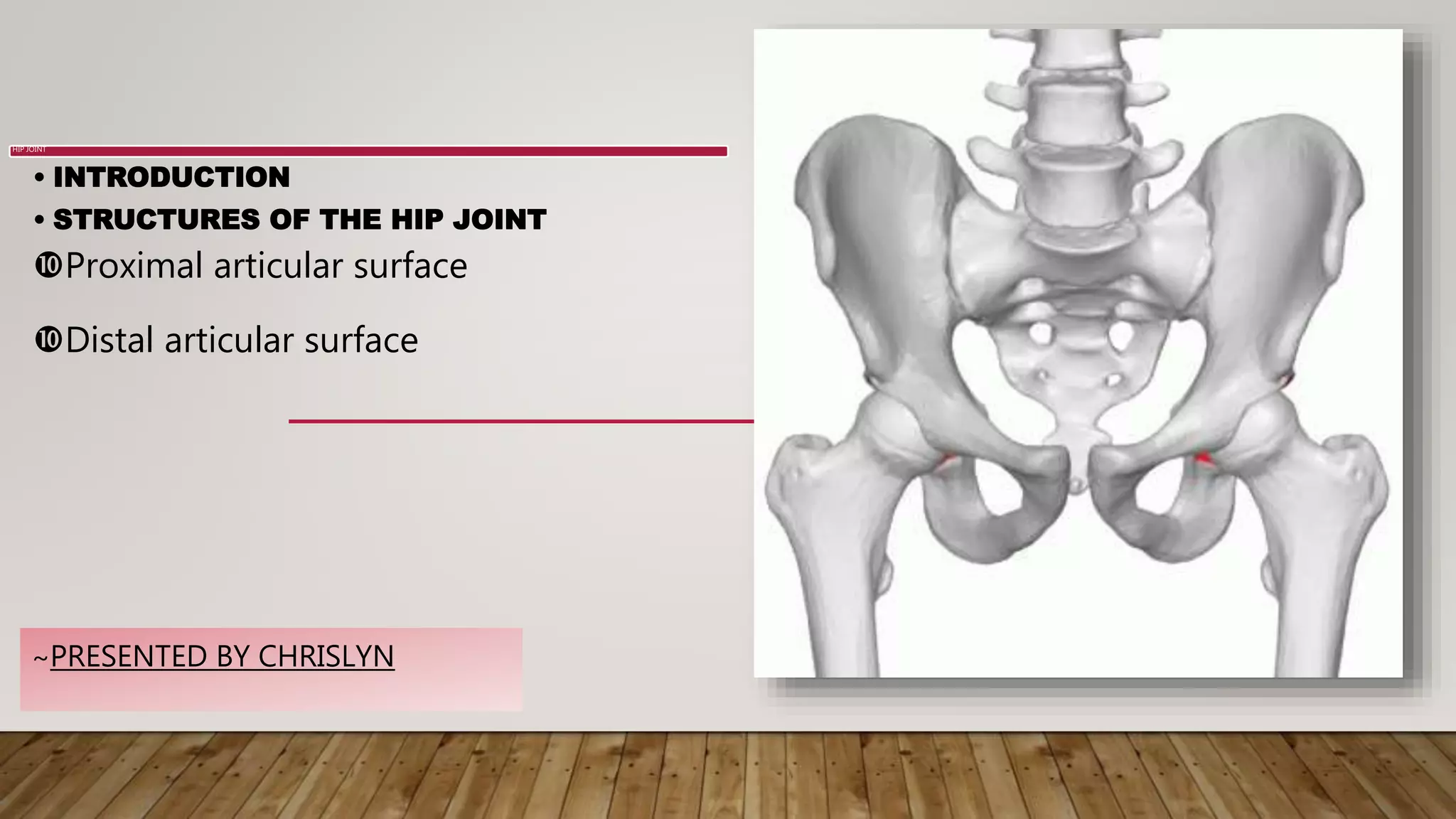
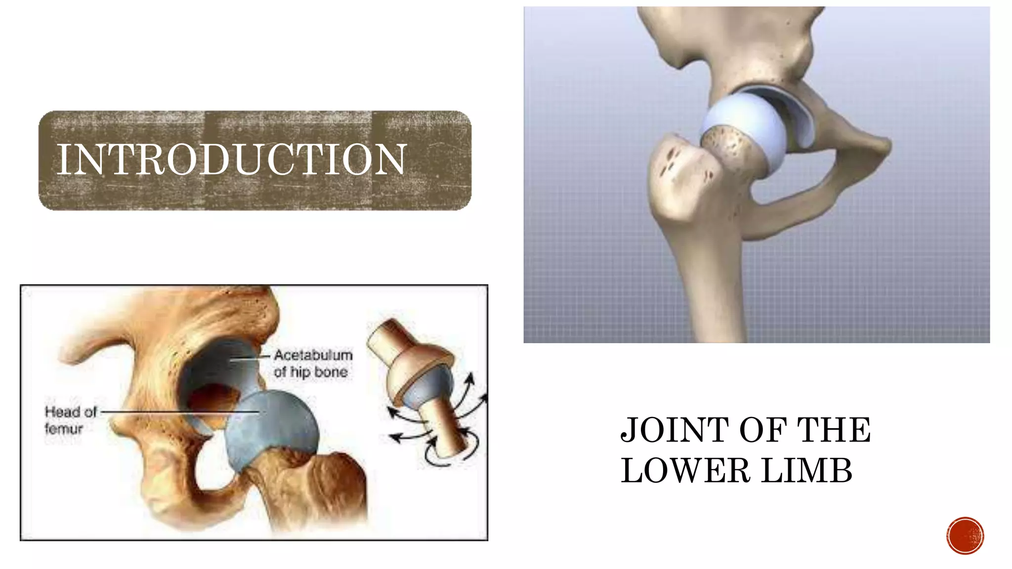
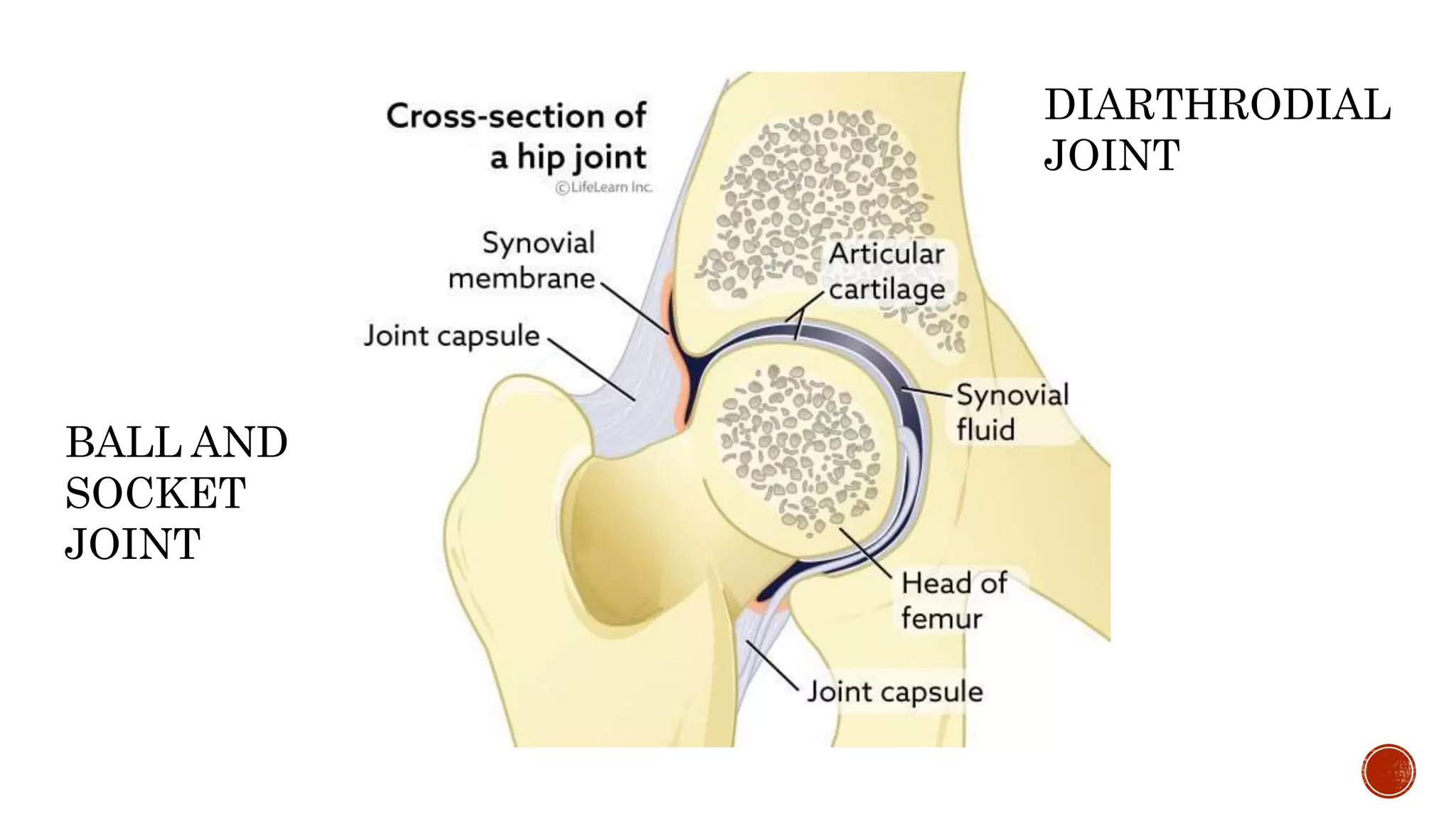
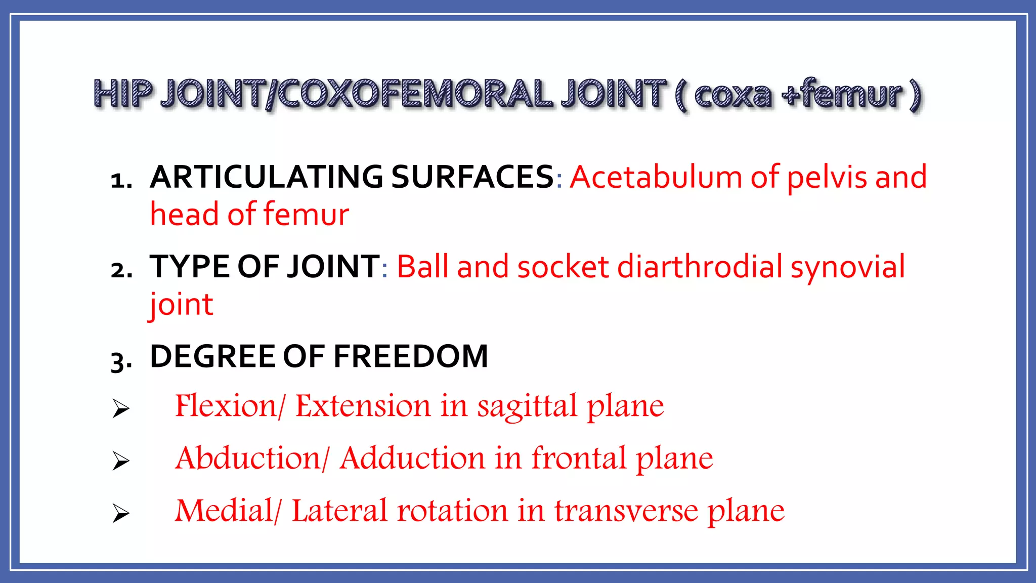
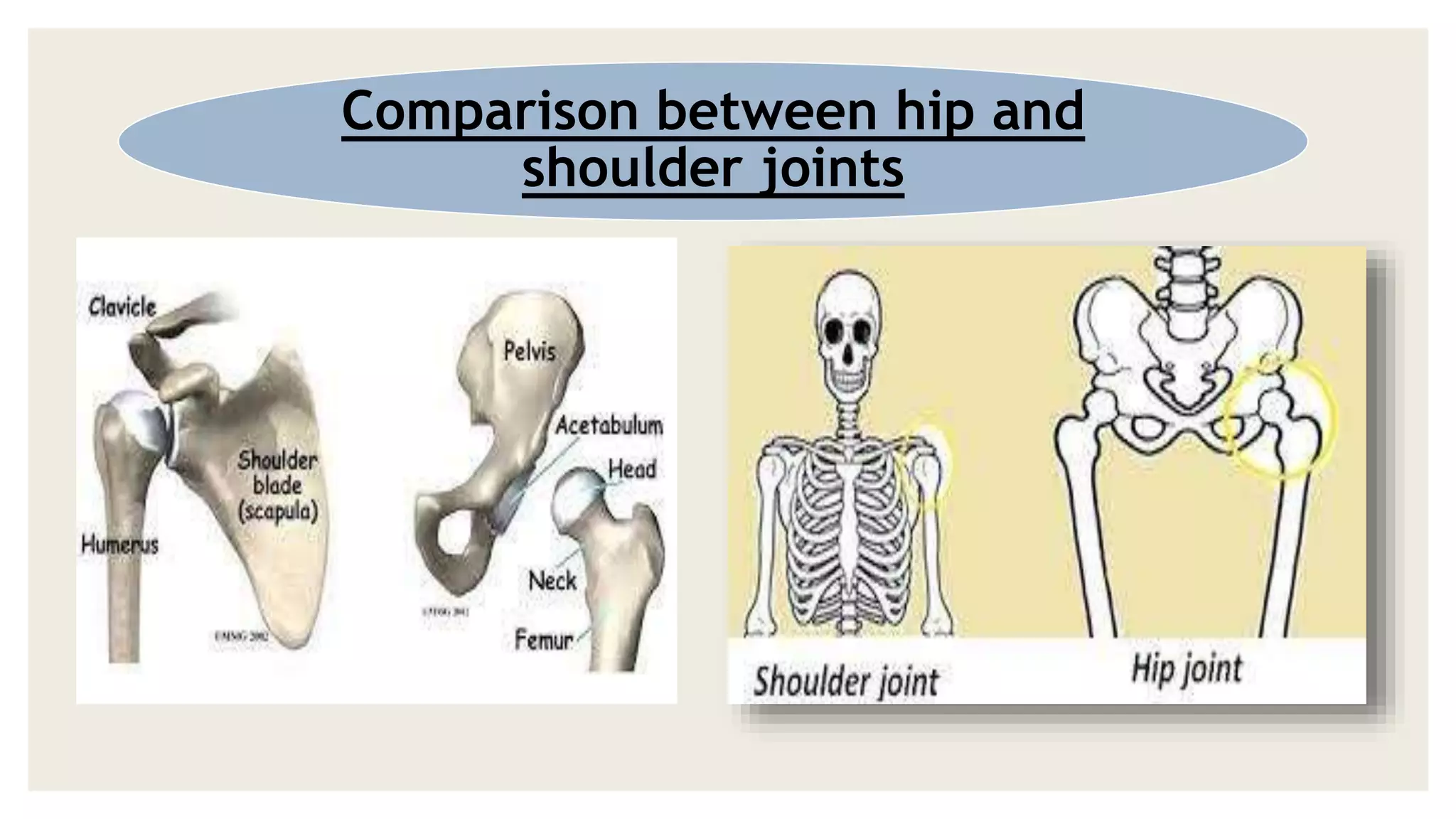
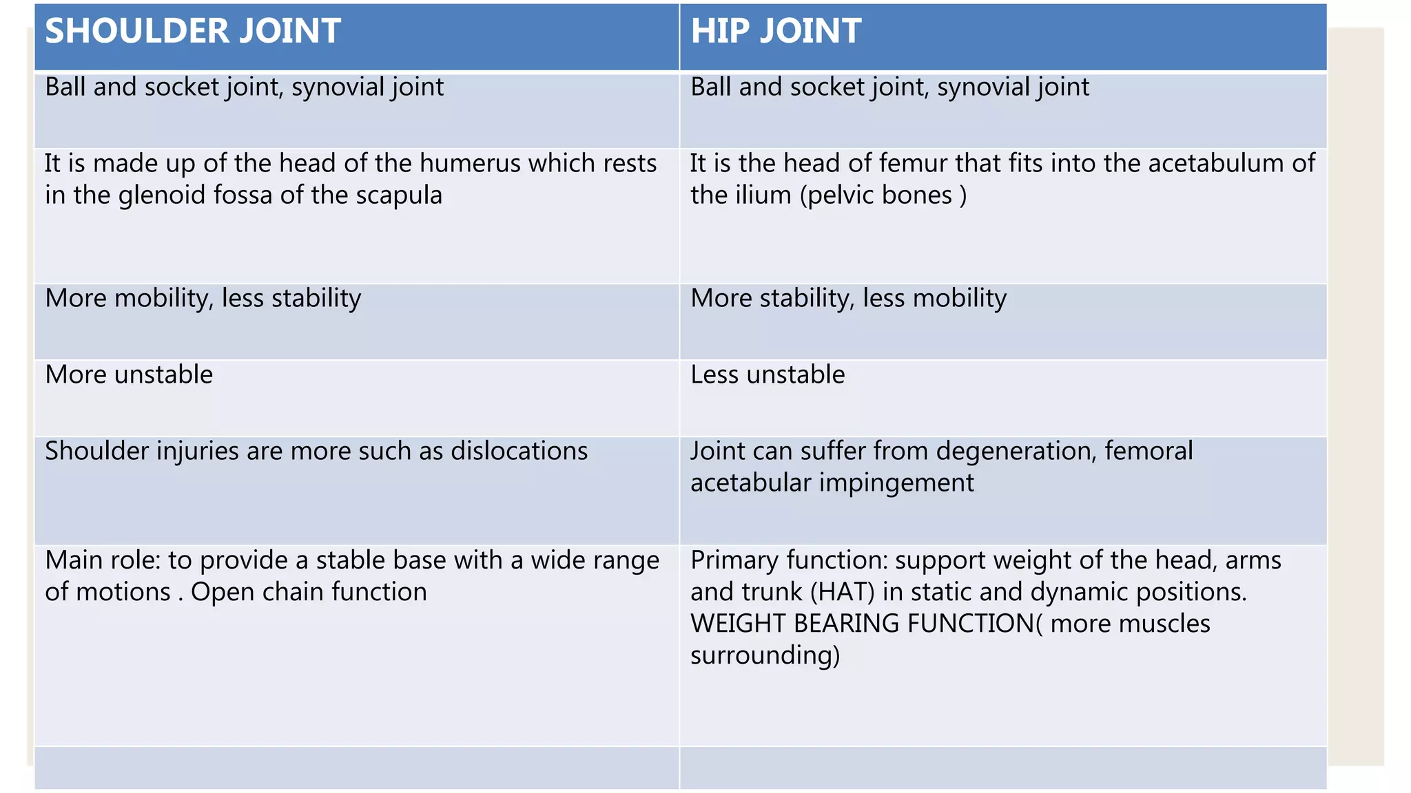
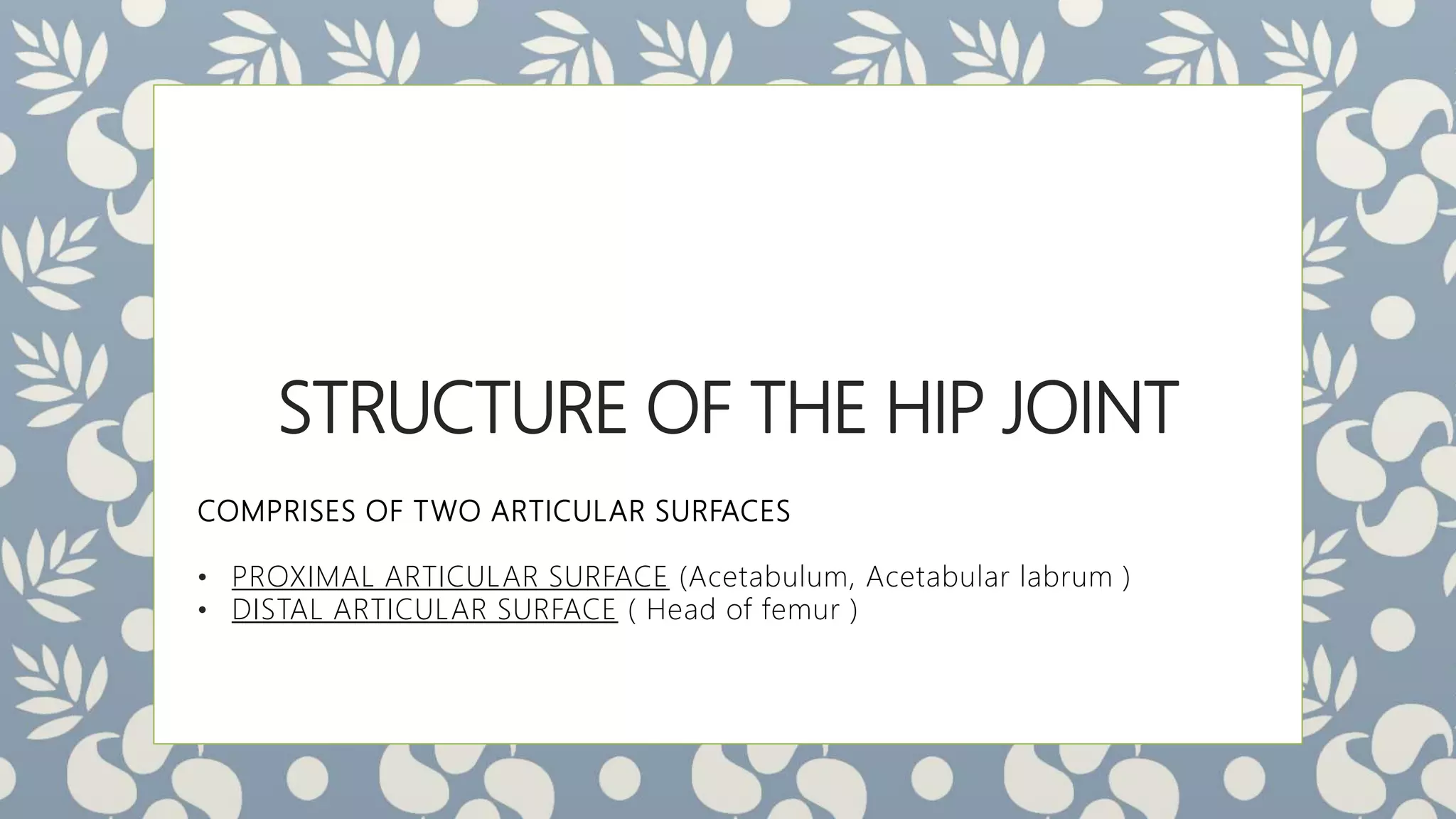
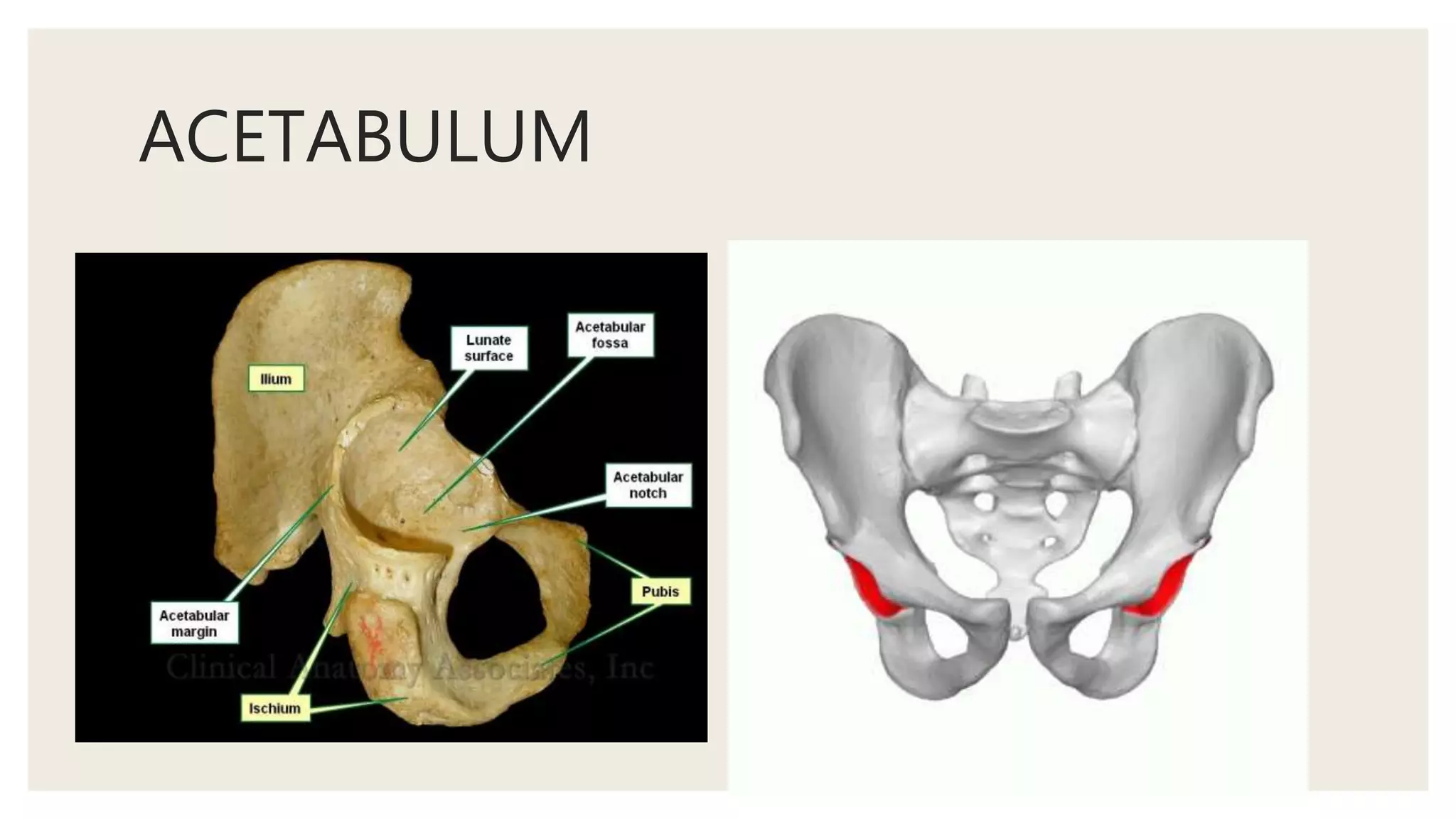
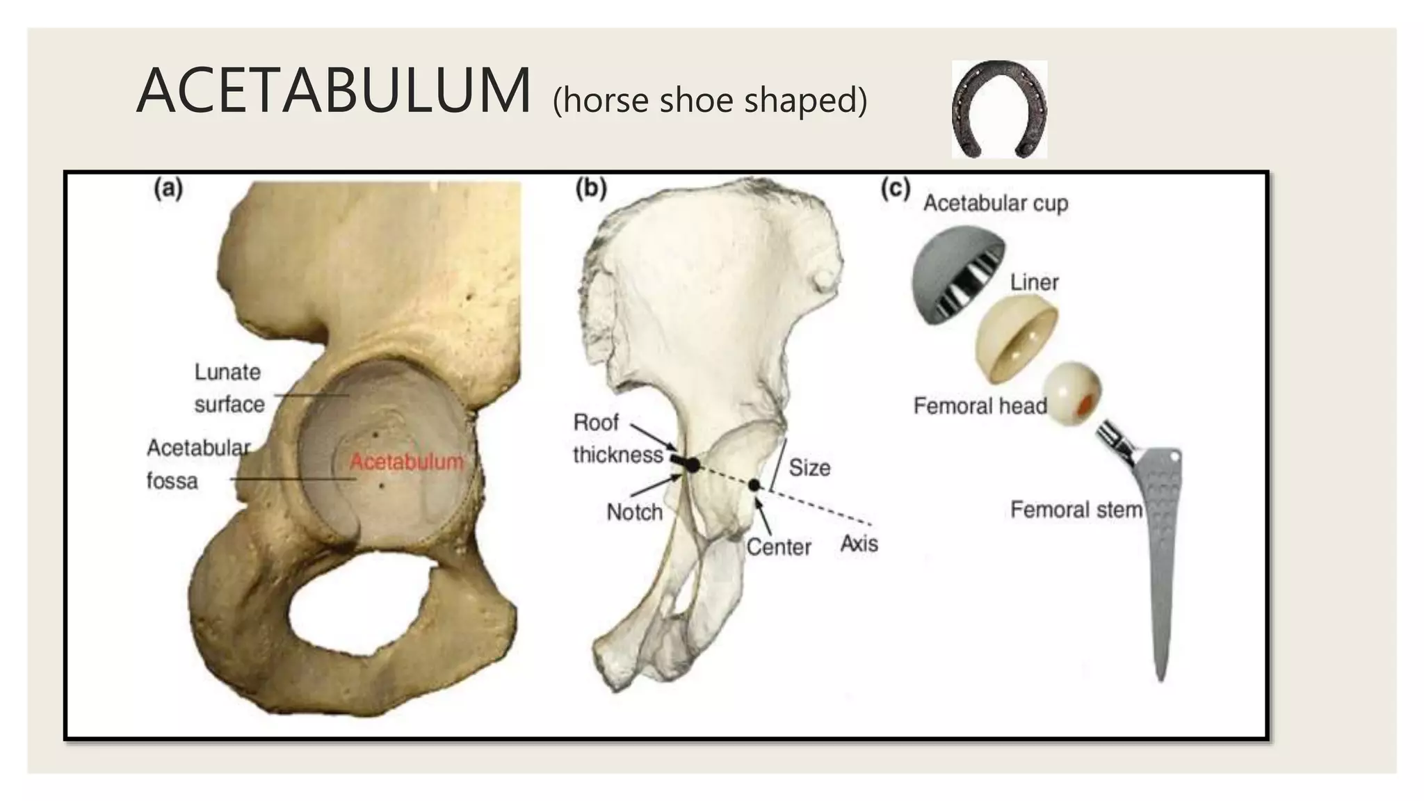
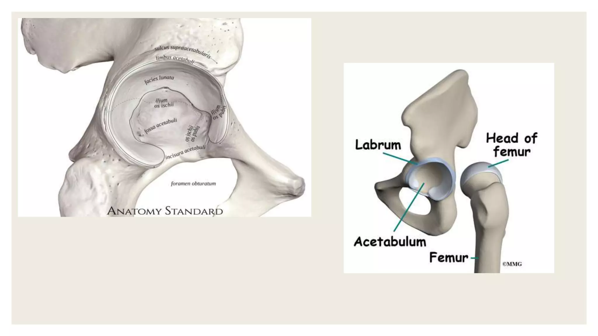
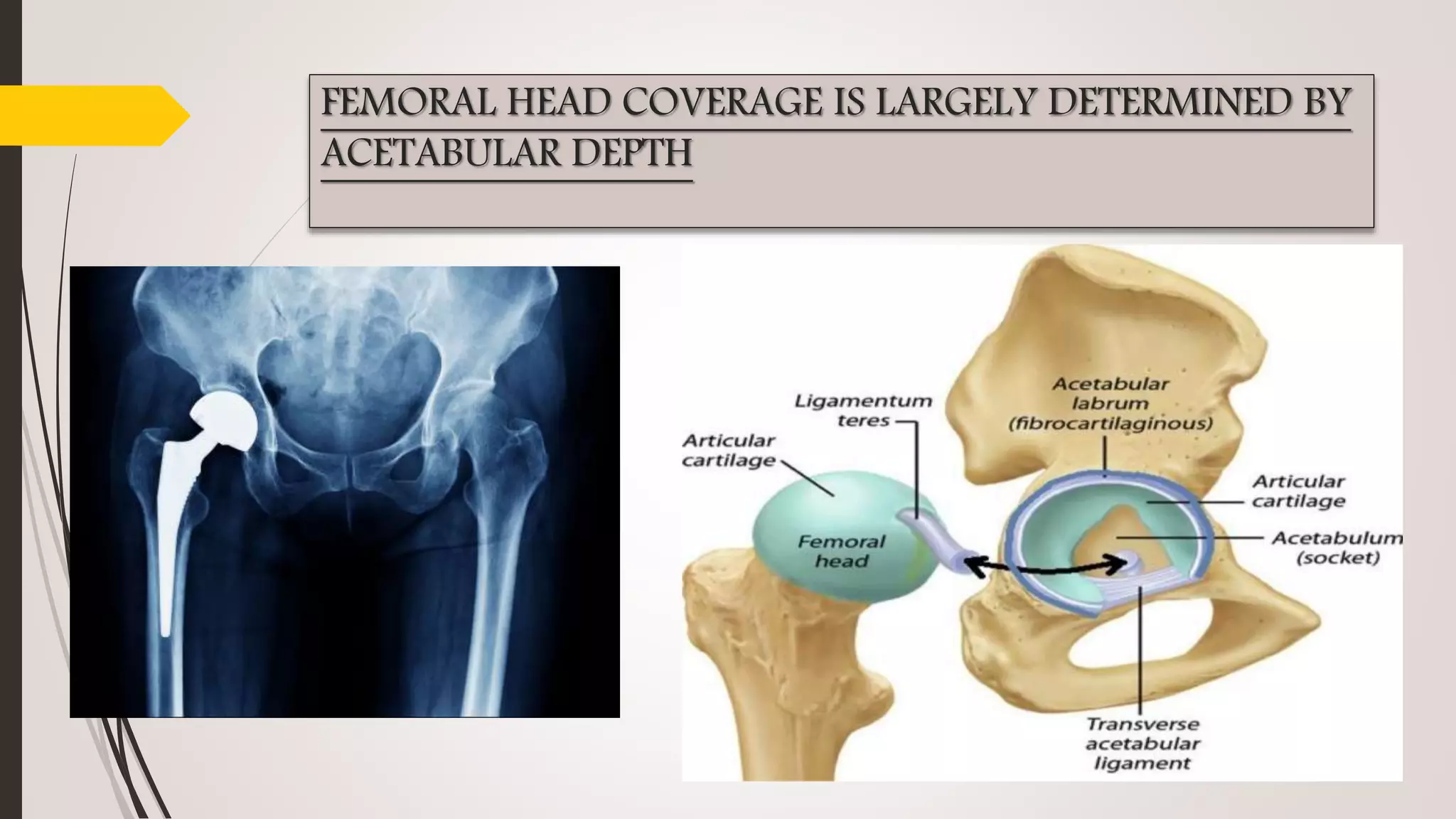
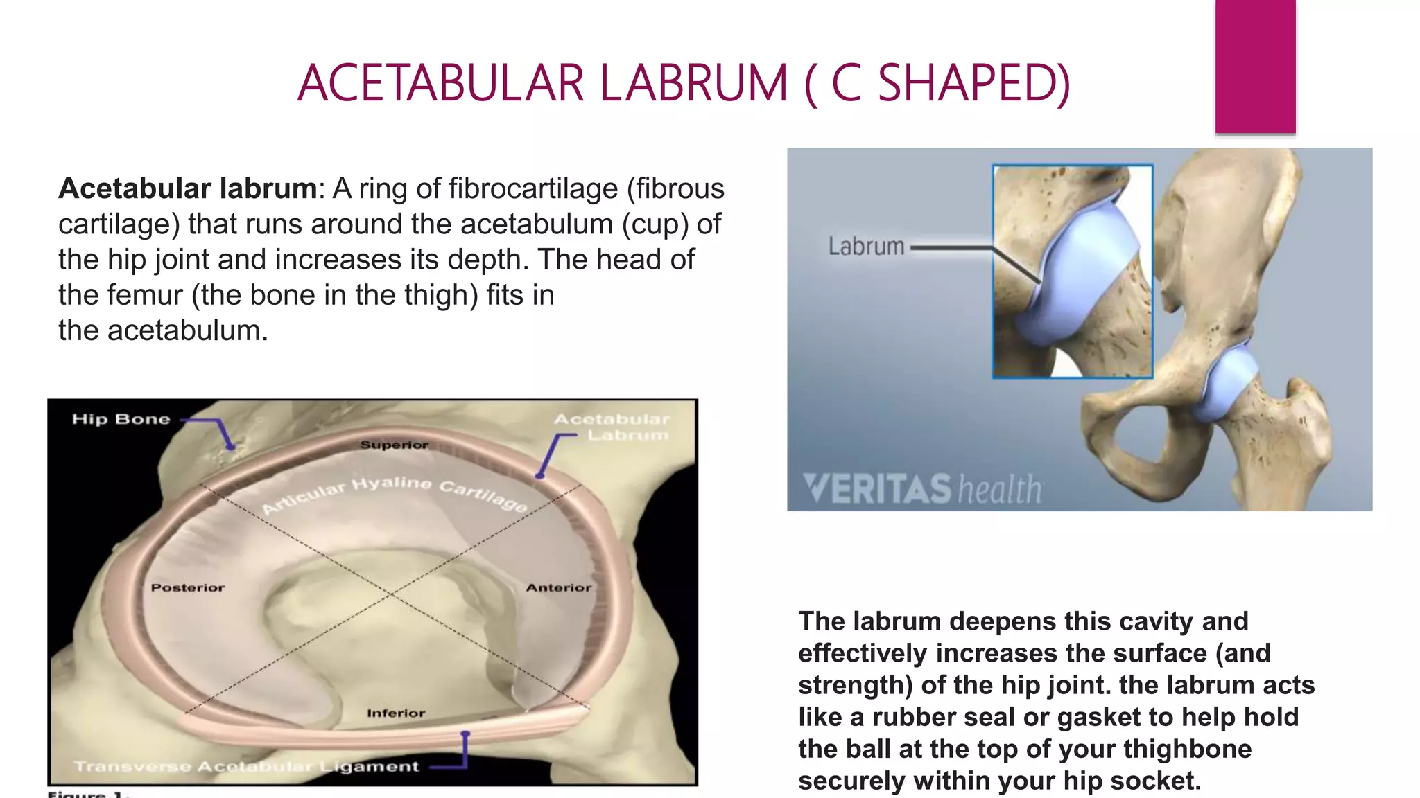
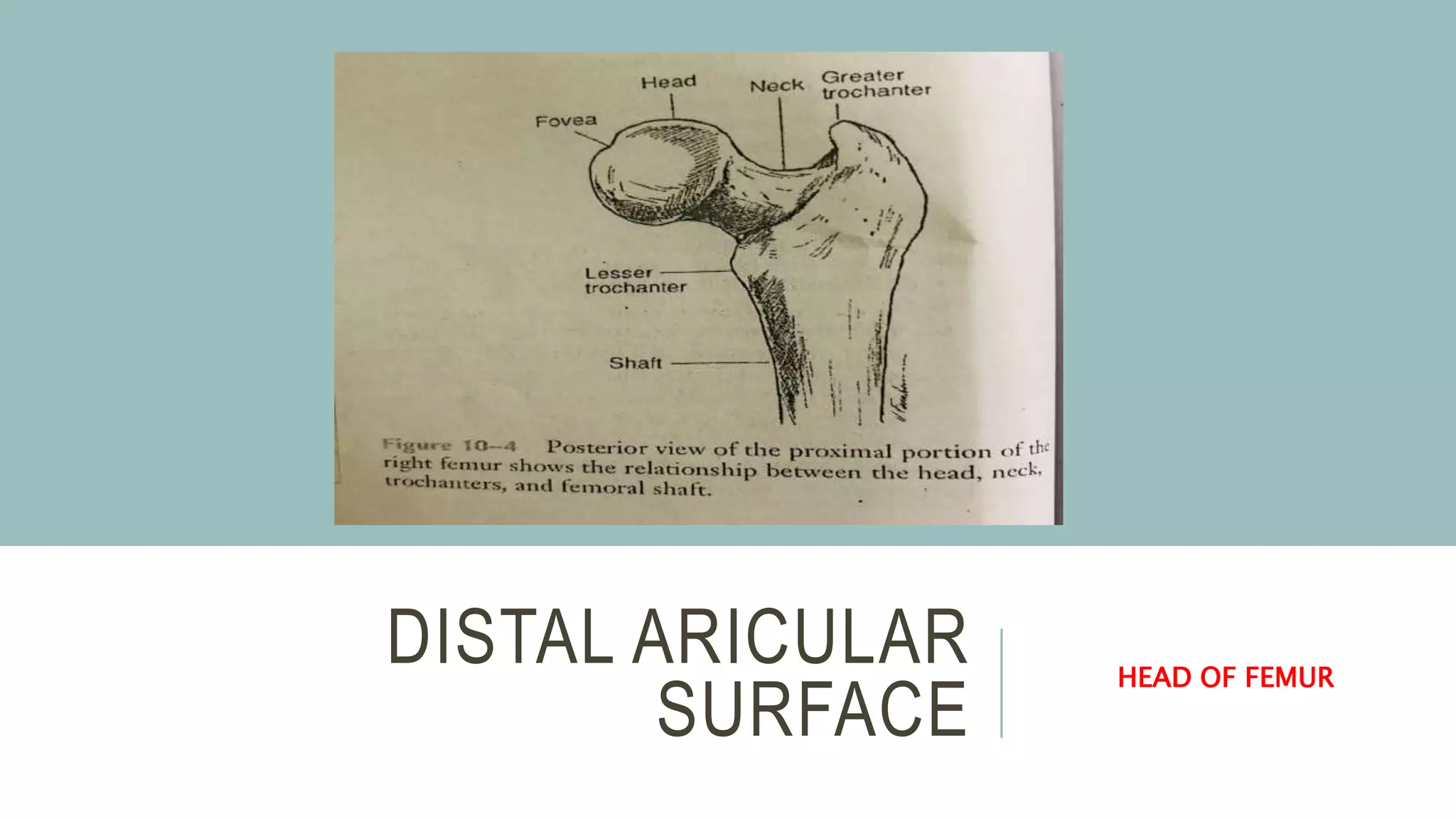
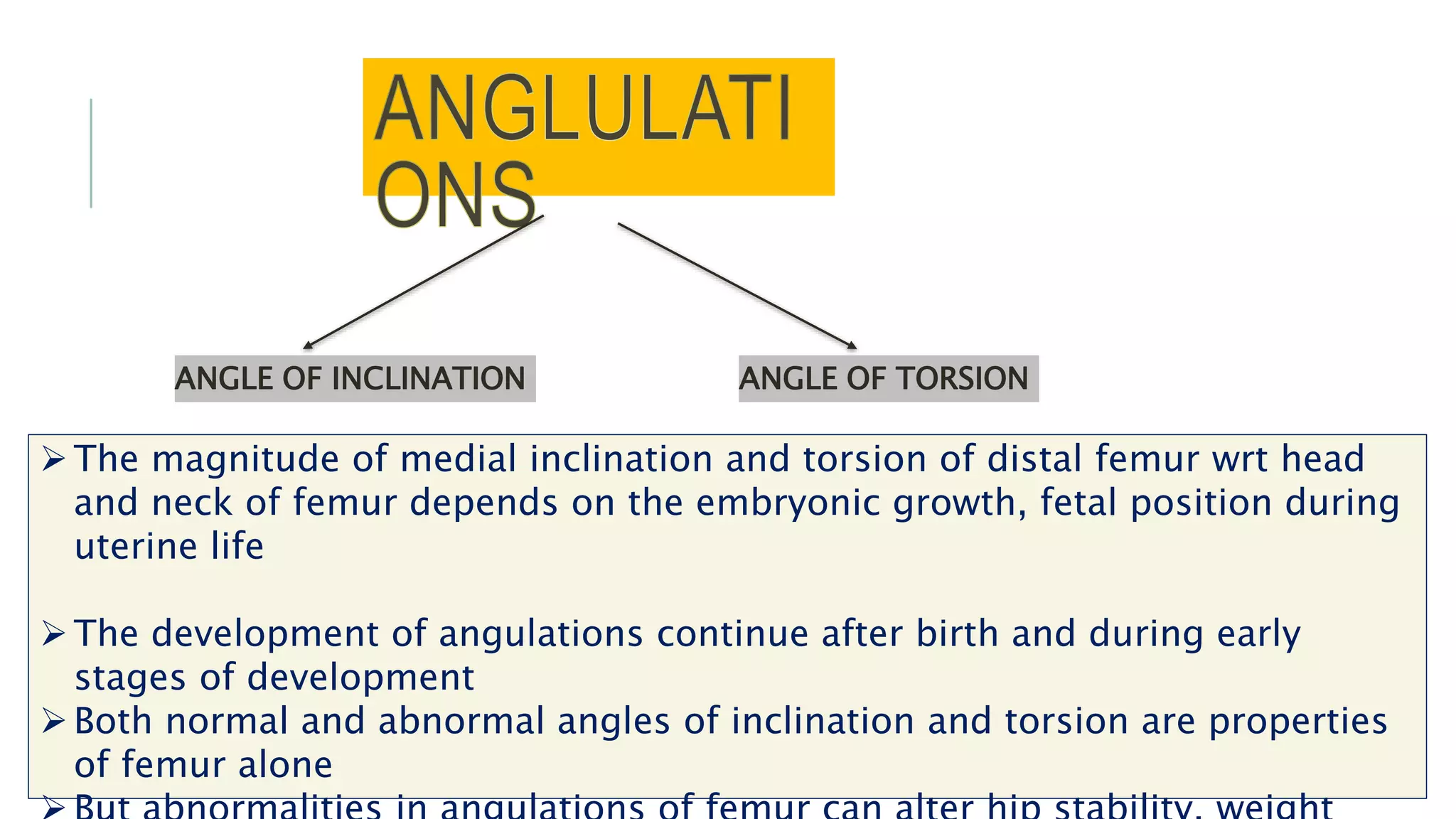
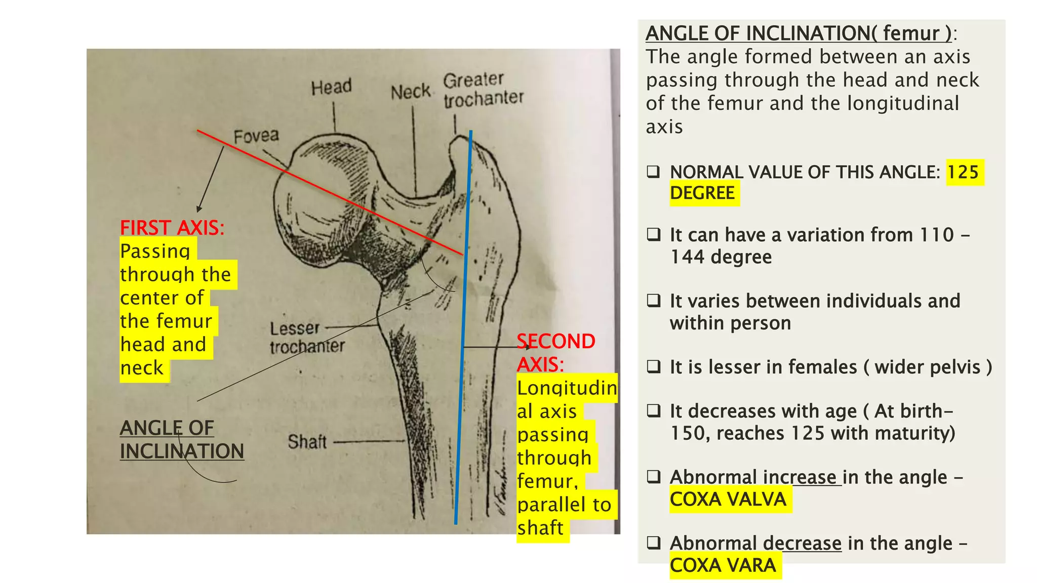
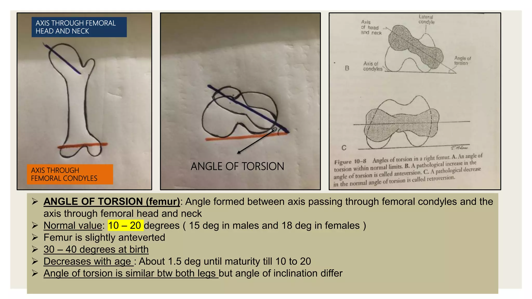
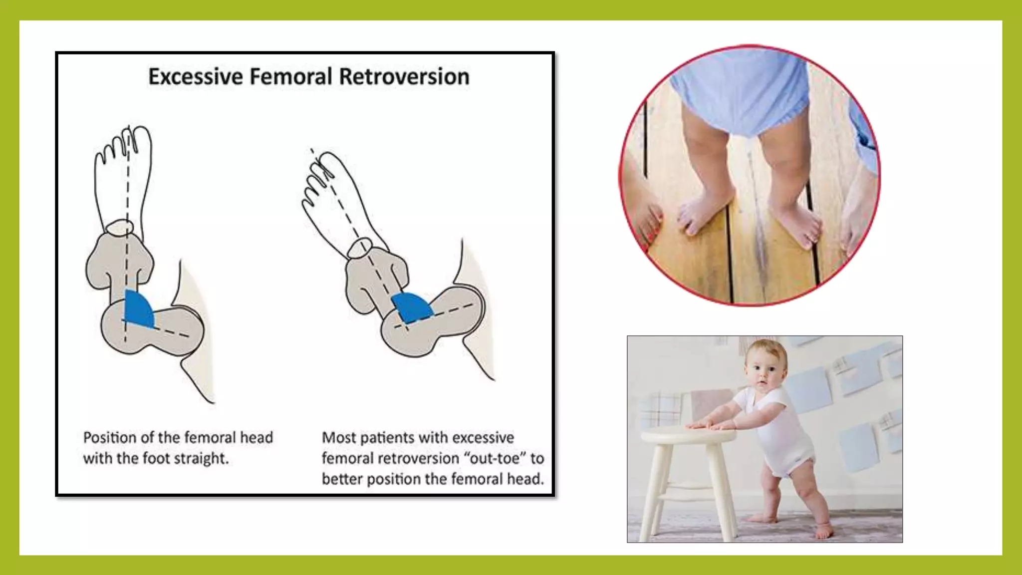
SlideShare a Scribd company logo
HIP JOINT
• INTRODUCTION
• STRUCTURES OF THE HIP JOINT
Proximal articular surface
Distal articular surface
~PRESENTED BY CHRISLYN
INTRODUCTION
JOINT OF THE
LOWER LIMB
DIARTHRODIAL
JOINT
BALL AND
SOCKET
JOINT
1. ARTICULATING SURFACES:Acetabulum of pelvis and
head of femur
2. TYPE OF JOINT: Ball and socket diarthrodial synovial
joint
3. DEGREE OF FREEDOM
 Flexion/ Extension in sagittal plane
 Abduction/ Adduction in frontal plane
 Medial/ Lateral rotation in transverse plane
Comparison between hip and
shoulder joints
SHOUDER JOINT
Ball and socket joint
It is made up of the head
of the humerus which rests
in the glenoid fossa of the
scapula
More mobility, less stability
More unstable
Shoulder injuries are more
such as dislocations
Main role: to provide a
stable base with a wide
range of motions . Open
change function
HIP JOINT
Ball and socket joint
It is the head of femur that fits
into the acetabulum of the
ilium (pelvic bones )
More stability, less mobility
Less unstable
Joint can suffer from
degeneration, femoral
acetabular impingement
Primary function: support
weight of the head, neck and
trunk (HAT) in static and
dynamic positions. WEIGHT
BEARING FUNCTION( more
muscles surrounding)
SHOULDER JOINT HIP JOINT
Ball and socket joint, synovial joint Ball and socket joint, synovial joint
It is made up of the head of the humerus which rests
in the glenoid fossa of the scapula
It is the head of femur that fits into the acetabulum of
the ilium (pelvic bones )
More mobility, less stability More stability, less mobility
More unstable Less unstable
Shoulder injuries are more such as dislocations Joint can suffer from degeneration, femoral
acetabular impingement
Main role: to provide a stable base with a wide range
of motions . Open chain function
Primary function: support weight of the head, arms
and trunk (HAT) in static and dynamic positions.
WEIGHT BEARING FUNCTION( more muscles
surrounding)
STRUCTURE OF THE HIP JOINT
COMPRISES OF TWO ARTICULAR SURFACES
• PROXIMAL ARTICULAR SURFACE (Acetabulum, Acetabular labrum )
• DISTAL ARTICULAR SURFACE ( Head of femur )
PROXIMAL
ARTICULAR
SURFACE
1. ACETABULUM
ACETABULUM
ACETABULUM (horse shoe shaped)
PROXIMAL ARTICULAR SURFACE
ACETABULUM OF PELVIC BONE FORMS PROXIMAL ARTICULAR SURFACE
1.Cuplike concave socket of the hip joint
2. Present on the lateral aspect of the pelvic bone
3. Three bones make up the pelvis that contribute to the structure of the acetabulum
a) Ischium (2/5 th of acetabulum)
b) Pubis (1/5 th of acetabulum)
c) Ilium ( remainder)
The periphery of the acetabulum called the lunate surface is covered with
hyaline cartilage
The acetabulum (horse shoe shaped) articulates with the head of femur
The inferior aspect of the lunate surface( ie base of horseshoe) has a notch
called acetabular notch. It is like a gap
The acetabular is spanned by a fibrous band called transverse acetabular
ligament that connects two ends of the horseshoe.
The transverse acetabular ligament creates a kind of tunnel under which
blood vessels pass beneath and reach the deepest of the acetabulum called
acetabulum fossa
The acetabular fossa ( innermost part) doesn’t take part in the articulation
The fossa contains fibroelastic fat covered with synovial membrane
The acetabular labrum deepens the acetabulum and surrounds its periphery.
POSITION: positioned laterally with inferior and anterior tilt
INCLINATIONS:
.50 degree laterally inclined
.20 degree anteriorly rotated (anteversion)
.20 degree anteriorly tilted in the frontal, transverse and sagittal
FEMORAL HEAD COVERAGE IS LARGELY DETERMINED BY
ACETABULAR DEPTH
ACETABULAR ABNORMALITIES that lead to
pathology including excessive cartilage wear
ACETABULAR DYSPLASIA
COXA PROFUNDA
ACETABULAR PROTRUSIO
ANTEVERION
RETROVERSION
ACETABULAR
ABNORMALITIES
ACETABULAR DYSPLASIA:
 Abnormally shallow acetabulum that results in lack of
coverage
 Dysplasia is the basic mechanical abnormality for instability
and disproportionate loading of the superior acetabular rim
COXA PROFUNDA AND ACETABULAR PROTRUSIO:
 Here the acetabulum excessively covers the femoral head
 Acetabular over coverage can lead to limited ROM and
internal impingement (thinning) between femoral and
acetabulum junction
ABNORMALITIES IN ACETABULAR DEPTH
ABNORMALITIES IN ACETABULAR POSTIONING
( INCLINATION AND VERSION- abnormal positioning in
the transverse plane)
ANTEVERSION
Anteversion of acetabulum exists when the acetabulum is
positioned too far anteriorly in the transverse plane
RETROVERSION
Retroversion of the acetabulum exists when the
is positioned too far posteriorly in the transverse plane
 ACETABULUM WITH
ANTEVERSION LESS INCLINATION LEAD TO INSTABILITY
INCLINATION/ RETROVERSION
OVER COVERAGE AND IMPINGEMENT
BETWEEN JOINT
CENTER EDGE ANGLE OF WIBERG
 Acetabular depth can be measured using Center Edge Angle of Wiberg
 It is the measure of depth of acetabulum with that of the femur head
 It is formed by a line connecting the lateral rim of the acetabulum and center of the
femoral head and a vertical line from the center of the femoral head
CENTER EDGE ANGLES CLASSIFIED AS FOLLOWS:
 DEFINITE DYSPLASIA: angle less than 16 degree
 POSSIBLE DYSPLASIA: 16-25 degree
 NORMAL: greater than 25 degree
 ABNORMAL OVERCOVERAGE: greater than normal. But not well defined
 EXCESSIVE ACETABULAR COVERAGE: greater than 40 degree
PROXIMAL
ARTICULAR SURFACE
2. ACETABULAR
LABRUM
Acetabular labrum: A ring of fibrocartilage (fibrous
cartilage) that runs around the acetabulum (cup) of
the hip joint and increases its depth. The head of
the femur (the bone in the thigh) fits in
the acetabulum.
ACETABULAR LABRUM ( C SHAPED)
The labrum deepens this cavity and
effectively increases the surface (and
strength) of the hip joint. the labrum acts
like a rubber seal or gasket to help hold
the ball at the top of your thighbone
securely within your hip socket.
The knee meniscus and glenoid labrum are anatomically distinct yet
analogous structures. Although the gross anatomy differs, both tissues are
composed of fibrocartilage with a complex but well-organized collagen
microstructure. The meniscus and labrum function to increase congruity and
stability, decrease contact stresses, and distribute load across their
respective joints.
Meniscus of knee
Labrum of glenohumeral joint
ANALOGOUS STRUCTURES ( same function, different structure)
2. ACETABULAR
LABRUM
 The entire periphery of the acetabulum is rimmed by a wedge shaped
fibrocartilage called acetabular labrum
 The labrum of the hip to a large extent is analogous to the meniscus of the knee
and labrum of the glenohumeral joint
 The labrum is attached to the periphery of the acetabulum by a zone of
calcified cartilage
 The labrum not only deepens the socket but also increases concavity of the
acetabulum through its triangular shape, grasping head of femur to maintain
contact with acetabulum
 It enhances joint stability by acting as a seal to maintain intra articular pressure
 It also decreases force transmitted to articular cartilage and provides
proprioceptive feedback
Nerve endings within the labrum not only provide proprioceptive
feedback but can also act as a source of pain
An abnormally shallow acetabulum will increase stress on the
surrounding capsule and labrum
The transverse acetabular ligament is considered to be a part of the
acetabulum labrum although unlike the labrum, it contains no
cartilage cells
Although it is positioned to protect the blood vessels travelling
beneath to reach the head of the femur, experimental data does
not support the notion of the transverse acetabular ligament as a
load bearing structure
The cause of a hip labral tear might be:
Trauma.
Injury to or dislocation of the hip joint
— which can occur during car accidents or from
playing contact sports such as football or hockey,
yoga not done properly
Degenerative health conditions: Osteoarthritis
is a chronic (long-term) wearing down of the
cartilage between the joints. As cartilage slowly
erodes over time, it becomes more prone to
tearing
The symptoms of a hip labral
tear include:
•Hip pain or stiffness
•Pain in the groin or buttocks
•A clicking or locking sound
hip area when you move
•Feeling unsteady on your
DISTAL ARICULAR
SURFACE
HEAD OF FEMUR
HEAD OF FEMUR
The distal articular surface is formed by the head of the femur.
The head of femur is spherical in shape covered by hyaline
cartilage
The articular area forms 2/3 rd of the sphere
Head of femur is connected to shaft by the neck
Inferior to the medial point of the head is a small depression
called fovea or fovea capitis.
The femoral neck is attached to the shaft of femur between
greater and lesser trochanters
The fovea is site of attachment of ligamentum teres.
ANGLE OF INCLINATION ANGLE OF TORSION
The magnitude of medial inclination and torsion of distal femur wrt head
and neck of femur depends on the embryonic growth, fetal position during
uterine life
The development of angulations continue after birth and during early
stages of development
Both normal and abnormal angles of inclination and torsion are properties
of femur alone
ANGLE OF INCLINATION
FIRST AXIS:
Passing
through the
center of
the femur
head and
neck
SECOND
AXIS:
Longitudin
al axis
passing
through
femur,
parallel to
shaft
ANGLE OF INCLINATION( femur ):
The angle formed between an axis
passing through the head and neck
of the femur and the longitudinal
axis
 NORMAL VALUE OF THIS ANGLE: 125
DEGREE
 It can have a variation from 110 -
144 degree
 It varies between individuals and
within person
 It is lesser in females ( wider pelvis )
 It decreases with age ( At birth-
150, reaches 125 with maturity)
 Abnormal increase in the angle -
COXA VALVA
 Abnormal decrease in the angle –
COXA VARA
ANGLE OF
INCLINATION
COXA VALGA
 It’s a condition in which angle of inclination increases
 The angle is >125
 The contact between articulating surfaces decreases ( exposure of
femoral head) therefore decrease in stability
 Trabecular system density decreases( line of weight bearing
changes, less strain on oblique axis, decrease in density).
Therefore weakness in neck of femur/ bone
 Moment arm of abductors ( lateral muscles )decreases ( as head
tilts upwards, distance decreases) , dec in muscles efficiency.. and
therefore muscle weakness ( stability )
 Gravitation adduction moment is unbalanced .. leads to instability
 Joint reaction force increases, leads to degenerative changes (
impingement, labral tear)
COXA VARA
 Pathological condition in which angle of inclination of femur
decreases
 Angle <120
 Increase in stability ( the head of femur sits inside completely)
 Increase in muscles efficiency ( increase in moment arm)
 Decrease in joint reaction forces ( less degenerative chances)
 But if it decreases too much then there are stability issues
 Increase in bending moment, lead to fracture of neck of femur
3 types of Coxa Vera
 CONGINETAL ( present since birth)
 DEVELOPMENTAL ( during bone fusing .. Developmental period)
 ACQUIRED ( due to diseases like rickets.. Vit D deficiency, calcium
def)
ANGLE OF TORSION
AXIS THROUGH
FEMORAL CONDYLES
AXIS THROUGH FEMORAL
HEAD AND NECK
ANGLE OF TORSION
 ANGLE OF TORSION (femur): Angle formed between axis passing through femoral condyles and the
axis through femoral head and neck
 Normal value: 10 – 20 degrees ( 15 deg in males and 18 deg in females )
 Femur is slightly anteverted
 30 – 40 degrees at birth
 Decreases with age : About 1.5 deg until maturity till 10 to 20
 Angle of torsion is similar btw both legs but angle of inclination differ
ANTEVERSION & RETROVERSION
◦ If the axis through femoral condyles lies in the frontal plane then the head and neck of the
femur are torsioned anteriorly, on the condyles.
◦ At birth it is 30-40 deg. This decreases about 1.5 per year until skeletal maturity
◦ Condition in which angle of anterior torsion increases is anteversion (>15 in males , >18 in
females)
◦ Articulating surfaces instable because large portion of femoral head is tilted outward, exposed
◦ When angle of torsion decrease..less than 15-20 it is called retroversion
◦ Variations in degree of Anteversion and retroversion also depends on assessing methods like CT
scan, radiograph, ultrasound to measure angle of femoral torsion
◦ Femoral anteversion is correlated with increase medial rotation ROM and decrease lateral
rotation so total hip rotation remains the same
◦ Femoral anteversion and coxa valga are commonly found togther but they function
independently
◦ can lead to dysfunction in distal and proximal parts of hip + knee +foot
◦ Other pathological angulations ( retroversion, coxa valga n coxa vara ) can also affect proximal
and distal parts of hip joint
RETROVERSION (AOT < 15-20)
………………………………………………………….
Retroversion refers to an abnormal backward rotation of the hip relative to the knee
This condition can affect patients of all ages and leads to abnormal stress in the low back, hip and
knee, as well as an abnormal gait (walking stance).
Femoral retroversion (also known as hip retroversion) is a rotational or torsional deformity in which
the femur (thighbone) twists backward (outward) in relation to the knee. Because the lower part of
the femur is connected to the knee, this also means that the knee is twisted outward relative to the
hip.
Femoral retroversion can occur in one or both legs
Femoral retroversion is often a congenital condition, meaning children are born with it. It also
appears to be related to the position of the baby as it grows in the womb. Torsional deformity can
also occur after a fracture, if a broken bone heals with incorrectly (called malunion).
Symptoms -out-toeing or "duck walk" – walking with the foot pointed outward instead of straight
ahead, learning to walk late (in children), flatfeet, difficulty with running, fatigues easily with physical
activity, poor balance or coordination ,hip and knee pain , low back pain degeneration or arthritis of
the hip
Diagnosis -The doctor will go through the developmental and family history and also observe the
patient’s gait (manner of walking) to look for signs of out-toeing or gait compensation. The physician
may also order an X ray or CT scan
Treatment - Many children born with femoral retroversion grow out it. An excessive femoral
retroversion can place stress on hip and knee joints, often leading to joint pain and abnormal wear..
labral tear. In these situations, a surgical procedure known as a femoral osteotomy may be used
Left: Position of an anteverted femoral head with the
foot facing straight forward. In this position, the femoral
head subluxes out of the front of the hip joint.
Right: Most patients with excessive hip anteversion
compensate by walking in-toed. This position keeps the
femoral head within the socket, which minimizes pain.
ANTEVERSION (AOT> 15-20)
…………………………………………………………………..
• Femoral anteversion is a forward (inward) rotation in the femur (thighbone), which connects to
the pelvis to form the hip joint. In other words the knee is excessively twisted inward relative to
the hip.
• Femoral anteversion can occur in one or both legs.
• Many children are born with femoral anteversions that they eventually grow out of. In people
who do not grow out of it, a mildly anteverted femoral head may cause no significant health
problems.
• But an excessive anteversion of the femur overloads the anterior (front) structures of the hip
joint, including the labrum and joint capsule.When the foot is positioned facing directly
forward, the femoral head may sublux (partially dislocate) from the socket of the hip joint,
called the acetabulum.This torsional malalignment places abnormal stress on both the hip and
knee joints, often leading to pain and abnormal joint wear.
•
• Symptoms- In-toeing, in which a person walks "pigeon-toed," with each foot
pointed slightly toward the other, Bowlegs (also called bowed legs), Keeping the
legs in this position often helps a patient maintain balance, Pain in the hips,
knees and/or ankles.
• Treatment- While many children grow out of their femoral anteversion
conditions, excessive anteversion may require surgical correction, as a
procedure known as a femoral osteotomy
Reduces hip stability
Articulating surfaces instable because large portion of femoral head is tilted outward,
exposed
Hip abductors fall more posteriorly to the joint, reducing moment arm for abduction
Affects the knee joint.When femoral head is anteverted, pressure from
capsuloligaments and ant musculature may push it into acetabulum causing entire
femur to medially rotate. Medial rotation of the condyles alters plane of knee flexion
therefore.. in toe gait. Abnormal position of knee joint axis ..medial femoral torsion.
Anteverted femur also affects biomechanics of patellofemoral joint at knee and
subtlar joint at foot
•INTRODUCTION- articulation, movements, comparison
between shoulder and hip joint
•ARTICULAR SURFACES
•Proximal articular surface- acetabulum( dysplasia, coxa
profunda, anteversion, retroversion), center edge angles
,labrum
•Distal articular surface- femur, angle of inclination( coxa
valga, coxa vara),angle of torsion (anteversion,
retroversion)
THANK YOU..

More Related Content

What's hot

Delorme boot
Delorme bootDelorme boot
Delorme boot
vrkv2007
 
Biomechanics of shoulder complex
Biomechanics of shoulder complexBiomechanics of shoulder complex
Biomechanics of shoulder complex
Vaibhavi Rathod
 
Glenohumeral joint-ppt.
Glenohumeral joint-ppt.Glenohumeral joint-ppt.
Glenohumeral joint-ppt.
Mohammad Akeel
 
Biomechanics of lumbar spine
Biomechanics of lumbar spineBiomechanics of lumbar spine
Biomechanics of lumbar spine
Venus Pagare
 
MWD
MWDMWD
Diadynamic currents
Diadynamic currentsDiadynamic currents
Diadynamic currents
Dr Usha (Physio)
 
Postural deviations
Postural deviationsPostural deviations
Postural deviations
Jayasri Prasanna
 
Low Frequency Currents SRS
Low Frequency Currents SRSLow Frequency Currents SRS
Low Frequency Currents SRS
Sreeraj S R
 
Electric shock and earthing
Electric shock and earthingElectric shock and earthing
Electric shock and earthing
Ramesh Adhikari
 
FARADIC CURRENT.pptx
FARADIC CURRENT.pptxFARADIC CURRENT.pptx
FARADIC CURRENT.pptx
PriyankaBhusal2
 
Russian current
Russian currentRussian current
Russian current
aditya romadhon
 
Biomechanics of wrist complex
Biomechanics of wrist complexBiomechanics of wrist complex
Biomechanics of wrist complex
Neeti Christian
 
BIOMECHANICS OF ELBOW COMPLEX
BIOMECHANICS OF ELBOW COMPLEXBIOMECHANICS OF ELBOW COMPLEX
BIOMECHANICS OF ELBOW COMPLEX
Muhammadasif909
 
anatomy and biomechanics of Shoulder joint
anatomy and biomechanics of Shoulder jointanatomy and biomechanics of Shoulder joint
anatomy and biomechanics of Shoulder joint
Harsha Nandini
 
Pathological gait
Pathological gaitPathological gait
Pathological gait
Neeti Christian
 
Hvpgs
HvpgsHvpgs
prehension.pptx
prehension.pptxprehension.pptx
prehension.pptx
ShiriShir
 
Biomechanics of elbow complex Dr Gurjant Singh.pptx
Biomechanics of elbow complex Dr Gurjant Singh.pptxBiomechanics of elbow complex Dr Gurjant Singh.pptx
Biomechanics of elbow complex Dr Gurjant Singh.pptx
Dr. Gurjant Singh
 
Subacromail bursitis
Subacromail bursitisSubacromail bursitis
Subacromail bursitis
Career Point University
 
Knee biomechanics
Knee biomechanicsKnee biomechanics
Knee biomechanics
Sreeraj S R
 

What's hot (20)

Delorme boot
Delorme bootDelorme boot
Delorme boot
 
Biomechanics of shoulder complex
Biomechanics of shoulder complexBiomechanics of shoulder complex
Biomechanics of shoulder complex
 
Glenohumeral joint-ppt.
Glenohumeral joint-ppt.Glenohumeral joint-ppt.
Glenohumeral joint-ppt.
 
Biomechanics of lumbar spine
Biomechanics of lumbar spineBiomechanics of lumbar spine
Biomechanics of lumbar spine
 
MWD
MWDMWD
MWD
 
Diadynamic currents
Diadynamic currentsDiadynamic currents
Diadynamic currents
 
Postural deviations
Postural deviationsPostural deviations
Postural deviations
 
Low Frequency Currents SRS
Low Frequency Currents SRSLow Frequency Currents SRS
Low Frequency Currents SRS
 
Electric shock and earthing
Electric shock and earthingElectric shock and earthing
Electric shock and earthing
 
FARADIC CURRENT.pptx
FARADIC CURRENT.pptxFARADIC CURRENT.pptx
FARADIC CURRENT.pptx
 
Russian current
Russian currentRussian current
Russian current
 
Biomechanics of wrist complex
Biomechanics of wrist complexBiomechanics of wrist complex
Biomechanics of wrist complex
 
BIOMECHANICS OF ELBOW COMPLEX
BIOMECHANICS OF ELBOW COMPLEXBIOMECHANICS OF ELBOW COMPLEX
BIOMECHANICS OF ELBOW COMPLEX
 
anatomy and biomechanics of Shoulder joint
anatomy and biomechanics of Shoulder jointanatomy and biomechanics of Shoulder joint
anatomy and biomechanics of Shoulder joint
 
Pathological gait
Pathological gaitPathological gait
Pathological gait
 
Hvpgs
HvpgsHvpgs
Hvpgs
 
prehension.pptx
prehension.pptxprehension.pptx
prehension.pptx
 
Biomechanics of elbow complex Dr Gurjant Singh.pptx
Biomechanics of elbow complex Dr Gurjant Singh.pptxBiomechanics of elbow complex Dr Gurjant Singh.pptx
Biomechanics of elbow complex Dr Gurjant Singh.pptx
 
Subacromail bursitis
Subacromail bursitisSubacromail bursitis
Subacromail bursitis
 
Knee biomechanics
Knee biomechanicsKnee biomechanics
Knee biomechanics
 

Similar to Hip joint - proximal and distal articular surface ppt

Hip joint biomechanics and pathomechanics
Hip joint biomechanics and pathomechanicsHip joint biomechanics and pathomechanics
Hip joint biomechanics and pathomechanics
Radhika Chintamani
 
biomechanics of hip joint
biomechanics of hip jointbiomechanics of hip joint
biomechanics of hip joint
MD FURQUAN
 
Hip joint biomecanics
Hip joint biomecanicsHip joint biomecanics
Hip joint biomecanics
Dr Chandan Verma
 
Pelvic girdle, Femur, Sacroiliac joint and Hip Joint
Pelvic girdle, Femur, Sacroiliac joint and Hip JointPelvic girdle, Femur, Sacroiliac joint and Hip Joint
Pelvic girdle, Femur, Sacroiliac joint and Hip Joint
Sado Anatomist
 
Applied and clinical anatomy of lower limb
Applied and clinical anatomy of lower limbApplied and clinical anatomy of lower limb
Applied and clinical anatomy of lower limb
drjabirwase
 
Cervical
CervicalCervical
Cervical
PhysioAid1
 
Kinetics and kimematics of the hip
Kinetics and kimematics of the hipKinetics and kimematics of the hip
Kinetics and kimematics of the hip
Kumarpal Singh
 
Atlantoaxial and occipital joint
Atlantoaxial and occipital jointAtlantoaxial and occipital joint
Atlantoaxial and occipital joint
ntkhab24
 
Hip joint
Hip jointHip joint
The hip complex
The hip complexThe hip complex
The hip complex
Dr.Rajal Sukhiyaji
 
Hip joint anatomy and its biomechanics
Hip joint anatomy and its biomechanicsHip joint anatomy and its biomechanics
Hip joint anatomy and its biomechanics
Gowtham Nallaiyan
 
Biomechanics of hip complex 1
Biomechanics of hip complex 1Biomechanics of hip complex 1
Biomechanics of hip complex 1
Dibyendunarayan Bid
 
Vertebral column... and Biomechanics.pptx
Vertebral column... and Biomechanics.pptxVertebral column... and Biomechanics.pptx
Vertebral column... and Biomechanics.pptx
sacootcbe
 
Assignment cervical spine
Assignment cervical spineAssignment cervical spine
Assignment cervical spine
Muhamamd Rizwan
 
09 Articulations Selected Articulations In Depth
09 Articulations   Selected Articulations In Depth09 Articulations   Selected Articulations In Depth
09 Articulations Selected Articulations In Depth
guest334add
 
09 Articulations Selected Articulations In Depth
09 Articulations   Selected Articulations In Depth09 Articulations   Selected Articulations In Depth
09 Articulations Selected Articulations In Depth
Kevin Young
 
Surgical approaches to hip joint
Surgical approaches to hip jointSurgical approaches to hip joint
Surgical approaches to hip joint
adityachakri
 
HIP JOINTS-1.pptx.........................
HIP JOINTS-1.pptx.........................HIP JOINTS-1.pptx.........................
HIP JOINTS-1.pptx.........................
IshaKanojiya1
 
hipjointanatomy
hipjointanatomyhipjointanatomy
hipjointanatomy
RathiNivedhana
 
Biomechanics of knee complex 1
Biomechanics of knee complex 1Biomechanics of knee complex 1
Biomechanics of knee complex 1
Dibyendunarayan Bid
 

Similar to Hip joint - proximal and distal articular surface ppt (20)

Hip joint biomechanics and pathomechanics
Hip joint biomechanics and pathomechanicsHip joint biomechanics and pathomechanics
Hip joint biomechanics and pathomechanics
 
biomechanics of hip joint
biomechanics of hip jointbiomechanics of hip joint
biomechanics of hip joint
 
Hip joint biomecanics
Hip joint biomecanicsHip joint biomecanics
Hip joint biomecanics
 
Pelvic girdle, Femur, Sacroiliac joint and Hip Joint
Pelvic girdle, Femur, Sacroiliac joint and Hip JointPelvic girdle, Femur, Sacroiliac joint and Hip Joint
Pelvic girdle, Femur, Sacroiliac joint and Hip Joint
 
Applied and clinical anatomy of lower limb
Applied and clinical anatomy of lower limbApplied and clinical anatomy of lower limb
Applied and clinical anatomy of lower limb
 
Cervical
CervicalCervical
Cervical
 
Kinetics and kimematics of the hip
Kinetics and kimematics of the hipKinetics and kimematics of the hip
Kinetics and kimematics of the hip
 
Atlantoaxial and occipital joint
Atlantoaxial and occipital jointAtlantoaxial and occipital joint
Atlantoaxial and occipital joint
 
Hip joint
Hip jointHip joint
Hip joint
 
The hip complex
The hip complexThe hip complex
The hip complex
 
Hip joint anatomy and its biomechanics
Hip joint anatomy and its biomechanicsHip joint anatomy and its biomechanics
Hip joint anatomy and its biomechanics
 
Biomechanics of hip complex 1
Biomechanics of hip complex 1Biomechanics of hip complex 1
Biomechanics of hip complex 1
 
Vertebral column... and Biomechanics.pptx
Vertebral column... and Biomechanics.pptxVertebral column... and Biomechanics.pptx
Vertebral column... and Biomechanics.pptx
 
Assignment cervical spine
Assignment cervical spineAssignment cervical spine
Assignment cervical spine
 
09 Articulations Selected Articulations In Depth
09 Articulations   Selected Articulations In Depth09 Articulations   Selected Articulations In Depth
09 Articulations Selected Articulations In Depth
 
09 Articulations Selected Articulations In Depth
09 Articulations   Selected Articulations In Depth09 Articulations   Selected Articulations In Depth
09 Articulations Selected Articulations In Depth
 
Surgical approaches to hip joint
Surgical approaches to hip jointSurgical approaches to hip joint
Surgical approaches to hip joint
 
HIP JOINTS-1.pptx.........................
HIP JOINTS-1.pptx.........................HIP JOINTS-1.pptx.........................
HIP JOINTS-1.pptx.........................
 
hipjointanatomy
hipjointanatomyhipjointanatomy
hipjointanatomy
 
Biomechanics of knee complex 1
Biomechanics of knee complex 1Biomechanics of knee complex 1
Biomechanics of knee complex 1
 

Recently uploaded

Compexometric titration/Chelatorphy titration/chelating titration
Compexometric titration/Chelatorphy titration/chelating titrationCompexometric titration/Chelatorphy titration/chelating titration
Compexometric titration/Chelatorphy titration/chelating titration
Vandana Devesh Sharma
 
Farming systems analysis: what have we learnt?.pptx
Farming systems analysis: what have we learnt?.pptxFarming systems analysis: what have we learnt?.pptx
Farming systems analysis: what have we learnt?.pptx
Frédéric Baudron
 
fermented food science of sauerkraut.pptx
fermented food science of sauerkraut.pptxfermented food science of sauerkraut.pptx
fermented food science of sauerkraut.pptx
ananya23nair
 
CLASS 12th CHEMISTRY SOLID STATE ppt (Animated)
CLASS 12th CHEMISTRY SOLID STATE ppt (Animated)CLASS 12th CHEMISTRY SOLID STATE ppt (Animated)
CLASS 12th CHEMISTRY SOLID STATE ppt (Animated)
eitps1506
 
Applied Science: Thermodynamics, Laws & Methodology.pdf
Applied Science: Thermodynamics, Laws & Methodology.pdfApplied Science: Thermodynamics, Laws & Methodology.pdf
Applied Science: Thermodynamics, Laws & Methodology.pdf
University of Hertfordshire
 
Immersive Learning That Works: Research Grounding and Paths Forward
Immersive Learning That Works: Research Grounding and Paths ForwardImmersive Learning That Works: Research Grounding and Paths Forward
Immersive Learning That Works: Research Grounding and Paths Forward
Leonel Morgado
 
23PH301 - Optics - Optical Lenses.pptx
23PH301 - Optics  -  Optical Lenses.pptx23PH301 - Optics  -  Optical Lenses.pptx
23PH301 - Optics - Optical Lenses.pptx
RDhivya6
 
Gadgets for management of stored product pests_Dr.UPR.pdf
Gadgets for management of stored product pests_Dr.UPR.pdfGadgets for management of stored product pests_Dr.UPR.pdf
Gadgets for management of stored product pests_Dr.UPR.pdf
PirithiRaju
 
Summary Of transcription and Translation.pdf
Summary Of transcription and Translation.pdfSummary Of transcription and Translation.pdf
Summary Of transcription and Translation.pdf
vadgavevedant86
 
MICROBIAL INTERACTION PPT/ MICROBIAL INTERACTION AND THEIR TYPES // PLANT MIC...
MICROBIAL INTERACTION PPT/ MICROBIAL INTERACTION AND THEIR TYPES // PLANT MIC...MICROBIAL INTERACTION PPT/ MICROBIAL INTERACTION AND THEIR TYPES // PLANT MIC...
MICROBIAL INTERACTION PPT/ MICROBIAL INTERACTION AND THEIR TYPES // PLANT MIC...
ABHISHEK SONI NIMT INSTITUTE OF MEDICAL AND PARAMEDCIAL SCIENCES , GOVT PG COLLEGE NOIDA
 
Tissue fluids_etiology_volume regulation_pressure.pptx
Tissue fluids_etiology_volume regulation_pressure.pptxTissue fluids_etiology_volume regulation_pressure.pptx
Tissue fluids_etiology_volume regulation_pressure.pptx
muralinath2
 
Juaristi, Jon. - El canon espanol. El legado de la cultura española a la civi...
Juaristi, Jon. - El canon espanol. El legado de la cultura española a la civi...Juaristi, Jon. - El canon espanol. El legado de la cultura española a la civi...
Juaristi, Jon. - El canon espanol. El legado de la cultura española a la civi...
frank0071
 
Travis Hills of MN is Making Clean Water Accessible to All Through High Flux ...
Travis Hills of MN is Making Clean Water Accessible to All Through High Flux ...Travis Hills of MN is Making Clean Water Accessible to All Through High Flux ...
Travis Hills of MN is Making Clean Water Accessible to All Through High Flux ...
Travis Hills MN
 
Evidence of Jet Activity from the Secondary Black Hole in the OJ 287 Binary S...
Evidence of Jet Activity from the Secondary Black Hole in the OJ 287 Binary S...Evidence of Jet Activity from the Secondary Black Hole in the OJ 287 Binary S...
Evidence of Jet Activity from the Secondary Black Hole in the OJ 287 Binary S...
Sérgio Sacani
 
Anti-Universe And Emergent Gravity and the Dark Universe
Anti-Universe And Emergent Gravity and the Dark UniverseAnti-Universe And Emergent Gravity and the Dark Universe
Anti-Universe And Emergent Gravity and the Dark Universe
Sérgio Sacani
 
Alternate Wetting and Drying - Climate Smart Agriculture
Alternate Wetting and Drying - Climate Smart AgricultureAlternate Wetting and Drying - Climate Smart Agriculture
Alternate Wetting and Drying - Climate Smart Agriculture
International Food Policy Research Institute- South Asia Office
 
EWOCS-I: The catalog of X-ray sources in Westerlund 1 from the Extended Weste...
EWOCS-I: The catalog of X-ray sources in Westerlund 1 from the Extended Weste...EWOCS-I: The catalog of X-ray sources in Westerlund 1 from the Extended Weste...
EWOCS-I: The catalog of X-ray sources in Westerlund 1 from the Extended Weste...
Sérgio Sacani
 
Sexuality - Issues, Attitude and Behaviour - Applied Social Psychology - Psyc...
Sexuality - Issues, Attitude and Behaviour - Applied Social Psychology - Psyc...Sexuality - Issues, Attitude and Behaviour - Applied Social Psychology - Psyc...
Sexuality - Issues, Attitude and Behaviour - Applied Social Psychology - Psyc...
PsychoTech Services
 
Describing and Interpreting an Immersive Learning Case with the Immersion Cub...
Describing and Interpreting an Immersive Learning Case with the Immersion Cub...Describing and Interpreting an Immersive Learning Case with the Immersion Cub...
Describing and Interpreting an Immersive Learning Case with the Immersion Cub...
Leonel Morgado
 
11.1 Role of physical biological in deterioration of grains.pdf
11.1 Role of physical biological in deterioration of grains.pdf11.1 Role of physical biological in deterioration of grains.pdf
11.1 Role of physical biological in deterioration of grains.pdf
PirithiRaju
 

Recently uploaded (20)

Compexometric titration/Chelatorphy titration/chelating titration
Compexometric titration/Chelatorphy titration/chelating titrationCompexometric titration/Chelatorphy titration/chelating titration
Compexometric titration/Chelatorphy titration/chelating titration
 
Farming systems analysis: what have we learnt?.pptx
Farming systems analysis: what have we learnt?.pptxFarming systems analysis: what have we learnt?.pptx
Farming systems analysis: what have we learnt?.pptx
 
fermented food science of sauerkraut.pptx
fermented food science of sauerkraut.pptxfermented food science of sauerkraut.pptx
fermented food science of sauerkraut.pptx
 
CLASS 12th CHEMISTRY SOLID STATE ppt (Animated)
CLASS 12th CHEMISTRY SOLID STATE ppt (Animated)CLASS 12th CHEMISTRY SOLID STATE ppt (Animated)
CLASS 12th CHEMISTRY SOLID STATE ppt (Animated)
 
Applied Science: Thermodynamics, Laws & Methodology.pdf
Applied Science: Thermodynamics, Laws & Methodology.pdfApplied Science: Thermodynamics, Laws & Methodology.pdf
Applied Science: Thermodynamics, Laws & Methodology.pdf
 
Immersive Learning That Works: Research Grounding and Paths Forward
Immersive Learning That Works: Research Grounding and Paths ForwardImmersive Learning That Works: Research Grounding and Paths Forward
Immersive Learning That Works: Research Grounding and Paths Forward
 
23PH301 - Optics - Optical Lenses.pptx
23PH301 - Optics  -  Optical Lenses.pptx23PH301 - Optics  -  Optical Lenses.pptx
23PH301 - Optics - Optical Lenses.pptx
 
Gadgets for management of stored product pests_Dr.UPR.pdf
Gadgets for management of stored product pests_Dr.UPR.pdfGadgets for management of stored product pests_Dr.UPR.pdf
Gadgets for management of stored product pests_Dr.UPR.pdf
 
Summary Of transcription and Translation.pdf
Summary Of transcription and Translation.pdfSummary Of transcription and Translation.pdf
Summary Of transcription and Translation.pdf
 
MICROBIAL INTERACTION PPT/ MICROBIAL INTERACTION AND THEIR TYPES // PLANT MIC...
MICROBIAL INTERACTION PPT/ MICROBIAL INTERACTION AND THEIR TYPES // PLANT MIC...MICROBIAL INTERACTION PPT/ MICROBIAL INTERACTION AND THEIR TYPES // PLANT MIC...
MICROBIAL INTERACTION PPT/ MICROBIAL INTERACTION AND THEIR TYPES // PLANT MIC...
 
Tissue fluids_etiology_volume regulation_pressure.pptx
Tissue fluids_etiology_volume regulation_pressure.pptxTissue fluids_etiology_volume regulation_pressure.pptx
Tissue fluids_etiology_volume regulation_pressure.pptx
 
Juaristi, Jon. - El canon espanol. El legado de la cultura española a la civi...
Juaristi, Jon. - El canon espanol. El legado de la cultura española a la civi...Juaristi, Jon. - El canon espanol. El legado de la cultura española a la civi...
Juaristi, Jon. - El canon espanol. El legado de la cultura española a la civi...
 
Travis Hills of MN is Making Clean Water Accessible to All Through High Flux ...
Travis Hills of MN is Making Clean Water Accessible to All Through High Flux ...Travis Hills of MN is Making Clean Water Accessible to All Through High Flux ...
Travis Hills of MN is Making Clean Water Accessible to All Through High Flux ...
 
Evidence of Jet Activity from the Secondary Black Hole in the OJ 287 Binary S...
Evidence of Jet Activity from the Secondary Black Hole in the OJ 287 Binary S...Evidence of Jet Activity from the Secondary Black Hole in the OJ 287 Binary S...
Evidence of Jet Activity from the Secondary Black Hole in the OJ 287 Binary S...
 
Anti-Universe And Emergent Gravity and the Dark Universe
Anti-Universe And Emergent Gravity and the Dark UniverseAnti-Universe And Emergent Gravity and the Dark Universe
Anti-Universe And Emergent Gravity and the Dark Universe
 
Alternate Wetting and Drying - Climate Smart Agriculture
Alternate Wetting and Drying - Climate Smart AgricultureAlternate Wetting and Drying - Climate Smart Agriculture
Alternate Wetting and Drying - Climate Smart Agriculture
 
EWOCS-I: The catalog of X-ray sources in Westerlund 1 from the Extended Weste...
EWOCS-I: The catalog of X-ray sources in Westerlund 1 from the Extended Weste...EWOCS-I: The catalog of X-ray sources in Westerlund 1 from the Extended Weste...
EWOCS-I: The catalog of X-ray sources in Westerlund 1 from the Extended Weste...
 
Sexuality - Issues, Attitude and Behaviour - Applied Social Psychology - Psyc...
Sexuality - Issues, Attitude and Behaviour - Applied Social Psychology - Psyc...Sexuality - Issues, Attitude and Behaviour - Applied Social Psychology - Psyc...
Sexuality - Issues, Attitude and Behaviour - Applied Social Psychology - Psyc...
 
Describing and Interpreting an Immersive Learning Case with the Immersion Cub...
Describing and Interpreting an Immersive Learning Case with the Immersion Cub...Describing and Interpreting an Immersive Learning Case with the Immersion Cub...
Describing and Interpreting an Immersive Learning Case with the Immersion Cub...
 
11.1 Role of physical biological in deterioration of grains.pdf
11.1 Role of physical biological in deterioration of grains.pdf11.1 Role of physical biological in deterioration of grains.pdf
11.1 Role of physical biological in deterioration of grains.pdf
 

Hip joint - proximal and distal articular surface ppt

  • 1. HIP JOINT • INTRODUCTION • STRUCTURES OF THE HIP JOINT Proximal articular surface Distal articular surface ~PRESENTED BY CHRISLYN
  • 4.
  • 5. 1. ARTICULATING SURFACES:Acetabulum of pelvis and head of femur 2. TYPE OF JOINT: Ball and socket diarthrodial synovial joint 3. DEGREE OF FREEDOM  Flexion/ Extension in sagittal plane  Abduction/ Adduction in frontal plane  Medial/ Lateral rotation in transverse plane
  • 6. Comparison between hip and shoulder joints
  • 7. SHOUDER JOINT Ball and socket joint It is made up of the head of the humerus which rests in the glenoid fossa of the scapula More mobility, less stability More unstable Shoulder injuries are more such as dislocations Main role: to provide a stable base with a wide range of motions . Open change function HIP JOINT Ball and socket joint It is the head of femur that fits into the acetabulum of the ilium (pelvic bones ) More stability, less mobility Less unstable Joint can suffer from degeneration, femoral acetabular impingement Primary function: support weight of the head, neck and trunk (HAT) in static and dynamic positions. WEIGHT BEARING FUNCTION( more muscles surrounding) SHOULDER JOINT HIP JOINT Ball and socket joint, synovial joint Ball and socket joint, synovial joint It is made up of the head of the humerus which rests in the glenoid fossa of the scapula It is the head of femur that fits into the acetabulum of the ilium (pelvic bones ) More mobility, less stability More stability, less mobility More unstable Less unstable Shoulder injuries are more such as dislocations Joint can suffer from degeneration, femoral acetabular impingement Main role: to provide a stable base with a wide range of motions . Open chain function Primary function: support weight of the head, arms and trunk (HAT) in static and dynamic positions. WEIGHT BEARING FUNCTION( more muscles surrounding)
  • 8. STRUCTURE OF THE HIP JOINT COMPRISES OF TWO ARTICULAR SURFACES • PROXIMAL ARTICULAR SURFACE (Acetabulum, Acetabular labrum ) • DISTAL ARTICULAR SURFACE ( Head of femur )
  • 12.
  • 13. PROXIMAL ARTICULAR SURFACE ACETABULUM OF PELVIC BONE FORMS PROXIMAL ARTICULAR SURFACE 1.Cuplike concave socket of the hip joint 2. Present on the lateral aspect of the pelvic bone 3. Three bones make up the pelvis that contribute to the structure of the acetabulum a) Ischium (2/5 th of acetabulum) b) Pubis (1/5 th of acetabulum) c) Ilium ( remainder)
  • 14. The periphery of the acetabulum called the lunate surface is covered with hyaline cartilage The acetabulum (horse shoe shaped) articulates with the head of femur The inferior aspect of the lunate surface( ie base of horseshoe) has a notch called acetabular notch. It is like a gap The acetabular is spanned by a fibrous band called transverse acetabular ligament that connects two ends of the horseshoe. The transverse acetabular ligament creates a kind of tunnel under which blood vessels pass beneath and reach the deepest of the acetabulum called acetabulum fossa The acetabular fossa ( innermost part) doesn’t take part in the articulation The fossa contains fibroelastic fat covered with synovial membrane The acetabular labrum deepens the acetabulum and surrounds its periphery.
  • 15. POSITION: positioned laterally with inferior and anterior tilt INCLINATIONS: .50 degree laterally inclined .20 degree anteriorly rotated (anteversion) .20 degree anteriorly tilted in the frontal, transverse and sagittal
  • 16. FEMORAL HEAD COVERAGE IS LARGELY DETERMINED BY ACETABULAR DEPTH
  • 17. ACETABULAR ABNORMALITIES that lead to pathology including excessive cartilage wear ACETABULAR DYSPLASIA COXA PROFUNDA ACETABULAR PROTRUSIO ANTEVERION RETROVERSION
  • 19.
  • 20. ACETABULAR DYSPLASIA:  Abnormally shallow acetabulum that results in lack of coverage  Dysplasia is the basic mechanical abnormality for instability and disproportionate loading of the superior acetabular rim COXA PROFUNDA AND ACETABULAR PROTRUSIO:  Here the acetabulum excessively covers the femoral head  Acetabular over coverage can lead to limited ROM and internal impingement (thinning) between femoral and acetabulum junction ABNORMALITIES IN ACETABULAR DEPTH
  • 21. ABNORMALITIES IN ACETABULAR POSTIONING ( INCLINATION AND VERSION- abnormal positioning in the transverse plane) ANTEVERSION Anteversion of acetabulum exists when the acetabulum is positioned too far anteriorly in the transverse plane RETROVERSION Retroversion of the acetabulum exists when the is positioned too far posteriorly in the transverse plane
  • 22.  ACETABULUM WITH ANTEVERSION LESS INCLINATION LEAD TO INSTABILITY INCLINATION/ RETROVERSION OVER COVERAGE AND IMPINGEMENT BETWEEN JOINT
  • 23.
  • 24.
  • 25. CENTER EDGE ANGLE OF WIBERG  Acetabular depth can be measured using Center Edge Angle of Wiberg  It is the measure of depth of acetabulum with that of the femur head  It is formed by a line connecting the lateral rim of the acetabulum and center of the femoral head and a vertical line from the center of the femoral head CENTER EDGE ANGLES CLASSIFIED AS FOLLOWS:  DEFINITE DYSPLASIA: angle less than 16 degree  POSSIBLE DYSPLASIA: 16-25 degree  NORMAL: greater than 25 degree  ABNORMAL OVERCOVERAGE: greater than normal. But not well defined  EXCESSIVE ACETABULAR COVERAGE: greater than 40 degree
  • 27. Acetabular labrum: A ring of fibrocartilage (fibrous cartilage) that runs around the acetabulum (cup) of the hip joint and increases its depth. The head of the femur (the bone in the thigh) fits in the acetabulum. ACETABULAR LABRUM ( C SHAPED) The labrum deepens this cavity and effectively increases the surface (and strength) of the hip joint. the labrum acts like a rubber seal or gasket to help hold the ball at the top of your thighbone securely within your hip socket.
  • 28. The knee meniscus and glenoid labrum are anatomically distinct yet analogous structures. Although the gross anatomy differs, both tissues are composed of fibrocartilage with a complex but well-organized collagen microstructure. The meniscus and labrum function to increase congruity and stability, decrease contact stresses, and distribute load across their respective joints. Meniscus of knee Labrum of glenohumeral joint ANALOGOUS STRUCTURES ( same function, different structure)
  • 29. 2. ACETABULAR LABRUM  The entire periphery of the acetabulum is rimmed by a wedge shaped fibrocartilage called acetabular labrum  The labrum of the hip to a large extent is analogous to the meniscus of the knee and labrum of the glenohumeral joint  The labrum is attached to the periphery of the acetabulum by a zone of calcified cartilage  The labrum not only deepens the socket but also increases concavity of the acetabulum through its triangular shape, grasping head of femur to maintain contact with acetabulum  It enhances joint stability by acting as a seal to maintain intra articular pressure  It also decreases force transmitted to articular cartilage and provides proprioceptive feedback
  • 30. Nerve endings within the labrum not only provide proprioceptive feedback but can also act as a source of pain An abnormally shallow acetabulum will increase stress on the surrounding capsule and labrum The transverse acetabular ligament is considered to be a part of the acetabulum labrum although unlike the labrum, it contains no cartilage cells Although it is positioned to protect the blood vessels travelling beneath to reach the head of the femur, experimental data does not support the notion of the transverse acetabular ligament as a load bearing structure
  • 31. The cause of a hip labral tear might be: Trauma. Injury to or dislocation of the hip joint — which can occur during car accidents or from playing contact sports such as football or hockey, yoga not done properly Degenerative health conditions: Osteoarthritis is a chronic (long-term) wearing down of the cartilage between the joints. As cartilage slowly erodes over time, it becomes more prone to tearing The symptoms of a hip labral tear include: •Hip pain or stiffness •Pain in the groin or buttocks •A clicking or locking sound hip area when you move •Feeling unsteady on your
  • 33. HEAD OF FEMUR The distal articular surface is formed by the head of the femur. The head of femur is spherical in shape covered by hyaline cartilage The articular area forms 2/3 rd of the sphere Head of femur is connected to shaft by the neck Inferior to the medial point of the head is a small depression called fovea or fovea capitis. The femoral neck is attached to the shaft of femur between greater and lesser trochanters The fovea is site of attachment of ligamentum teres.
  • 34. ANGLE OF INCLINATION ANGLE OF TORSION The magnitude of medial inclination and torsion of distal femur wrt head and neck of femur depends on the embryonic growth, fetal position during uterine life The development of angulations continue after birth and during early stages of development Both normal and abnormal angles of inclination and torsion are properties of femur alone
  • 36. FIRST AXIS: Passing through the center of the femur head and neck SECOND AXIS: Longitudin al axis passing through femur, parallel to shaft ANGLE OF INCLINATION( femur ): The angle formed between an axis passing through the head and neck of the femur and the longitudinal axis  NORMAL VALUE OF THIS ANGLE: 125 DEGREE  It can have a variation from 110 - 144 degree  It varies between individuals and within person  It is lesser in females ( wider pelvis )  It decreases with age ( At birth- 150, reaches 125 with maturity)  Abnormal increase in the angle - COXA VALVA  Abnormal decrease in the angle – COXA VARA ANGLE OF INCLINATION
  • 37. COXA VALGA  It’s a condition in which angle of inclination increases  The angle is >125  The contact between articulating surfaces decreases ( exposure of femoral head) therefore decrease in stability  Trabecular system density decreases( line of weight bearing changes, less strain on oblique axis, decrease in density). Therefore weakness in neck of femur/ bone  Moment arm of abductors ( lateral muscles )decreases ( as head tilts upwards, distance decreases) , dec in muscles efficiency.. and therefore muscle weakness ( stability )  Gravitation adduction moment is unbalanced .. leads to instability  Joint reaction force increases, leads to degenerative changes ( impingement, labral tear)
  • 38. COXA VARA  Pathological condition in which angle of inclination of femur decreases  Angle <120  Increase in stability ( the head of femur sits inside completely)  Increase in muscles efficiency ( increase in moment arm)  Decrease in joint reaction forces ( less degenerative chances)  But if it decreases too much then there are stability issues  Increase in bending moment, lead to fracture of neck of femur 3 types of Coxa Vera  CONGINETAL ( present since birth)  DEVELOPMENTAL ( during bone fusing .. Developmental period)  ACQUIRED ( due to diseases like rickets.. Vit D deficiency, calcium def)
  • 40. AXIS THROUGH FEMORAL CONDYLES AXIS THROUGH FEMORAL HEAD AND NECK ANGLE OF TORSION  ANGLE OF TORSION (femur): Angle formed between axis passing through femoral condyles and the axis through femoral head and neck  Normal value: 10 – 20 degrees ( 15 deg in males and 18 deg in females )  Femur is slightly anteverted  30 – 40 degrees at birth  Decreases with age : About 1.5 deg until maturity till 10 to 20  Angle of torsion is similar btw both legs but angle of inclination differ
  • 41. ANTEVERSION & RETROVERSION ◦ If the axis through femoral condyles lies in the frontal plane then the head and neck of the femur are torsioned anteriorly, on the condyles. ◦ At birth it is 30-40 deg. This decreases about 1.5 per year until skeletal maturity ◦ Condition in which angle of anterior torsion increases is anteversion (>15 in males , >18 in females) ◦ Articulating surfaces instable because large portion of femoral head is tilted outward, exposed ◦ When angle of torsion decrease..less than 15-20 it is called retroversion ◦ Variations in degree of Anteversion and retroversion also depends on assessing methods like CT scan, radiograph, ultrasound to measure angle of femoral torsion ◦ Femoral anteversion is correlated with increase medial rotation ROM and decrease lateral rotation so total hip rotation remains the same ◦ Femoral anteversion and coxa valga are commonly found togther but they function independently ◦ can lead to dysfunction in distal and proximal parts of hip + knee +foot ◦ Other pathological angulations ( retroversion, coxa valga n coxa vara ) can also affect proximal and distal parts of hip joint
  • 42. RETROVERSION (AOT < 15-20) …………………………………………………………. Retroversion refers to an abnormal backward rotation of the hip relative to the knee This condition can affect patients of all ages and leads to abnormal stress in the low back, hip and knee, as well as an abnormal gait (walking stance). Femoral retroversion (also known as hip retroversion) is a rotational or torsional deformity in which the femur (thighbone) twists backward (outward) in relation to the knee. Because the lower part of the femur is connected to the knee, this also means that the knee is twisted outward relative to the hip. Femoral retroversion can occur in one or both legs Femoral retroversion is often a congenital condition, meaning children are born with it. It also appears to be related to the position of the baby as it grows in the womb. Torsional deformity can also occur after a fracture, if a broken bone heals with incorrectly (called malunion).
  • 43. Symptoms -out-toeing or "duck walk" – walking with the foot pointed outward instead of straight ahead, learning to walk late (in children), flatfeet, difficulty with running, fatigues easily with physical activity, poor balance or coordination ,hip and knee pain , low back pain degeneration or arthritis of the hip Diagnosis -The doctor will go through the developmental and family history and also observe the patient’s gait (manner of walking) to look for signs of out-toeing or gait compensation. The physician may also order an X ray or CT scan Treatment - Many children born with femoral retroversion grow out it. An excessive femoral retroversion can place stress on hip and knee joints, often leading to joint pain and abnormal wear.. labral tear. In these situations, a surgical procedure known as a femoral osteotomy may be used
  • 44. Left: Position of an anteverted femoral head with the foot facing straight forward. In this position, the femoral head subluxes out of the front of the hip joint. Right: Most patients with excessive hip anteversion compensate by walking in-toed. This position keeps the femoral head within the socket, which minimizes pain.
  • 45. ANTEVERSION (AOT> 15-20) ………………………………………………………………….. • Femoral anteversion is a forward (inward) rotation in the femur (thighbone), which connects to the pelvis to form the hip joint. In other words the knee is excessively twisted inward relative to the hip. • Femoral anteversion can occur in one or both legs. • Many children are born with femoral anteversions that they eventually grow out of. In people who do not grow out of it, a mildly anteverted femoral head may cause no significant health problems. • But an excessive anteversion of the femur overloads the anterior (front) structures of the hip joint, including the labrum and joint capsule.When the foot is positioned facing directly forward, the femoral head may sublux (partially dislocate) from the socket of the hip joint, called the acetabulum.This torsional malalignment places abnormal stress on both the hip and knee joints, often leading to pain and abnormal joint wear. •
  • 46. • Symptoms- In-toeing, in which a person walks "pigeon-toed," with each foot pointed slightly toward the other, Bowlegs (also called bowed legs), Keeping the legs in this position often helps a patient maintain balance, Pain in the hips, knees and/or ankles. • Treatment- While many children grow out of their femoral anteversion conditions, excessive anteversion may require surgical correction, as a procedure known as a femoral osteotomy Reduces hip stability Articulating surfaces instable because large portion of femoral head is tilted outward, exposed Hip abductors fall more posteriorly to the joint, reducing moment arm for abduction Affects the knee joint.When femoral head is anteverted, pressure from capsuloligaments and ant musculature may push it into acetabulum causing entire femur to medially rotate. Medial rotation of the condyles alters plane of knee flexion therefore.. in toe gait. Abnormal position of knee joint axis ..medial femoral torsion. Anteverted femur also affects biomechanics of patellofemoral joint at knee and subtlar joint at foot
  • 47.
  • 48. •INTRODUCTION- articulation, movements, comparison between shoulder and hip joint •ARTICULAR SURFACES •Proximal articular surface- acetabulum( dysplasia, coxa profunda, anteversion, retroversion), center edge angles ,labrum •Distal articular surface- femur, angle of inclination( coxa valga, coxa vara),angle of torsion (anteversion, retroversion)