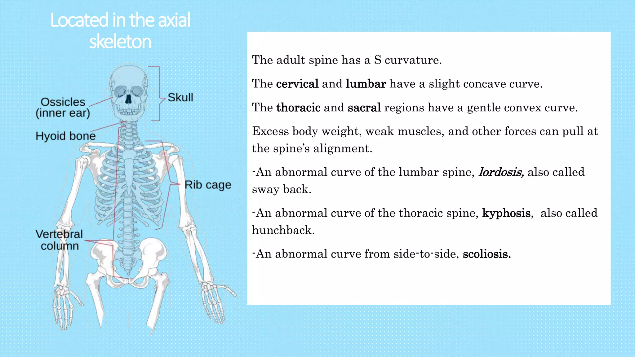
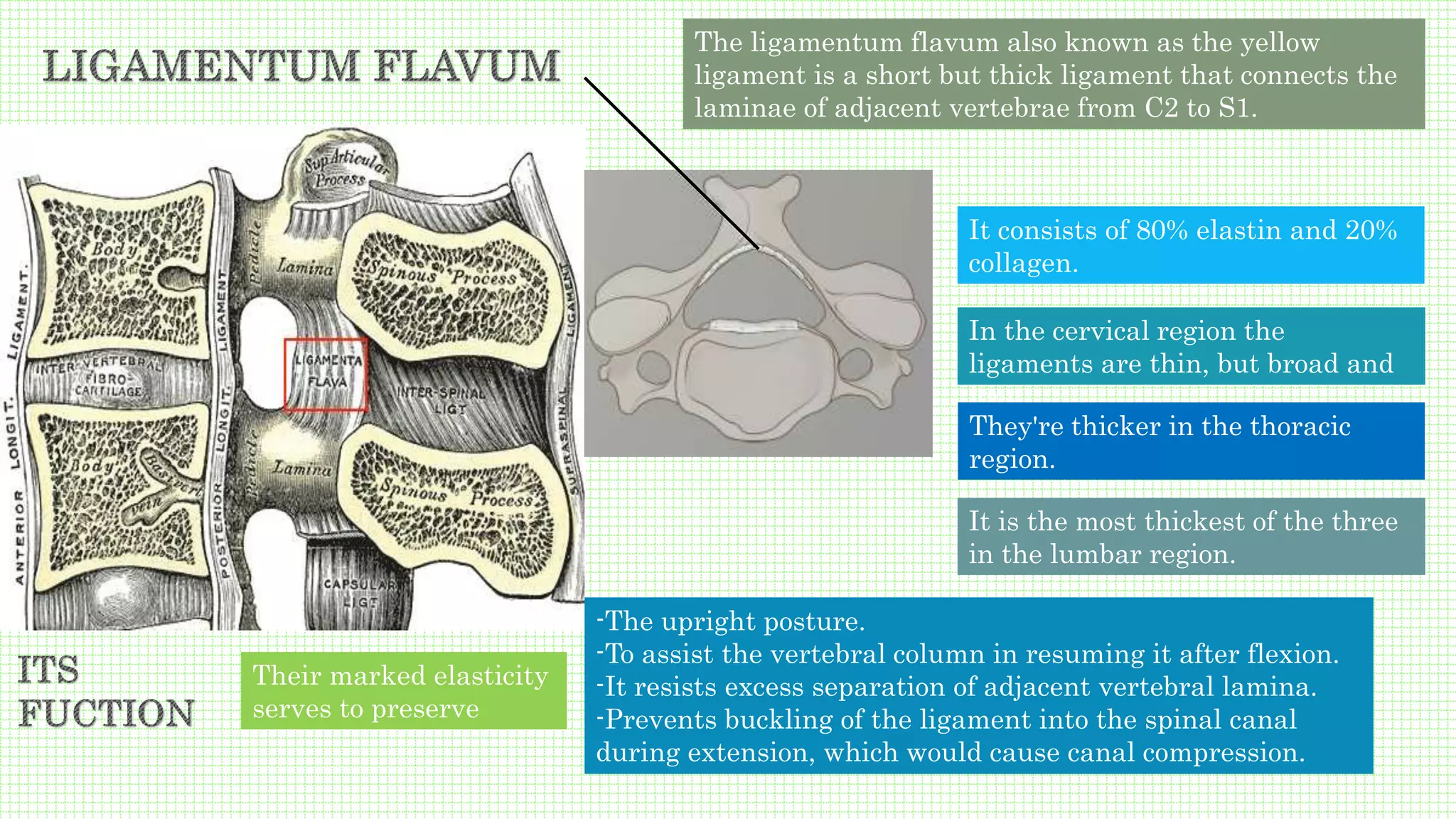
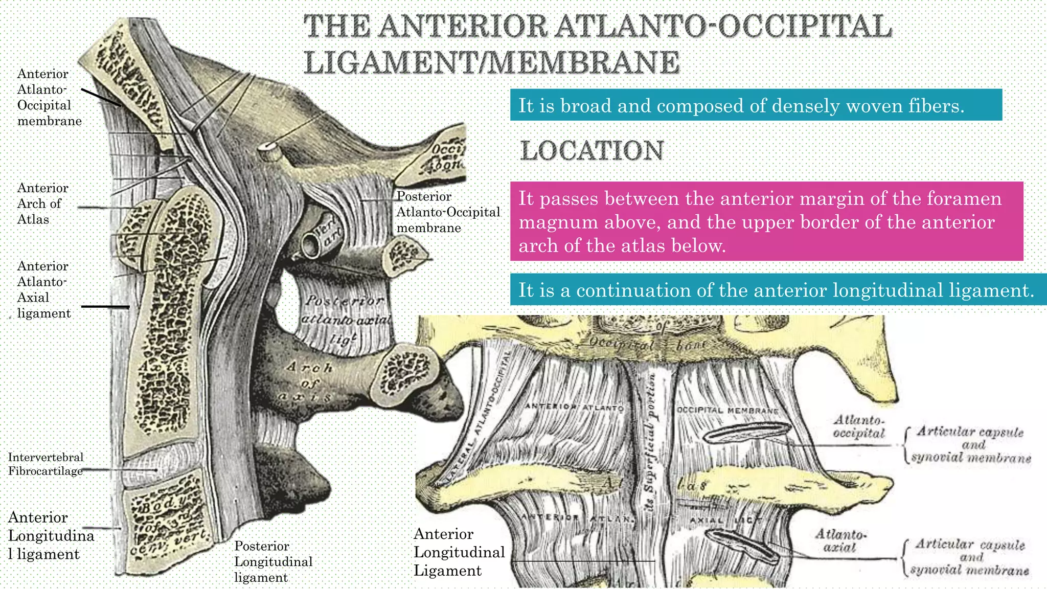
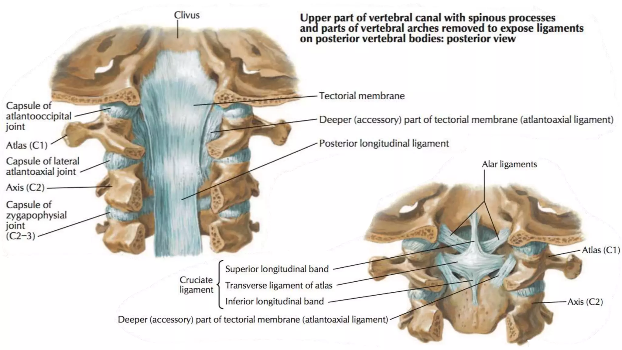
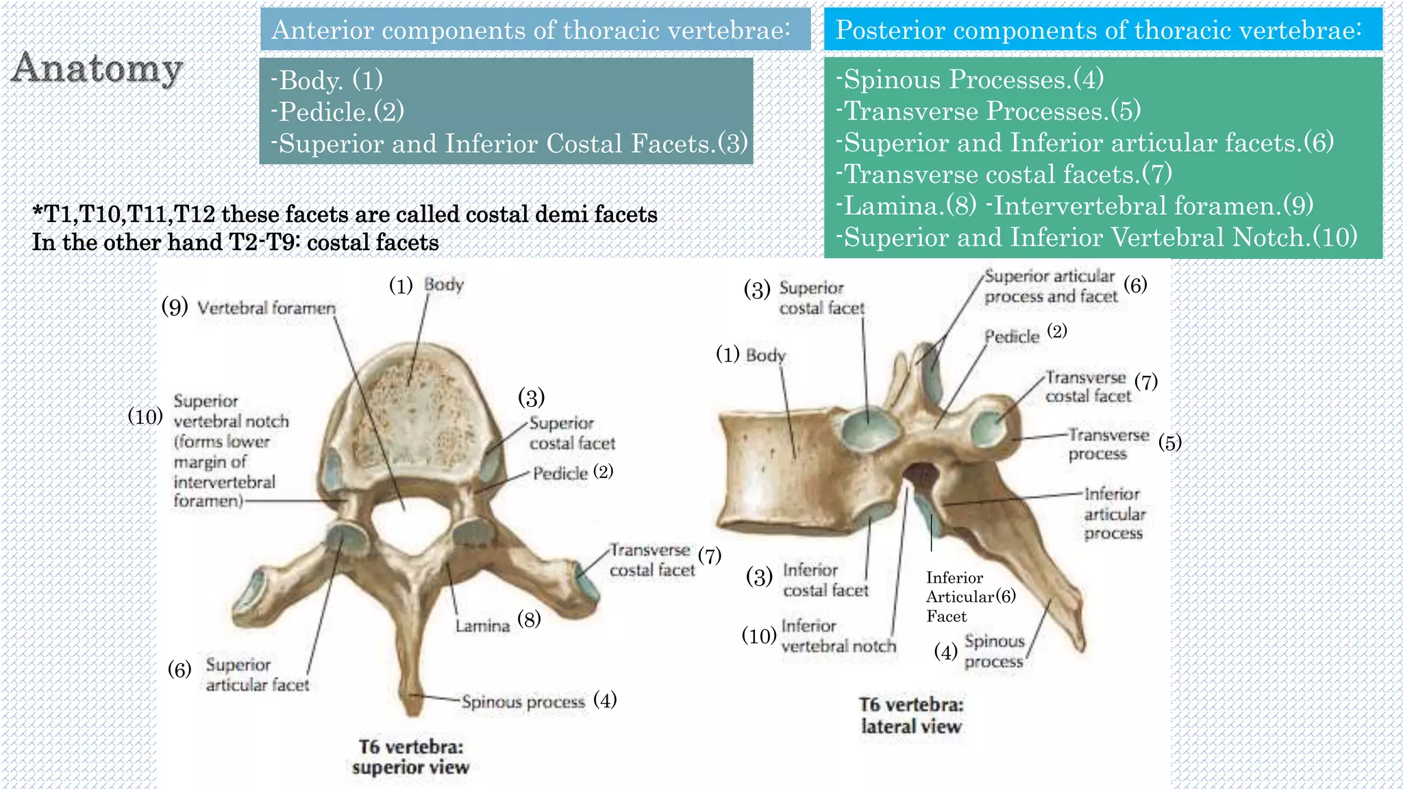
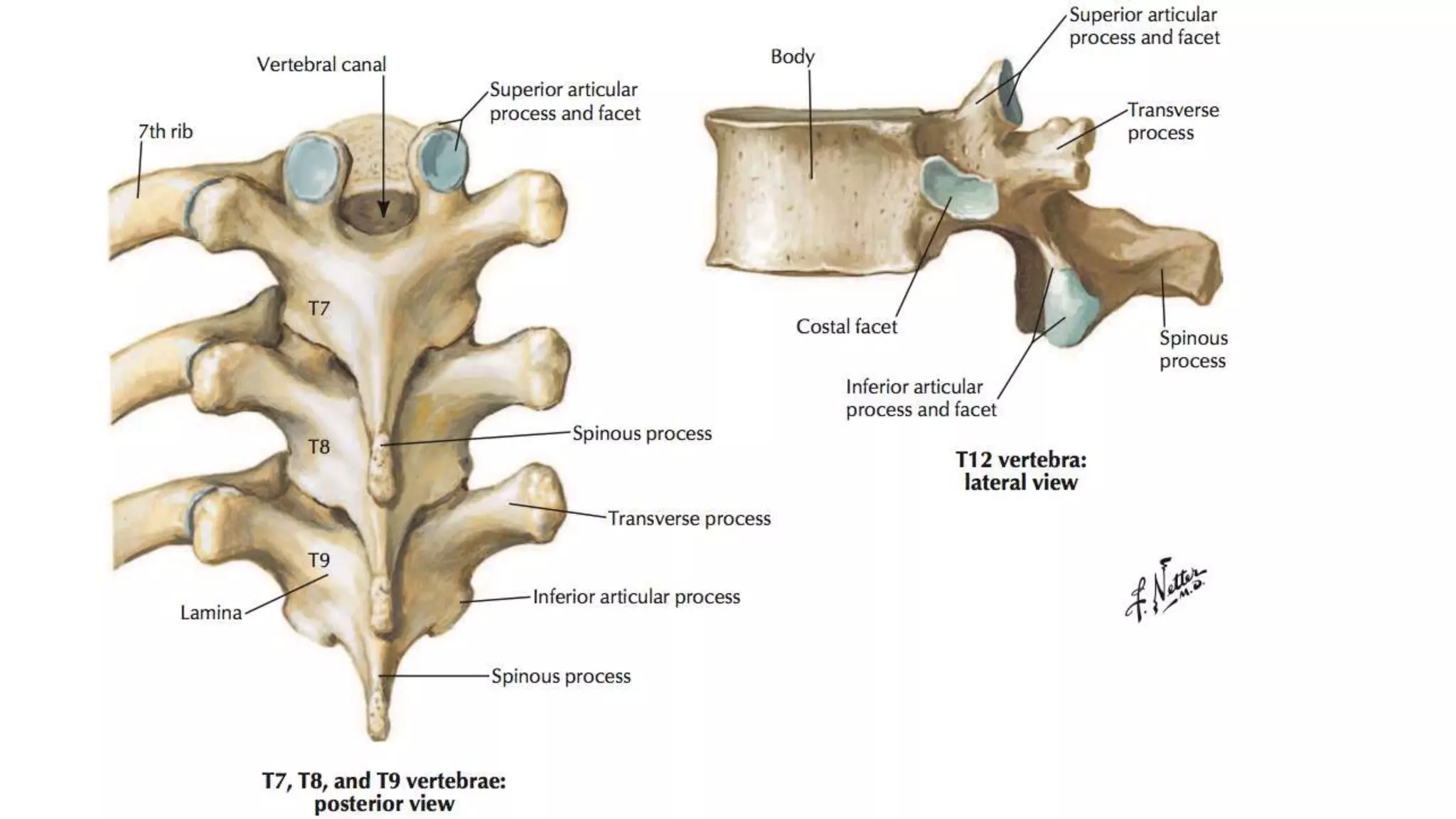
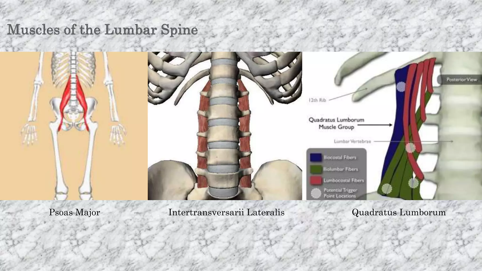
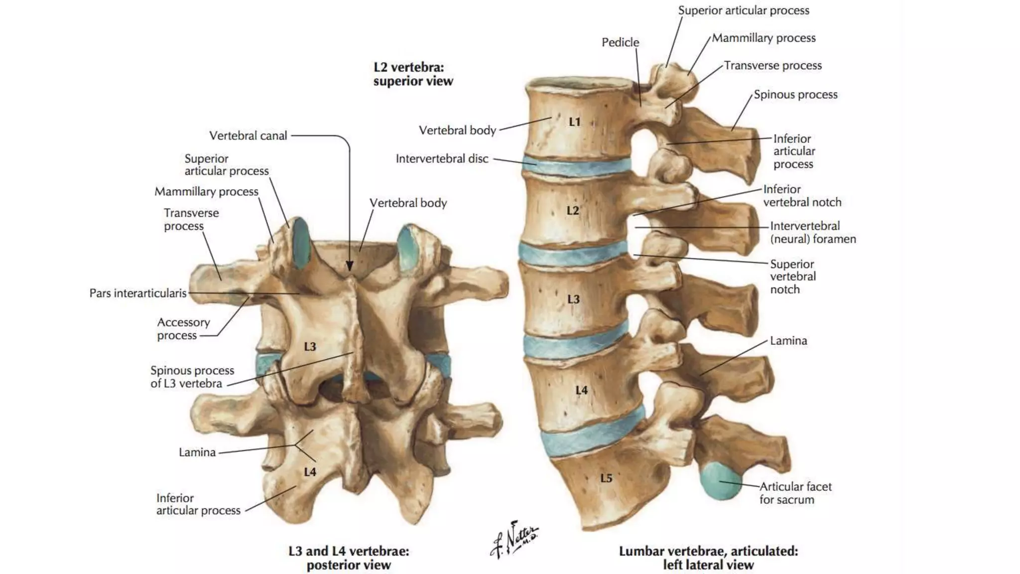
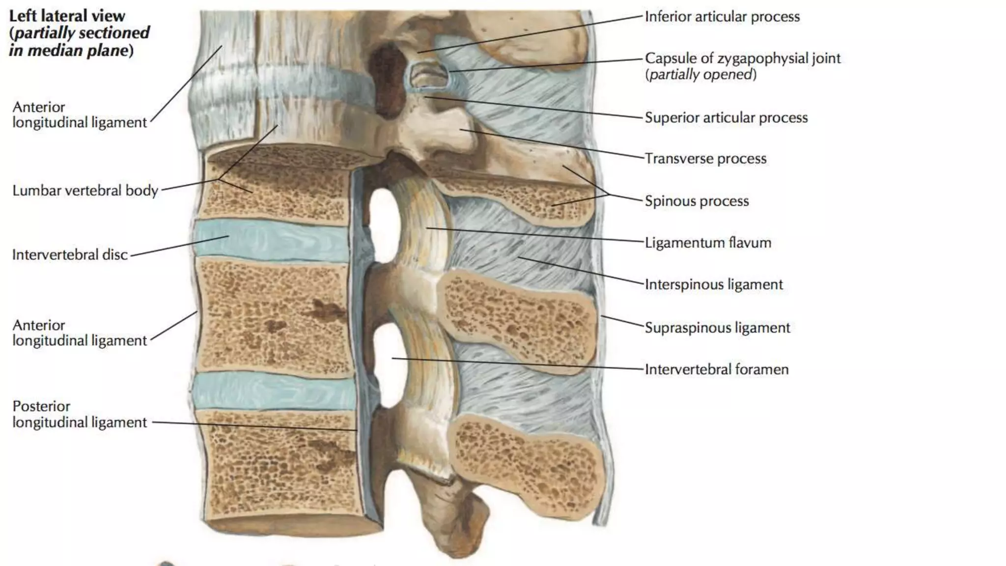
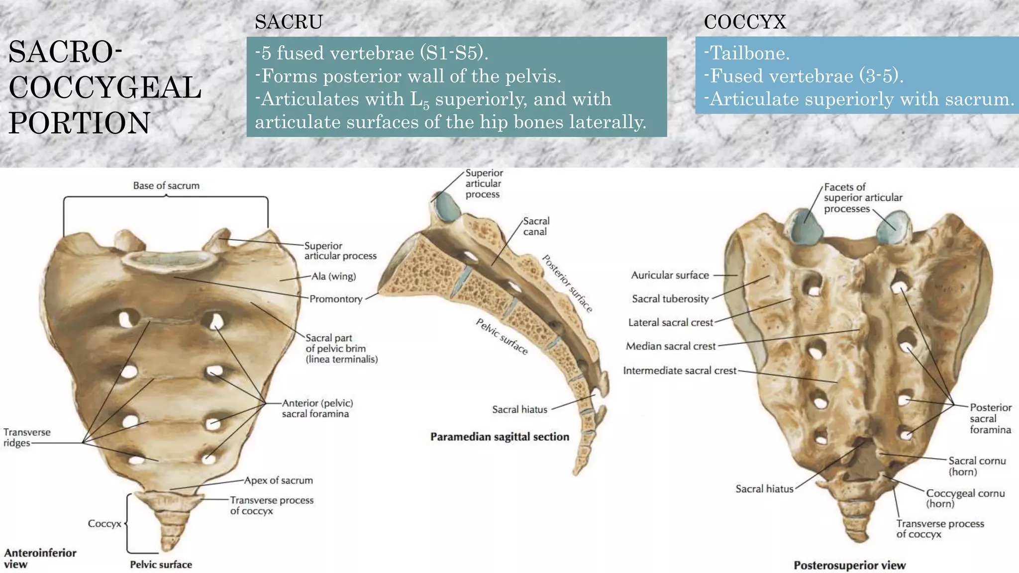
The vertebral column
-Anatomy descripcion
-Its function
-Joints
SpinalColumn
The vertebral column is generally composed of 33
vertebral segments.
Only the top 24 are moveable.
The sacral and coccygeal are fused.
Five parts:
-Cervical (The neck): 7 vertebraes
-Thoracic (The chest and ribs): 12 vertebraes
-Lumbar (The lower back): 5 vertebraes
-Sacral (Pelvic girdle): 5 vertebraes
-Coccygeal (Caudal): 4 vertebraes
It provides the main support
for your body, allowing you to
stand upright, bend, and
twist, while protecting the
spinal cord from injuries.
Descripcion:
Locatedintheaxial
skeleton
The adult spine has a S curvature.
The cervical and lumbar have a slight concave curve.
The thoracic and sacral regions have a gentle convex curve.
Excess body weight, weak muscles, and other forces can pull at
the spine’s alignment.
-An abnormal curve of the lumbar spine, lordosis, also called
sway back.
-An abnormal curve of the thoracic spine, kyphosis, also called
hunchback.
-An abnormal curve from side-to-side, scoliosis.
Movements in
sagittal view
(Flexion-
Lateral
Movements
Lateral
movements
Head view
In summary:
Flexion
Extension
Hyper-Extension
Rotation
Lateral Movements (Left-Right)
Most vertebrae are composed of:
-A body: Notes that C1 has lots its body
-A vertebrae arch (also called the neural arch or sometimes vertebral foramen):
made of pedicles (wall) and laminas (roof), that form a vertebral canal (also called a
neural canal) within wich lies the spinal cord.
-Processes: on the dorsal portion of most vertebrae that provides sites for muscle
attachment, articulation with other vertebrae and sometimes articulation with ribs.
Two types of vertebrae:
TYPICAL ATYPICAL
A basic anatomical structure composed of
all components.
The vertebrae whose structure is a little
bit different from that of the basic
anatomy due to vertebra function and
position.
Cervical Vertebrae
C3 to C6 C1,C2 and C7
Thoracic Vertebrae
T2 to T8 T1 and T9 to T12
Lumbar Vertebrae
L1 to L4 L5
Moreover, most vertebrae in an adults’ vertebral
column are typical while few are atypical.
CERVICAL
PORTION
The main function of the cervical spine is to support de weight of the head. (About 10
pounds).
The seven cervical vertebraes are numbered from C1 to C7.
The cervical part lets the skull realized different movements.
In Greek Mythology, Atlas was
a TITAN who was responsible for
bearing the weight of the
heavens on his shoulders, a
punishment bestowed on him
by ZEUS.
ATLAS
A ring-like structure and the absence of a body.
With the cervical axis, those two are called craniovertebral vertebrae.
The joints between the vertebral arches are known as a type of synovial joint.
Thus, there are 4 main features we need to consider about the Atlas:
-Short Anterior Arch -Long Posterior Arch -Lateral Masses -Transverse Processes
The most anterior part of the anterior
arch forms the anterior tubercle.
The anterior surface is convex and
projects outwards, in an anterior
direction.
The anterior tubercle
The anterior tubercle is extremely
important for the attachment of a
ligament, the anterior longitudinal
ligament.
Inferior view
THE ANTERIOR ARCH
The anterior longitudinal ligament is a strong, fibrous band that runs along
the anterolateral aspects of vertebrae and IV discs.
The anterior longitudinal ligament is thin over the
vertebrae, but thick over the IV discs.
It’s the only ligament to prevent
hyperextension of the vertebral
column.
It extends superiorly from the
pelvic (anterior surface) of the
sacrum to the anterior tubercle
of the Atlas.
Also extends superiorly above
the anterior tubercle, to connect
to the anterior surface of the
foramen magnum
Foramen magnum
Anterior
surface
Anterior
surface’s
sacrum
Either side of the midline, the anterior tubercle also
gives attachment to the superior oblique part of the
longus colli muscle.
Longus Colli Muscle is Greek
for long muscle of the neck.
It appears to be
somewhat
fusiform shaped
The longus colli muscle spans
superiorly from the anterior tubercle
of the atlas, as stated before,
inferiorly until the T3 vertebra.
The Superior Oblique Portion or
Upper Part of the Longus
Colli originates from the anterior
tubercles of the Atlas to the
transverse processes of the C3,
C4 and C5 vertebrae.
Inferior Oblique Portion or
Lower Part of the Longus
Colli originates from the
front of the bodies of T1, T2
and sometimes T3 and It
ascends obliquely
and laterally inserts to
the transverse processes of
C5 and C6.
Vertical Portion or Middle Part
of the Longus Colli originates
from the anterior arch of the T3-
T1/C7-C6 to insert in the
anterior tubercle of the C4-C2.
The Longus Colli
Muscle is involved in
moving the neck or
vertebral column in the
neck.
Neck flexion: bending the neck/head straight down
Neck rotation: turning the head to the left and right
Neck ipsilateral rotation/flexion: combining neck
flexion and rotation, allows a person to turn their
head to the side while their neck is bent forward
Neck Flexion
Neck extension
It extends posteriorly until the posterior most tubercle, the posterior tubercle of the atlas.
Posterior
Tubercle
Serves as a rudimentary spinous
process of the C1, since no
proper spinous process exists.
The posterior tubercle gives
attachment to a ligament,
known as the ligamentum
nuchae.
THE POSTERIOR ARCH
Is a large median ligament composed of
tendons and fascia located between the
posterior muscles of the neck.
It is a superior and posterior extension
of the supraspinous ligament.
It extends
from
the occipital
protuberance
to the spinous
process of
the C7
vertebra.
Limits flexion of the neck.
To sustain the weight of the head.
Provides an attachment for Trapezius and Splenius capitis.
Occipital
protuberance
Vertebrae CVII
The superior part consists
of medial fibers from the
cervical trapezius.
The part of the
ligamentum
nuchae that
attaches to muscles
The inferior part consists of
interweaving tendons from splenius
capitis and rhomboid minor.
Splenius Capitis BASE
APEX
Spine
Post tubercles
of C1-C6
Spine of C7
External Occipital Crest
Free
border
The supraspinous ligament is a thick, cord
like fibrous band that connects the apices of
the spinous processes.
It from the vertebrae
C7 to the S1
It is continuous with the ligamentum nuchae
above C7, and it continuous
with interspinous ligaments between the
spinous processes.
They’re thin, fibrous membranes that connect adjacent spinous
processes together
The interspinous ligaments are narrow in the thoracic region,
but thicker in the lumbar region.
Both the supraspinous
ligament and the interspinous
ligament prevent hyperflexion
of the vertebral column.
In the cervical region, they are very underdeveloped, and are
considered part of the ligamentum nuchae.
From C1 to the
S1
The ligamentum flavum also known as the yellow
ligament is a short but thick ligament that connects the
laminae of adjacent vertebrae from C2 to S1.
It consists of 80% elastin and 20%
collagen.
In the cervical region the
ligaments are thin, but broad and
long.
They're thicker in the thoracic
region.
It is the most thickest of the three
in the lumbar region.
Their marked elasticity
serves to preserve
-The upright posture.
-To assist the vertebral column in resuming it after flexion.
-It resists excess separation of adjacent vertebral lamina.
-Prevents buckling of the ligament into the spinal canal
during extension, which would cause canal compression.
The facet joint capsule is an area of
connective tissue that covers and
closes the facet joint.
Also known as the
zygapophysis or
zygapophyseal joint.
This connective tissue creates a sort of bulky seam that
holds the two bones forming the facet joint together.
The facet joint capsule resists forces of tension that are
developed across the joint when it rotates.
Each vertebra has four facet joints.
Two on the top (called superior facets) and two on the
bottom (called inferior facets).
On the inside of the joint cavity is a lining (called the
synovial lining) that makes synovial fluid.
It lubricates the bones that make up the facet joint, as
well as the space between them, which helps make for
smooth, frictionless motion in the joint.
The face joint capsule is richly supplied with nerves
which play a role in pain perception.
The American Journal of Neuroradiology say that
facet joints may be responsible for up to 45% of low
back pain cases and up to 55% cases of chronic neck
pain without disc herniation.
The next continuation we’ll talk in more detail
about the disc herniation and synovial fluid.
The Posterior Longitudinal
Ligament runs up and down
behind (posterior) the spine
and inside the spinal canal.
This ligament is composed of
smooth, shining, longitudinal
fibers, denser and more compact
than those of the anterior
ligament
The posterior longitudinal ligament, anterior
longitudinal ligament and ligamentum flavum are three
extremely important components of the spinal anatomy
because they prevent the hyper-flexion of the body
together.
It prevents the excesive
flexion of the body.
It becomes tectorial
membrane in the
craniovertebral region much
broader and stronger.
The Posterior Tubercle also brings attachment to some muscles
The Rectus Capitis Posterior Minor The Interspinalis Cervicis
The superior surface of the posterior arch provides attachment to the posterior atlanto-occipital membrane.
The Rectus Capitis Posterior Major
It is a broad, thin fibrous membrane.
It extends from the superior surface of the posterior arch,
superiorly till the posterior margin of the foramen magnum.
Both the posterior atlanto-occipital membrane and posterior atlanto-axial
ligament are continuations of the ligamentum flavum.
In the inferolateral
part of the
membrane allows
the passage of the
C1 spinal nerve
and vertebral
Anterior
Atlanto-
Occipital
membrane
Anterior
Arch of
Atlas
Anterior
Atlanto-
Axial
ligament
Intervertebral
Fibrocartilage
Anterior
Longitudina
l ligament
Posterior
Longitudinal
ligament
Posterior
Atlanto-Occipital
membrane
It is broad and composed of densely woven fibers.
It passes between the anterior margin of the foramen
magnum above, and the upper border of the anterior
arch of the atlas below.
It is a continuation of the anterior longitudinal ligament.
Anterior
Longitudinal
Ligament
The most bulky part of the Atlas.
To support the weight of the globe of the head.
They articulate with the occipital condyles of the base of the
skull, to form the atlanto-occipital joint.
LATERAL
MASSES
Occipital Condyles
Stuff that supports the joint
FIBROUS CAPSULE
Surround the joint
Thick posterolaterally and anteromedially
ANTERIOR ATLANTO OCCIPITAL
LIGAMENT/MEMBRANE
Anterior margin of foramen above to upper border of
anterior arch of atlas below.
Anteriorly strengthened by anterior longitudinal ligament.
Laterally continuous with the anterior part of capsular
ligament.
Blood supply:
Vertebral artery
Nerve supply:
C1 nerve
TRANSVERS
E
PROCESSES
They’re the lateral most structures of the Atlas.
They contain the most important part, the foramen
transversarium, that transmits the vertebral artery.
It also provides attachment to a large number of muscles.
They’re the point of articulation of the ribs (in the thoracic spine).
The transverse processes help
reinforce the framework of the
spinal canal, which is a long
space via which the spinal cord
goes through the base of the
skull till the lower back.
Rectus Capitis Lateralis Obliqus Capitis
Superior
Obliqus Capitis Inferior
Levator Scapulae Splenius Cervicis Rectus Capitis Anterior
The Longus Capitis
Movement Muscles Involved
Flexion Longus Capitis
Rectus Capitis Anterior
Extension
Rectus Capitis Posterior Major and
Minor
Oblique Capitis Superior
Semispinalis Capitis
Splenius Capitis
Trapezius
Lateral Flexion
Rectus Capitis Lateralis
Semispinalis Capitis
Splenius Capitis
Sternomastoid
Trapezius
*I will check these muscles in the muscles of the neck
The others muscles aren’t attach on the Atlas.
Inferior View
AXI
S
It is the largest and heaviest cervical segment.
The odontoid process (dens) articulates with the anterior arch of the atlas, around which the
atlas rotates and bends laterally.
In contrast to the other cervical vertebrae, C2 does not have a discrete pedicle.
It has a very large and prominent spinous process.
A superior, tooth-like projection.
The rotation of the head side-to-side allows that the atlas
bone rotates around the peg-like odontoid processes.
This is the reason why the second cervical vertebra is also
called the axis bone.
Axis (An imaginary center line which something rotates).
This structure and his movement made by the Atlanto-Axial
joint
The ligaments that surround these bones are:
-Articular Capsules.
-Posterior Atlanto-Axial ligament.
-Anterior Atlanto-Axial ligament.
-Transverse ligament of the atlas.
-Alar ligament.
-Apical ligament.
-Tectorial membrane.
It is a complex joint made up of three synovial joints and
constitutes the most mobile articulation of the spine.
The two alar ligaments and transverse ligaments are the three
synovial joints.
Two capsules. They’re thick and loose.
They connect the margins of the Lateral
Masses of the atlas with those of the
Posterior Articular Surfaces of the Axis.
Each is strengthened
A broad, thin membrane.
It’s attached, above, to the lower border of the posterior
arch of the atlas; below, to the upper edges of the
lamina of the axis.
It is a continuation of the Ligamentum Flavum.
It is in relation, behind, with the obliqus
capitis inferior muscle.
The posterior
Atlanto-Axial
ligament
It contributes the stabilization of the atlanto-axial joint
movement that overall allows 10-15° of
flexion/extension and 30° of axial rotation.
A strong membrane, fixed.
It is above, to the lower border of the anterior
arch of the atlas; below, to the front of the body
of the axis.
It is a continuation upward of
the Anterior longitudinal ligament.Anterior
Atlanto-Axial
ligament The ligament is in relation, in front, with
the longi capitis.
The Anterior Atlanto-Axial Ligament
The Posterior Atlanto-Axial Ligament
They formed a complex structure called Atlanto-Axial
Ligament Complex (Axis).
It is actually a collection of 3 ligaments.
-The transverse ligament of the atlas.
-Superior. longitudinal band.
-Inferior longitudinal band.
The superior and inferior longitudinal bands are simply
superior and inferior extensions from the middle of the
transverse ligament of the atlas.
A cross-shaped structure of ligaments, and hence the
name, cruciate (cross like).
The superior longitudinal band extends superiorly from the
transverse ligament of the atlas, attaching on the clivus deeper
through the foramen magnum than the apical ligament.
The inferior longitudinal ligament extends inferiorly from the inferior border of the transverse ligament of
the atlas towards the body of the axis.
The lateral mass contains a small tubercle for the attachment of a
ligament known as the transverse ligament of the atlas.
It extends from the medial surface of one lateral mass to the medial surface of the
lateral mass on the opposite side.
The transverse ligament of the atlas bounds
the odontoid process posteriorly.
This is known as the median atlanto-axial
joint, formed between the odontoid process.
Non-elastic, fails suddenly with quick force.
Prevents excesive anterior shift.
A pair strong of cord like
ligaments.
Arise from either side of the odontoid process and attach to
the medial aspect of the occipital condyles.
Taut in flexion, limit rotation
and side flexion to the opposite
side.
Play a role in stabilizing C1 and
C2, especially in rotation.
A fibrous cord in the triangular interval between
the alar ligaments.
Arises from the tip of the odontoid process on the
Axis (usually from a small coronal groove located
there) and inserts into the anterior rim of the
foramen magnum at the basion (mid-point).
A vestigial structure
Being intimately blended with the deep portion
of the anterior atlanto-occipital membrane and
superior crus of the transverse ligament of the
atlas.
They are interposed
between the transverse
processes.
Cervical region: They consist of a few irregular,
scattered fibers.
Thoracic region: They are rounded cords intimately
connected with the deep muscles of the back.
Lumbar region: They are thin and membranous.
Limit lateral
flexion of the spine.
Strong broad structure which covers the Odontoid process and it's ligaments.
Continuous with the Posterior longitudinal ligament and found on the
internal surface of the vertebral canal.
Arises from the posterior surface of the body of the Axis and, expanding as it
ascends, is attached to the basilar groove of the occipital bone, in front of the
foramen magnum.
Its anterior surface is in relation with the Transverse ligament of the atlas
and its posterior surface with the dura mater. As it enters the cranial cavity it
becomes continuous with the dura mater.
Contributes to the stability of the upper cervical spine. Limits Flexion C0-C1
and C1-C2 and rotation C0-C1.
There are six cervical discs.
The first disc is between the axis (C2) and C3.
The disc, comprised of an annulus fibrosus, a
nucleus pulposus and two cartilaginous endplates.
To serve as the spine's shock absorbing system, which protect
the vertebrae, brain, and other structures (i.e. nerves).
The discs allow some vertebral motion: extension and flexion,
and lateral bending.
Individual disc movement is very limited.Links adjacent vertebral bodies together.
A type of cartilaginous joint, known as a symphysis.
The annulus fibrosus
encloses the nucleus
pulposus.
Outer structure that
encases the nucleus
pulposus.
Characterized by high tensile
strength and its ability to
prevent intervertebral
distraction.
Remains flexible enough
to allow for motion.
Composed of type I collagen that is obliquely oriented, water, and proteoglycans.
Fibroblast-like cells
Responsible for producing type I collagen and proteoglycans
It contains a hydrated gel–like matter that resists compression.
Composed of type II collagen (50%
approximately) type I (20%)
, water, and proteoglycans.
Aggrecan is a proteoglycan primarily
responsible for maintaining water content
of the disc interact with water and resist
compression.
Chondrocyte-like cells
Responsible for producing type II collagen and proteoglycans survive in hypoxic conditions.
Approximately 88% water
Most flexion–extension takes place
at the joints C3–C4, C4–C5 and
especially C5–C6.
Lateral flexion and axial
rotation occur mainly at C2–C3,
C3–C4, C4–C5.
Here some pictures of the
anatomy in this portion that
is called the cervical spine.
Also known as Joints of Luschka.
The uncovertebral joints are present from C3 to C7. Formed between uncinate processes below, and the
uncus above.
They allow for flexion and extension and limit lateral flexion in the cervical spine.
Thought to reinforce the intervertebral disc postero -laterally and therefore provide protection for structures
at risk of disc herniation.
Prevents posterior linear translation movements of the vertebral bodies.
Important in providing stability and guiding the motion of the cervical spine.
Uncinate processes
THORACIC PORTION
12 vertebrae connected with
intervertebral discs. T1-T12.
Presence of facets for the joints with
the ribs (all except the 11th and 12th
vertebra).
The main function of the thoracic
spine is to hold the rib cage and
protect the heart, lungs and
oesophagus.
The typical: T2 to T9. The atypical: T1, T10, T11, T12.
Has on either side of the body, an entire articular fact for the head of the first rib,
and a demi-facet for the upper half of the head of the second rib.
The spinous process is thick, long, and almost horizontal.
The transverse processes are long, with the upper vertebral notches deeper than
any of those found on the other thoracic vertebrae.
A single pair of whole facets is present which articulate with the 10th rib.
Each have a single pair of entire costal facets, which are located on the pedicles.
It is larger than the 1st Thoracic vertebrae.
The thoracic spinal nerve 2 passes through underneath T2.
The thoracic spinal nerve 3 passes through underneath T3.
The thoracic spinal nerve 4 passes through underneath T4.
The trachea branches into the two main bronchi at the level of T5.
The thoracic spinal nerve 5 passes through underneath T5.
The thoracic spinal nerve 6 passes through underneath T6.
The thoracic spinal nerve 7 passes through underneath T7.
The T8 and T9 vertebrae are found at the same level as the
xiphoid process.
The thoracic spinal nerve 8 passes through underneath T8.
The thoracic spinal nerve 9 passes through underneath T9.
Anterior components of thoracic vertebrae:
-Body. (1)
-Pedicle.(2)
-Superior and Inferior Costal Facets.(3)
Posterior components of thoracic vertebrae:
-Spinous Processes.(4)
-Transverse Processes.(5)
-Superior and Inferior articular facets.(6)
-Transverse costal facets.(7)
-Lamina.(8) -Intervertebral foramen.(9)
-Superior and Inferior Vertebral Notch.(10)
(2)
(1)
(1)
(2)
*T1,T10,T11,T12 these facets are called costal demi facets
In the other hand T2-T9: costal facets
(3)
(3)
(3)
(4)
(4)
(5)
(7)
(7)
(8)
(9)
(6)
(6)
(10)
(10)
Inferior
Articular
Facet
(6)
With the ribs:
Each vertebra contains three points articulations with ribs.
The superior demi facet that is called the costovertebral joint.
The transverse costal facet articulates again with the same rib
of the superior demi facet with the costovertebral joint.
The inferior demi facet articulates with the rib below.
For example, the superior demi facet and costal facet on
the transverse process of T5 will articulate with the fifth
rib.
The inferior demi facet of T5 will articulate with the
sixth rib.
The articulations that connect the heads of the ribs with the
bodies of the thoracic vertebrae.
Joining of ribs to the vertebrae occurs at two places, the
head and the tubercle of the rib.
A number of small ligaments also support
the costovertebral joints.
At the same time we’ll see the two types of joints that are attach with the thoracic spine.
Those that are present throughout the vertebral column:
Between vertebral bodies – adjacent vertebral bodies
are joined by intervertebral discs.
Between vertebral arches – formed by the articulation
of superior and inferior articular processes from
adjacent vertebrae.
For Example:
-Anterior and Posterior Longitudinal Ligaments.
-Ligamentum Flavum.
-Interspinous Ligament.
-Supraspinous Ligament.
-The Facet Joint Capsule.
Radiate ligament of head of rib – three
bands which connect the rib head to the two
vertebral bodies and intervertebral disc.
Costotransverse ligament – Connects the
neck of the rib and the transverse process.
Those unique of the thoracic spine:
Lateral costotransverse ligament –
Extends from the transverse process
to the tubercle of the rib.
Superior costotransverse ligament –
Passes from the upper border of the
neck of the rib to the transverse
process of the vertebra superior to it.
Here some pictures of the thoracic
spine.
LUMBAR
PORTION
Most people have five bones (vertebrae) in the lumbar
spine, although it is not unusual to have six.
Is the third major region of the spine.
They are the largest
of the vertebrae
because of their
weight-bearing
function supporting
the torso and head.
A strong band of connective tissue.
One of the three vertebropelvic ligaments.
They join the 4th and 5th
lumbar vertebrae (L4 and
L5) to the iliac bone crest
at the back of the pelvis.
The portion of the iliolumbar ligament originating from the L-5
transverse process is made up of two bands (anterior and posterior).
It’s broad and flat and has two different anatomic varieties.
It has two different anatomic varieties.
Type 1 originates from the anterior aspect of the inferolateral portion of the L-5 transverse
process and fans out widely before inserting on the anterior portion of the iliac tuberosity.
Type 2 originates anteriorly, laterally, and posteriorly from inferolateral aspect of the L-5
transverse process and fans out before inserting on the anterior portion of the iliac tuberosity.
It originates from the apex of the L-5 transverse process and is
fusiform.
It’s thinner than the anterior, with a smaller insertional base on the iliac crest,
which explains its lesser resistance to torsional overloading.
It plays an important role in restraining
movement in lumbosacral and the
sacroiliac joints.
The ililumbar ligament strengthen the
lumbosacral joint assisted by:
-The lateral lumbosacral ligament vertebral
joint.
-The posterior and anterior longitudinal
ligaments.
-The ligamenta flava.
-The interspinous and supraspinous
ligaments.
Iliolumbar ligament
Psoas Major Intertransversarii Lateralis Quadratus Lumborum
Interspinales Lumborum Multifidus Rotadores
SpinalisLongissimus LumborumIliocostalis
-Posterior longitudinal
ligaments.
-Anterior longitudinal
ligaments.
-Intertransverse
ligaments.
-Interspinous ligaments.
-Supraspinous
ligaments.
Here some images of the
Lumbar Spine.
SACRO-
COCCYGEAL
PORTION
SACRU
M-5 fused vertebrae (S1-S5).
-Forms posterior wall of the pelvis.
-Articulates with L5 superiorly, and with
articulate surfaces of the hip bones laterally.
COCCYX
-Tailbone.
-Fused vertebrae (3-5).
-Articulate superiorly with sacrum.
The superior lateral surface on either side of the
sacrum articulates with the inner aspects of the pelvis.
This area forms the capsular, synovial
Sacroiliac joint.
It transmits all the forces of the upper body to the pelvis and legs.
There are many more ligaments in the back of the
sacroiliac joint than in the front.
In back, the ligaments are:
-The interosseous sacroiliac ligament.
-The posterior sacroiliac ligament.
-The iliolumbar ligament.
-The sacrotuberous ligament.
-The sacrospinous ligament.
In front:
-The anterior longitudinal ligament.
Located between the ilium and sacrum.
It helps maintain joint congruency and is located where
most of the weight transfer from the spine to the hip
(and lower extremity) occurs.
Short, strong, fibers connecting tuberosities
of the sacrum and ilium.
There are two parts:
Short sacroiliac ligaments:
Composed of horizontal fibers extending
from the sacrum to the posterior part
of the iliac bone.
Long sacroiliac ligaments:
Composed of fibers extending vertically
from the sacrum to the posterior
superior iliac spine.
Series of flat band.
It stabilizes sacroiliac joint.
It limits anterior rotation/displacement of
pelvic girdle.
It extends from the lateral part of the
Sacrum and Coccyx and the posterior
inferior iliac spine.
It is largely comprised of strong collagen fibers.
It diminishes the range of
sacroiliac joint motion.
It is attached to the ischial spine (a bone prominence in the
lower pelvis) and the lateral (side) regions of the sacrum (at
the bottom of the spine) and coccyx, or tailbone.
The sacrospinous ligament borders the sacro tuberous
ligament and shares fiber space with this ligament.
The sacrospinous and sacro tuberous ligaments work together
to limit the upward tilt of the sacrum, called nutation.
Here some pictures.
The vertebral Column Descripcion
Movements
Spinal Overview
Types of vertebrae
Typical Atypical
Cervical Portion
C1 or Atlas
Anatomy
Short Anterior Arch
Long Posterior Arch
Lateral Masses Transverse Processes
Anterior Tubercle
Anterior Longitudinal Ligament
Longus Colli Muscle
Posterior Tubercle
Ligamentum Nuchae
Supraspinous Ligament
Interspinous Ligament
Ligamentum Flavum
Facet Joint Capsule
Posterior Longitudinal
Ligament
The Rectus Capitis
Posterior Minor
Muscle
The Interspinalis Cervicis
The Rectus Capitis
Posterior Major Muscle
The Anterior Atlanto-Occipital
Ligament Membrane The Posterior Atlanto-
Occipital
Ligament MembraneAtlanto-
Occipital
Joint
Rectus Capitis
Lateralis Muscle
Obliqus Capitis
Superior Muscle
Obliqus Capitis
Inferior Muscle
Levator Scapulae
Splenius Cervicis
Rectus Capitis Anterior
The Longus Capitis
C2 or Axis
Anatomy
Dens
Atlanto-Axial Joint
Articular Capsule
Posterior Atlanto-Axial Ligament
Anterior Atlanto-Axial Ligament
The Cruciate Ligament
The Transverse Ligament
Alar Ligament Apical Ligament
Intertransversal Ligament
Tectorial Membrane
Intervertebral Discs (Cervical Portion)
Annulus Fibrous
Nucleus Pulposus
C3-C7 Spinal Column Uncovertebral Joints Summary Typical C.V.
Atypical C.V.
C3-C6
C1-C2 and C7
Thoracic Portion
Lumbar Portion
Typical T.V
Atypical T.V 1st Thoracic Vertebrae 10th Thoracic Vertebrae 11th 12th Thoracic
Vertebrae
2nd Thoracic Vertebrae 3th Thoracic Vertebrae 4th Thoracic Vertebrae
5th Thoracic Vertebrae 6th 7th Thoracic
Vertebrae
8th 9th Thoracic
Vertebrae
Anatomy
Joints Costovertebral
Joints
Summary
Iliolumbar Ligament
Psoas Major Muscle
Intertransversarii
Lateralis Muscle
Quadratus
Lumborum Muscle
Interspinales
Lumborum Muscle
Multifudus MuscleRotadores Muscle
Iliocostalis Muscle
Longissimus
Lumborum Muscle
Spinalis Muscle
Posterior
Longitudinal
Ligament
Anterior
Longitudinal
Ligament
Interspinous
Ligament
Supraspinous
Ligament
Intertransverse
Ligament
Summary
Typical
Atypical
L1-L4
L5
-Anterior and Posterior Longitudinal Ligaments.
-Ligamentum Flavum -Interspinous Ligament –Intervertebral
Discs
-Supraspinous Ligament -The Facet Joint Capsule.
-Radiate Ligament -Costotransverse Ligament
SacroCoccygeal Portion
Sacrum
Coccyx
Sacro-Iliac Joint
The Intersseous Sacro-Iliac Ligament
Posterior Sacro-Iliac Ligament
Sacrotuberous Ligament
Sacrospinous Ligament
Summary

Vertebral column

  • 1.
    The vertebral column -Anatomydescripcion -Its function -Joints
  • 2.
    SpinalColumn The vertebral columnis generally composed of 33 vertebral segments. Only the top 24 are moveable. The sacral and coccygeal are fused. Five parts: -Cervical (The neck): 7 vertebraes -Thoracic (The chest and ribs): 12 vertebraes -Lumbar (The lower back): 5 vertebraes -Sacral (Pelvic girdle): 5 vertebraes -Coccygeal (Caudal): 4 vertebraes It provides the main support for your body, allowing you to stand upright, bend, and twist, while protecting the spinal cord from injuries. Descripcion:
  • 3.
    Locatedintheaxial skeleton The adult spinehas a S curvature. The cervical and lumbar have a slight concave curve. The thoracic and sacral regions have a gentle convex curve. Excess body weight, weak muscles, and other forces can pull at the spine’s alignment. -An abnormal curve of the lumbar spine, lordosis, also called sway back. -An abnormal curve of the thoracic spine, kyphosis, also called hunchback. -An abnormal curve from side-to-side, scoliosis.
  • 5.
  • 6.
  • 7.
  • 8.
    Most vertebrae arecomposed of: -A body: Notes that C1 has lots its body -A vertebrae arch (also called the neural arch or sometimes vertebral foramen): made of pedicles (wall) and laminas (roof), that form a vertebral canal (also called a neural canal) within wich lies the spinal cord. -Processes: on the dorsal portion of most vertebrae that provides sites for muscle attachment, articulation with other vertebrae and sometimes articulation with ribs.
  • 9.
    Two types ofvertebrae: TYPICAL ATYPICAL A basic anatomical structure composed of all components. The vertebrae whose structure is a little bit different from that of the basic anatomy due to vertebra function and position. Cervical Vertebrae C3 to C6 C1,C2 and C7 Thoracic Vertebrae T2 to T8 T1 and T9 to T12 Lumbar Vertebrae L1 to L4 L5 Moreover, most vertebrae in an adults’ vertebral column are typical while few are atypical.
  • 10.
    CERVICAL PORTION The main functionof the cervical spine is to support de weight of the head. (About 10 pounds). The seven cervical vertebraes are numbered from C1 to C7. The cervical part lets the skull realized different movements.
  • 12.
    In Greek Mythology,Atlas was a TITAN who was responsible for bearing the weight of the heavens on his shoulders, a punishment bestowed on him by ZEUS.
  • 14.
    ATLAS A ring-like structureand the absence of a body. With the cervical axis, those two are called craniovertebral vertebrae. The joints between the vertebral arches are known as a type of synovial joint. Thus, there are 4 main features we need to consider about the Atlas: -Short Anterior Arch -Long Posterior Arch -Lateral Masses -Transverse Processes The most anterior part of the anterior arch forms the anterior tubercle. The anterior surface is convex and projects outwards, in an anterior direction. The anterior tubercle The anterior tubercle is extremely important for the attachment of a ligament, the anterior longitudinal ligament. Inferior view THE ANTERIOR ARCH
  • 15.
    The anterior longitudinalligament is a strong, fibrous band that runs along the anterolateral aspects of vertebrae and IV discs. The anterior longitudinal ligament is thin over the vertebrae, but thick over the IV discs. It’s the only ligament to prevent hyperextension of the vertebral column. It extends superiorly from the pelvic (anterior surface) of the sacrum to the anterior tubercle of the Atlas. Also extends superiorly above the anterior tubercle, to connect to the anterior surface of the foramen magnum Foramen magnum Anterior surface Anterior surface’s sacrum
  • 16.
    Either side ofthe midline, the anterior tubercle also gives attachment to the superior oblique part of the longus colli muscle. Longus Colli Muscle is Greek for long muscle of the neck. It appears to be somewhat fusiform shaped The longus colli muscle spans superiorly from the anterior tubercle of the atlas, as stated before, inferiorly until the T3 vertebra.
  • 17.
    The Superior ObliquePortion or Upper Part of the Longus Colli originates from the anterior tubercles of the Atlas to the transverse processes of the C3, C4 and C5 vertebrae. Inferior Oblique Portion or Lower Part of the Longus Colli originates from the front of the bodies of T1, T2 and sometimes T3 and It ascends obliquely and laterally inserts to the transverse processes of C5 and C6. Vertical Portion or Middle Part of the Longus Colli originates from the anterior arch of the T3- T1/C7-C6 to insert in the anterior tubercle of the C4-C2.
  • 18.
    The Longus Colli Muscleis involved in moving the neck or vertebral column in the neck. Neck flexion: bending the neck/head straight down Neck rotation: turning the head to the left and right Neck ipsilateral rotation/flexion: combining neck flexion and rotation, allows a person to turn their head to the side while their neck is bent forward Neck Flexion Neck extension
  • 19.
    It extends posteriorlyuntil the posterior most tubercle, the posterior tubercle of the atlas. Posterior Tubercle Serves as a rudimentary spinous process of the C1, since no proper spinous process exists. The posterior tubercle gives attachment to a ligament, known as the ligamentum nuchae. THE POSTERIOR ARCH
  • 20.
    Is a largemedian ligament composed of tendons and fascia located between the posterior muscles of the neck. It is a superior and posterior extension of the supraspinous ligament.
  • 21.
    It extends from the occipital protuberance tothe spinous process of the C7 vertebra. Limits flexion of the neck. To sustain the weight of the head. Provides an attachment for Trapezius and Splenius capitis. Occipital protuberance Vertebrae CVII
  • 22.
    The superior partconsists of medial fibers from the cervical trapezius. The part of the ligamentum nuchae that attaches to muscles The inferior part consists of interweaving tendons from splenius capitis and rhomboid minor. Splenius Capitis BASE APEX Spine Post tubercles of C1-C6 Spine of C7 External Occipital Crest Free border
  • 23.
    The supraspinous ligamentis a thick, cord like fibrous band that connects the apices of the spinous processes. It from the vertebrae C7 to the S1 It is continuous with the ligamentum nuchae above C7, and it continuous with interspinous ligaments between the spinous processes.
  • 24.
    They’re thin, fibrousmembranes that connect adjacent spinous processes together The interspinous ligaments are narrow in the thoracic region, but thicker in the lumbar region. Both the supraspinous ligament and the interspinous ligament prevent hyperflexion of the vertebral column. In the cervical region, they are very underdeveloped, and are considered part of the ligamentum nuchae. From C1 to the S1
  • 25.
    The ligamentum flavumalso known as the yellow ligament is a short but thick ligament that connects the laminae of adjacent vertebrae from C2 to S1. It consists of 80% elastin and 20% collagen. In the cervical region the ligaments are thin, but broad and long. They're thicker in the thoracic region. It is the most thickest of the three in the lumbar region. Their marked elasticity serves to preserve -The upright posture. -To assist the vertebral column in resuming it after flexion. -It resists excess separation of adjacent vertebral lamina. -Prevents buckling of the ligament into the spinal canal during extension, which would cause canal compression.
  • 26.
    The facet jointcapsule is an area of connective tissue that covers and closes the facet joint. Also known as the zygapophysis or zygapophyseal joint. This connective tissue creates a sort of bulky seam that holds the two bones forming the facet joint together. The facet joint capsule resists forces of tension that are developed across the joint when it rotates. Each vertebra has four facet joints. Two on the top (called superior facets) and two on the bottom (called inferior facets). On the inside of the joint cavity is a lining (called the synovial lining) that makes synovial fluid. It lubricates the bones that make up the facet joint, as well as the space between them, which helps make for smooth, frictionless motion in the joint.
  • 27.
    The face jointcapsule is richly supplied with nerves which play a role in pain perception. The American Journal of Neuroradiology say that facet joints may be responsible for up to 45% of low back pain cases and up to 55% cases of chronic neck pain without disc herniation. The next continuation we’ll talk in more detail about the disc herniation and synovial fluid.
  • 28.
    The Posterior Longitudinal Ligamentruns up and down behind (posterior) the spine and inside the spinal canal. This ligament is composed of smooth, shining, longitudinal fibers, denser and more compact than those of the anterior ligament The posterior longitudinal ligament, anterior longitudinal ligament and ligamentum flavum are three extremely important components of the spinal anatomy because they prevent the hyper-flexion of the body together. It prevents the excesive flexion of the body. It becomes tectorial membrane in the craniovertebral region much broader and stronger.
  • 29.
    The Posterior Tuberclealso brings attachment to some muscles The Rectus Capitis Posterior Minor The Interspinalis Cervicis The superior surface of the posterior arch provides attachment to the posterior atlanto-occipital membrane. The Rectus Capitis Posterior Major
  • 30.
    It is abroad, thin fibrous membrane. It extends from the superior surface of the posterior arch, superiorly till the posterior margin of the foramen magnum. Both the posterior atlanto-occipital membrane and posterior atlanto-axial ligament are continuations of the ligamentum flavum. In the inferolateral part of the membrane allows the passage of the C1 spinal nerve and vertebral
  • 31.
    Anterior Atlanto- Occipital membrane Anterior Arch of Atlas Anterior Atlanto- Axial ligament Intervertebral Fibrocartilage Anterior Longitudina l ligament Posterior Longitudinal ligament Posterior Atlanto-Occipital membrane Itis broad and composed of densely woven fibers. It passes between the anterior margin of the foramen magnum above, and the upper border of the anterior arch of the atlas below. It is a continuation of the anterior longitudinal ligament. Anterior Longitudinal Ligament
  • 32.
    The most bulkypart of the Atlas. To support the weight of the globe of the head. They articulate with the occipital condyles of the base of the skull, to form the atlanto-occipital joint. LATERAL MASSES Occipital Condyles Stuff that supports the joint FIBROUS CAPSULE Surround the joint Thick posterolaterally and anteromedially ANTERIOR ATLANTO OCCIPITAL LIGAMENT/MEMBRANE Anterior margin of foramen above to upper border of anterior arch of atlas below. Anteriorly strengthened by anterior longitudinal ligament. Laterally continuous with the anterior part of capsular ligament. Blood supply: Vertebral artery Nerve supply: C1 nerve
  • 33.
    TRANSVERS E PROCESSES They’re the lateralmost structures of the Atlas. They contain the most important part, the foramen transversarium, that transmits the vertebral artery. It also provides attachment to a large number of muscles. They’re the point of articulation of the ribs (in the thoracic spine). The transverse processes help reinforce the framework of the spinal canal, which is a long space via which the spinal cord goes through the base of the skull till the lower back.
  • 34.
    Rectus Capitis LateralisObliqus Capitis Superior Obliqus Capitis Inferior
  • 35.
    Levator Scapulae SpleniusCervicis Rectus Capitis Anterior
  • 36.
    The Longus Capitis MovementMuscles Involved Flexion Longus Capitis Rectus Capitis Anterior Extension Rectus Capitis Posterior Major and Minor Oblique Capitis Superior Semispinalis Capitis Splenius Capitis Trapezius Lateral Flexion Rectus Capitis Lateralis Semispinalis Capitis Splenius Capitis Sternomastoid Trapezius *I will check these muscles in the muscles of the neck The others muscles aren’t attach on the Atlas.
  • 37.
  • 38.
    AXI S It is thelargest and heaviest cervical segment. The odontoid process (dens) articulates with the anterior arch of the atlas, around which the atlas rotates and bends laterally. In contrast to the other cervical vertebrae, C2 does not have a discrete pedicle. It has a very large and prominent spinous process. A superior, tooth-like projection. The rotation of the head side-to-side allows that the atlas bone rotates around the peg-like odontoid processes. This is the reason why the second cervical vertebra is also called the axis bone. Axis (An imaginary center line which something rotates). This structure and his movement made by the Atlanto-Axial joint
  • 39.
    The ligaments thatsurround these bones are: -Articular Capsules. -Posterior Atlanto-Axial ligament. -Anterior Atlanto-Axial ligament. -Transverse ligament of the atlas. -Alar ligament. -Apical ligament. -Tectorial membrane. It is a complex joint made up of three synovial joints and constitutes the most mobile articulation of the spine. The two alar ligaments and transverse ligaments are the three synovial joints.
  • 40.
    Two capsules. They’rethick and loose. They connect the margins of the Lateral Masses of the atlas with those of the Posterior Articular Surfaces of the Axis. Each is strengthened
  • 41.
    A broad, thinmembrane. It’s attached, above, to the lower border of the posterior arch of the atlas; below, to the upper edges of the lamina of the axis. It is a continuation of the Ligamentum Flavum. It is in relation, behind, with the obliqus capitis inferior muscle. The posterior Atlanto-Axial ligament It contributes the stabilization of the atlanto-axial joint movement that overall allows 10-15° of flexion/extension and 30° of axial rotation.
  • 42.
    A strong membrane,fixed. It is above, to the lower border of the anterior arch of the atlas; below, to the front of the body of the axis. It is a continuation upward of the Anterior longitudinal ligament.Anterior Atlanto-Axial ligament The ligament is in relation, in front, with the longi capitis. The Anterior Atlanto-Axial Ligament The Posterior Atlanto-Axial Ligament They formed a complex structure called Atlanto-Axial Ligament Complex (Axis).
  • 43.
    It is actuallya collection of 3 ligaments. -The transverse ligament of the atlas. -Superior. longitudinal band. -Inferior longitudinal band. The superior and inferior longitudinal bands are simply superior and inferior extensions from the middle of the transverse ligament of the atlas. A cross-shaped structure of ligaments, and hence the name, cruciate (cross like). The superior longitudinal band extends superiorly from the transverse ligament of the atlas, attaching on the clivus deeper through the foramen magnum than the apical ligament. The inferior longitudinal ligament extends inferiorly from the inferior border of the transverse ligament of the atlas towards the body of the axis.
  • 44.
    The lateral masscontains a small tubercle for the attachment of a ligament known as the transverse ligament of the atlas. It extends from the medial surface of one lateral mass to the medial surface of the lateral mass on the opposite side. The transverse ligament of the atlas bounds the odontoid process posteriorly. This is known as the median atlanto-axial joint, formed between the odontoid process. Non-elastic, fails suddenly with quick force. Prevents excesive anterior shift.
  • 45.
    A pair strongof cord like ligaments. Arise from either side of the odontoid process and attach to the medial aspect of the occipital condyles. Taut in flexion, limit rotation and side flexion to the opposite side. Play a role in stabilizing C1 and C2, especially in rotation.
  • 46.
    A fibrous cordin the triangular interval between the alar ligaments. Arises from the tip of the odontoid process on the Axis (usually from a small coronal groove located there) and inserts into the anterior rim of the foramen magnum at the basion (mid-point). A vestigial structure Being intimately blended with the deep portion of the anterior atlanto-occipital membrane and superior crus of the transverse ligament of the atlas.
  • 47.
    They are interposed betweenthe transverse processes. Cervical region: They consist of a few irregular, scattered fibers. Thoracic region: They are rounded cords intimately connected with the deep muscles of the back. Lumbar region: They are thin and membranous. Limit lateral flexion of the spine.
  • 48.
    Strong broad structurewhich covers the Odontoid process and it's ligaments. Continuous with the Posterior longitudinal ligament and found on the internal surface of the vertebral canal. Arises from the posterior surface of the body of the Axis and, expanding as it ascends, is attached to the basilar groove of the occipital bone, in front of the foramen magnum. Its anterior surface is in relation with the Transverse ligament of the atlas and its posterior surface with the dura mater. As it enters the cranial cavity it becomes continuous with the dura mater. Contributes to the stability of the upper cervical spine. Limits Flexion C0-C1 and C1-C2 and rotation C0-C1.
  • 50.
    There are sixcervical discs. The first disc is between the axis (C2) and C3. The disc, comprised of an annulus fibrosus, a nucleus pulposus and two cartilaginous endplates. To serve as the spine's shock absorbing system, which protect the vertebrae, brain, and other structures (i.e. nerves). The discs allow some vertebral motion: extension and flexion, and lateral bending. Individual disc movement is very limited.Links adjacent vertebral bodies together. A type of cartilaginous joint, known as a symphysis.
  • 51.
    The annulus fibrosus enclosesthe nucleus pulposus. Outer structure that encases the nucleus pulposus. Characterized by high tensile strength and its ability to prevent intervertebral distraction. Remains flexible enough to allow for motion. Composed of type I collagen that is obliquely oriented, water, and proteoglycans. Fibroblast-like cells Responsible for producing type I collagen and proteoglycans
  • 52.
    It contains ahydrated gel–like matter that resists compression. Composed of type II collagen (50% approximately) type I (20%) , water, and proteoglycans. Aggrecan is a proteoglycan primarily responsible for maintaining water content of the disc interact with water and resist compression. Chondrocyte-like cells Responsible for producing type II collagen and proteoglycans survive in hypoxic conditions. Approximately 88% water
  • 53.
    Most flexion–extension takesplace at the joints C3–C4, C4–C5 and especially C5–C6. Lateral flexion and axial rotation occur mainly at C2–C3, C3–C4, C4–C5.
  • 54.
    Here some picturesof the anatomy in this portion that is called the cervical spine. Also known as Joints of Luschka. The uncovertebral joints are present from C3 to C7. Formed between uncinate processes below, and the uncus above. They allow for flexion and extension and limit lateral flexion in the cervical spine. Thought to reinforce the intervertebral disc postero -laterally and therefore provide protection for structures at risk of disc herniation. Prevents posterior linear translation movements of the vertebral bodies. Important in providing stability and guiding the motion of the cervical spine. Uncinate processes
  • 61.
    THORACIC PORTION 12 vertebraeconnected with intervertebral discs. T1-T12. Presence of facets for the joints with the ribs (all except the 11th and 12th vertebra). The main function of the thoracic spine is to hold the rib cage and protect the heart, lungs and oesophagus. The typical: T2 to T9. The atypical: T1, T10, T11, T12.
  • 62.
    Has on eitherside of the body, an entire articular fact for the head of the first rib, and a demi-facet for the upper half of the head of the second rib. The spinous process is thick, long, and almost horizontal. The transverse processes are long, with the upper vertebral notches deeper than any of those found on the other thoracic vertebrae. A single pair of whole facets is present which articulate with the 10th rib. Each have a single pair of entire costal facets, which are located on the pedicles.
  • 63.
    It is largerthan the 1st Thoracic vertebrae. The thoracic spinal nerve 2 passes through underneath T2. The thoracic spinal nerve 3 passes through underneath T3. The thoracic spinal nerve 4 passes through underneath T4. The trachea branches into the two main bronchi at the level of T5. The thoracic spinal nerve 5 passes through underneath T5. The thoracic spinal nerve 6 passes through underneath T6. The thoracic spinal nerve 7 passes through underneath T7. The T8 and T9 vertebrae are found at the same level as the xiphoid process. The thoracic spinal nerve 8 passes through underneath T8. The thoracic spinal nerve 9 passes through underneath T9.
  • 64.
    Anterior components ofthoracic vertebrae: -Body. (1) -Pedicle.(2) -Superior and Inferior Costal Facets.(3) Posterior components of thoracic vertebrae: -Spinous Processes.(4) -Transverse Processes.(5) -Superior and Inferior articular facets.(6) -Transverse costal facets.(7) -Lamina.(8) -Intervertebral foramen.(9) -Superior and Inferior Vertebral Notch.(10) (2) (1) (1) (2) *T1,T10,T11,T12 these facets are called costal demi facets In the other hand T2-T9: costal facets (3) (3) (3) (4) (4) (5) (7) (7) (8) (9) (6) (6) (10) (10) Inferior Articular Facet (6)
  • 65.
    With the ribs: Eachvertebra contains three points articulations with ribs. The superior demi facet that is called the costovertebral joint. The transverse costal facet articulates again with the same rib of the superior demi facet with the costovertebral joint. The inferior demi facet articulates with the rib below. For example, the superior demi facet and costal facet on the transverse process of T5 will articulate with the fifth rib. The inferior demi facet of T5 will articulate with the sixth rib.
  • 66.
    The articulations thatconnect the heads of the ribs with the bodies of the thoracic vertebrae. Joining of ribs to the vertebrae occurs at two places, the head and the tubercle of the rib. A number of small ligaments also support the costovertebral joints. At the same time we’ll see the two types of joints that are attach with the thoracic spine. Those that are present throughout the vertebral column: Between vertebral bodies – adjacent vertebral bodies are joined by intervertebral discs. Between vertebral arches – formed by the articulation of superior and inferior articular processes from adjacent vertebrae. For Example: -Anterior and Posterior Longitudinal Ligaments. -Ligamentum Flavum. -Interspinous Ligament. -Supraspinous Ligament. -The Facet Joint Capsule.
  • 67.
    Radiate ligament ofhead of rib – three bands which connect the rib head to the two vertebral bodies and intervertebral disc. Costotransverse ligament – Connects the neck of the rib and the transverse process. Those unique of the thoracic spine:
  • 68.
    Lateral costotransverse ligament– Extends from the transverse process to the tubercle of the rib. Superior costotransverse ligament – Passes from the upper border of the neck of the rib to the transverse process of the vertebra superior to it. Here some pictures of the thoracic spine.
  • 71.
    LUMBAR PORTION Most people havefive bones (vertebrae) in the lumbar spine, although it is not unusual to have six. Is the third major region of the spine. They are the largest of the vertebrae because of their weight-bearing function supporting the torso and head.
  • 72.
    A strong bandof connective tissue. One of the three vertebropelvic ligaments. They join the 4th and 5th lumbar vertebrae (L4 and L5) to the iliac bone crest at the back of the pelvis. The portion of the iliolumbar ligament originating from the L-5 transverse process is made up of two bands (anterior and posterior). It’s broad and flat and has two different anatomic varieties. It has two different anatomic varieties. Type 1 originates from the anterior aspect of the inferolateral portion of the L-5 transverse process and fans out widely before inserting on the anterior portion of the iliac tuberosity. Type 2 originates anteriorly, laterally, and posteriorly from inferolateral aspect of the L-5 transverse process and fans out before inserting on the anterior portion of the iliac tuberosity.
  • 73.
    It originates fromthe apex of the L-5 transverse process and is fusiform. It’s thinner than the anterior, with a smaller insertional base on the iliac crest, which explains its lesser resistance to torsional overloading. It plays an important role in restraining movement in lumbosacral and the sacroiliac joints. The ililumbar ligament strengthen the lumbosacral joint assisted by: -The lateral lumbosacral ligament vertebral joint. -The posterior and anterior longitudinal ligaments. -The ligamenta flava. -The interspinous and supraspinous ligaments. Iliolumbar ligament
  • 74.
    Psoas Major IntertransversariiLateralis Quadratus Lumborum
  • 75.
  • 76.
    SpinalisLongissimus LumborumIliocostalis -Posterior longitudinal ligaments. -Anteriorlongitudinal ligaments. -Intertransverse ligaments. -Interspinous ligaments. -Supraspinous ligaments. Here some images of the Lumbar Spine.
  • 80.
    SACRO- COCCYGEAL PORTION SACRU M-5 fused vertebrae(S1-S5). -Forms posterior wall of the pelvis. -Articulates with L5 superiorly, and with articulate surfaces of the hip bones laterally. COCCYX -Tailbone. -Fused vertebrae (3-5). -Articulate superiorly with sacrum.
  • 81.
    The superior lateralsurface on either side of the sacrum articulates with the inner aspects of the pelvis. This area forms the capsular, synovial Sacroiliac joint. It transmits all the forces of the upper body to the pelvis and legs. There are many more ligaments in the back of the sacroiliac joint than in the front. In back, the ligaments are: -The interosseous sacroiliac ligament. -The posterior sacroiliac ligament. -The iliolumbar ligament. -The sacrotuberous ligament. -The sacrospinous ligament. In front: -The anterior longitudinal ligament.
  • 82.
    Located between theilium and sacrum. It helps maintain joint congruency and is located where most of the weight transfer from the spine to the hip (and lower extremity) occurs. Short, strong, fibers connecting tuberosities of the sacrum and ilium.
  • 83.
    There are twoparts: Short sacroiliac ligaments: Composed of horizontal fibers extending from the sacrum to the posterior part of the iliac bone. Long sacroiliac ligaments: Composed of fibers extending vertically from the sacrum to the posterior superior iliac spine. Series of flat band. It stabilizes sacroiliac joint. It limits anterior rotation/displacement of pelvic girdle.
  • 84.
    It extends fromthe lateral part of the Sacrum and Coccyx and the posterior inferior iliac spine. It is largely comprised of strong collagen fibers. It diminishes the range of sacroiliac joint motion.
  • 85.
    It is attachedto the ischial spine (a bone prominence in the lower pelvis) and the lateral (side) regions of the sacrum (at the bottom of the spine) and coccyx, or tailbone. The sacrospinous ligament borders the sacro tuberous ligament and shares fiber space with this ligament. The sacrospinous and sacro tuberous ligaments work together to limit the upward tilt of the sacrum, called nutation. Here some pictures.
  • 87.
    The vertebral ColumnDescripcion Movements Spinal Overview Types of vertebrae Typical Atypical Cervical Portion C1 or Atlas Anatomy Short Anterior Arch Long Posterior Arch Lateral Masses Transverse Processes Anterior Tubercle Anterior Longitudinal Ligament Longus Colli Muscle Posterior Tubercle Ligamentum Nuchae Supraspinous Ligament Interspinous Ligament Ligamentum Flavum Facet Joint Capsule Posterior Longitudinal Ligament The Rectus Capitis Posterior Minor Muscle The Interspinalis Cervicis The Rectus Capitis Posterior Major Muscle The Anterior Atlanto-Occipital Ligament Membrane The Posterior Atlanto- Occipital Ligament MembraneAtlanto- Occipital Joint Rectus Capitis Lateralis Muscle Obliqus Capitis Superior Muscle Obliqus Capitis Inferior Muscle Levator Scapulae Splenius Cervicis Rectus Capitis Anterior The Longus Capitis
  • 88.
    C2 or Axis Anatomy Dens Atlanto-AxialJoint Articular Capsule Posterior Atlanto-Axial Ligament Anterior Atlanto-Axial Ligament The Cruciate Ligament The Transverse Ligament Alar Ligament Apical Ligament Intertransversal Ligament Tectorial Membrane Intervertebral Discs (Cervical Portion) Annulus Fibrous Nucleus Pulposus C3-C7 Spinal Column Uncovertebral Joints Summary Typical C.V. Atypical C.V. C3-C6 C1-C2 and C7
  • 89.
    Thoracic Portion Lumbar Portion TypicalT.V Atypical T.V 1st Thoracic Vertebrae 10th Thoracic Vertebrae 11th 12th Thoracic Vertebrae 2nd Thoracic Vertebrae 3th Thoracic Vertebrae 4th Thoracic Vertebrae 5th Thoracic Vertebrae 6th 7th Thoracic Vertebrae 8th 9th Thoracic Vertebrae Anatomy Joints Costovertebral Joints Summary Iliolumbar Ligament Psoas Major Muscle Intertransversarii Lateralis Muscle Quadratus Lumborum Muscle Interspinales Lumborum Muscle Multifudus MuscleRotadores Muscle Iliocostalis Muscle Longissimus Lumborum Muscle Spinalis Muscle Posterior Longitudinal Ligament Anterior Longitudinal Ligament Interspinous Ligament Supraspinous Ligament Intertransverse Ligament Summary Typical Atypical L1-L4 L5 -Anterior and Posterior Longitudinal Ligaments. -Ligamentum Flavum -Interspinous Ligament –Intervertebral Discs -Supraspinous Ligament -The Facet Joint Capsule. -Radiate Ligament -Costotransverse Ligament
  • 90.
    SacroCoccygeal Portion Sacrum Coccyx Sacro-Iliac Joint TheIntersseous Sacro-Iliac Ligament Posterior Sacro-Iliac Ligament Sacrotuberous Ligament Sacrospinous Ligament Summary